![]()
![]()
![]()
Use LEFT and RIGHT arrow keys to navigate between flashcards;
Use UP and DOWN arrow keys to flip the card;
H to show hint;
A reads text to speech;
54 Cards in this Set
- Front
- Back
|
Gastrulation |
Go from bilaminar to trilaminar structure. Epiblast cells invaginate inward to form endoderm and mesoderm, leaving the ectoderm. |
|
|
Neural material germ layer |
Ectoderm |
|
|
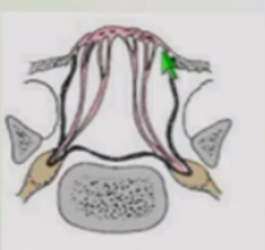
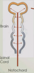
Neurulation |
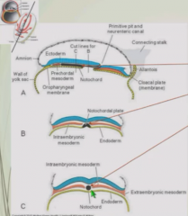
At primitive node, prenotochordal cells invaginate. Initially interdigitate with endoderm, then detach and proliferate to form solid rod ,the notochord. |
|
|
Notochord function |
Primary organizing center for nervous system but none contribute to nervous cells - become vertebrae. |
|
|
Sequence of events in forming neural tube |
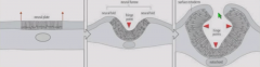
Notochord sends molecular signals that induce ectoderm to become neural plate. Floor plate (right above notochord) forms hinge point. As cells divide, neural plate pushed into two folds. As neural tube continues to grow, secondary hinge point forms half way up so neural folds meet in middle, forming roof plate. |
|
|
Closure of neural tube |
Begins at middle of back. Zippers anteriorly and posteriorly. At a given point of development, neural tube is at different stages down length. |
|
|
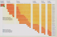
Neural tube closure points and neuropores |
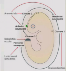
Primary closure point in center of back. Secondary closure point near head. Tertiary closure point in forebrain neuropore that closes off cranial region. Results in posterior neuropore, anterior neuropore, and hindbrain neuropore (between primary and secondary closure points) |
|
|
Anencephaly |
Errors in closure points 2 and 3 or anterior neuropore. |
|
|
Cranial rachiscisis |
Failure of closure of cranial neuropore. |
|
|
Spina bifida |
Normally, vertebral structures are closed off and muscle and skin are over them. |
|
|
Spinal Rachischisis |
Complete failure of skin and muscles to close causing neural tissue to be exposed to external world. Severe condition, possibly fatal. Treat by bringing skin from surrounding areas up and over |
|
|
Spina bifida occulta |
Neural arch is incomplete and spinal tissue is closer to skin surface as should be. Asymptomatic - still have thoracolumbar fascia and deep back muscles protecting the region. Prevalence - 10% of people |
|
|
Classes of spina bifida cystica |
Meningocele Meningomyelocele |
|
|
Meningocele |
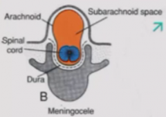
Skin closed over but CSF in subarachnoid space is bulging out, forming bulge in space
Treatment - remove cyst, close opening |
|
|
Meningomyelocele |
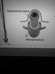
Skin closed over but CSF in subarachnoid space and neural tissue is bulging out. Treatment - remove cyst but restore neural tissue first |
|
|
Neural crest migration |
As neural tube closes, neural crest cells escape from the tube. Continue to migrate thorugh brain, gives rise to PNS elements, etc: Dorsal root ganglia, sympathetic chain ganglia, enteric nervous system, adrenal medulla, cardiac valves Cranial ganglia, meninges, craniofacial skeleton and connective tissue, and melanocytes |
|
|
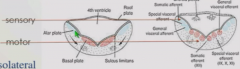
Dorsal/ventral axis patterning |
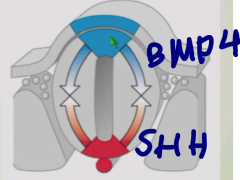
Notochord and floor plate secretes Sonic hedgehog - ventral fate Roof plate secretes BMP4 - dorsal fate Work in reciprocal gradient. |
|
|
What specific structures do BMP4 and SHH specify in spinal cord? |
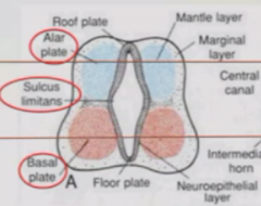
BMP4 specifies alar plate, which becomes sensory domain of spinal cord SHH specifies basal plate, which becomes motor regions of spinal cord |
|
|
Sulcus limitans |

Bump between basal and alar plate that separates motor and sensory regions of spinal cord. Continues into adulthood. |
|
|
Alar plate and basal plate derivatives in brainstem |
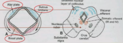
Alar plate specifies sensory nuclei of cranial nerves, which are dorsal to sulcus limitans Basal plate specifies motor nuclei of cranial nerves, which emerge from various parts of brainstem |
|
|
Alar plate and basal plate derivatives in fourth ventricle |
Fourth ventricle roof that closes ove rpushes alar plates to lateral side. Sensory region is dorsolateral Motor domainsi is ventromedial. |
|
|
Neural tube radial patterning |
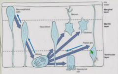
Neuroepithelial stemm cells shrink down to ventricular surface. One of daughter cells replenishes stem cells and other becomes neuron or neuroglia. Later on in development, radioglia take over stem cell role. |
|
|
Neural tube Rostral/Caudal patterning |
In caudal region, most of Hox genes are expressed. In cranial, fewer and fewer Hox genes are expressed. |
|
|
Hox genes |
Highly conserved, driven by retinoic acid. Gene order on chromosome matches anatomic order of expression. |
|
|
Neural tube medial and lateral patterning |
Close to SHH - medial Far from SHH - lateral Especially in brain |
|
|
Holoprosencephaly |
Error in cranial lateralization so lateralize frontal regions of brain. Leads to single cerebral hemistphere and craniofacial structure. Clinically nonsurvivable. |
|
|
Molecular mechanism of brain regionalizatoin |
Homeobox genes |
|
|
Three broad regions of brain |
Prosencephalon (forebrain) Mesencephalon (midbrain) Rhomencephalon (hindbrain) Also called primary brain vesicles or swellings |
|
|
Primary flexure |
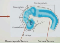
Forms cervical flexure is posterior and mesencephalic flexure which is more anterior. Bend tube almost 90 degrees so what was dorsal and posterior becomes dorsal and superior. |
|
|
Secondary flexure |
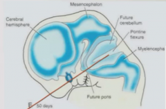
Forms in pointine-medullary region. Called pontine flexure. Tucks developing brainstem underneath cerebellum so end up with adult organization of cerebral cortex, midbrain, cerebellum, and hindbrain. |
|
|
Dividing forebrain (prosencephalon) |
Telencephalon (cerebral cortices and hippocampus) Diencephalon (thalamus and hypothalamic regions) |
|
|
Dividing midbrain (mesencephalon) |
Remains mesencephalon/midbrain forever. Functions as fiber tracts between anterior and posterior brain and various structures. |
|
|
Dividing hindbrain (rhombencephalon) |
Metencephalon - cerebellum and pons - important for fine motor control and carrying fiber tracts Myencephalon - medulla - key controller of autonomic structures Important for autonomic control. Becomes continuous with spinal cord. |
|
|
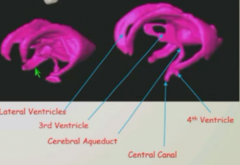
Formation of ventricles |
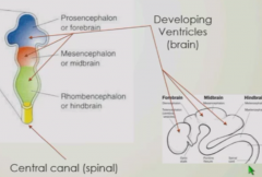
As forebrain, midbrain, and hindbrain grow, form ventricles. Ventricles are continuous with central canal of spinal cord which it uses to communicate, drainage, support material, and provides CSF fluid-filled bath for buoyancy, pressure, position. |
|
|
Formation of ventricle |

Forebrain -> Ventricle expands to follow left and right cerebral cortices to make lateral ventricles. Expands into telencephalon region. |
|
|
Interventricular foramen |

Allows lateral ventricles to communicate. |
|
|
Formation of third ventricle |

Space in diencephalon expands a little. |
|
|
Cerebral aqueduct/aqueduct of Sylvius |
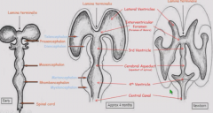
Goes through midbrain. Narrow ventricular tube that allows communicate between third and fourth ventricle. |
|
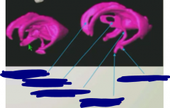
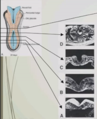
Label the structures |
|
|
|
Hydrocephalus |
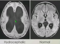
Buildup of CSF pressure in lateral and third ventricle. Expands brain. May be due to stenosis of cerebral aqueduct. |
|
|
Treatment of hydrocephalus |
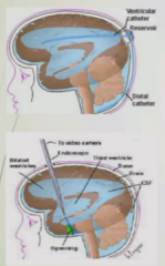
Cerebral shunt - access lateral ventricle through posterior cranium. Drain fluid to abdomen. Endoscopic third ventriculostomy - Drill a hole in third ventricle, allowing CSF flow to ventral subarachnoid space. |
|
|
Growth cone |
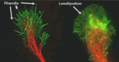
Leadin gprocess of axon with flat foot (lamellipodium) which has filopodia, finger-like extensions that sample the local environment. |
|
|
Growth cone movement |
Chemoattraction - can turn towards or away from molecules due to diffusible cues. Contact-mediated signaling - same happens if it contacts a "guide post cell" |
|
|
Nerve or tract formation. |
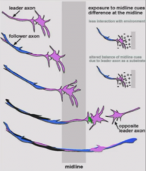
Pioneer axon samples environment and makes initial pathway Follower axons track along pioneer axons resulting in bundle of axons - nerve or tract. When cross midline, growth cone of one side meets growth cone of other side, forming tract. |
|
|
Peripheral nerve formation |
Pioneer axon goes out and followers track alongside. |
|
|
Central tract formation |
Use pioneer and follower mechanism. |
|
|
Corpus callosoum formation |
Formed by axons from one side crossing to other side. |
|
|
Agenesis of Corpus Callosum |
Incomplete formation of corpus callosum, resulting in absence of collosum. No communication between left and right brain = significant mental outcomes. Partial defect in corpus callosum is present in 5-6% of individuals. May be asymptomatic or lead to delayed milestones and cognitive/social/behavioral impairment. |
|
|
Neural migration in cerebral cortex |
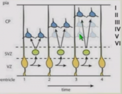
Cells divide in ventricular layer and migrate outwards. First set of cells stop in layer 6, next set stops in layer 5, and so on. Forms from inside-out or deep to superficial pattern. |
|
|
Source of inhibitory neurons in cerebral cortex |
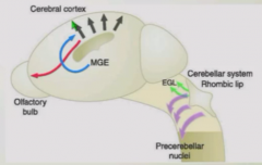
Cells from ganglionic eminence undergo long-range migration into cerebral cortex - provides all inhibitory neurons in cerebral cortex. Mistargeting of inhibitory neurons might be involved in autism, other neurocognitive diseases. |
|
|
Formation of gyri/sulci |
Massive increase in amount of neurons migrating to cerebral cortex causes brain to be thrown up into series of folds, gyri, and grooves, sulci. Dramatically increases cortical surface area. Deep sulci are called fissures. |
|
|
First fissures to form |
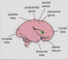
Lateral fissure - separates frontal from temporal Central sulcus - separates frontal from parietal regions |
|
|
Pachygria |
Do not make enough gyri. Results in cognitive impairment because not as much neural tissue available. |
|
|
Lissencephaly |
Absence of gyri - completely smooth bra1in. May have just central sulcus and lateral fissures. Results in huge reduction in neural processing power. Since founded in faulty migration of neurons or inadequate cell division, no way to correct problem. |