Reading...
![]()
Play button
![]()
Play button
![]()
Use LEFT and RIGHT arrow keys to navigate between flashcards;
Use UP and DOWN arrow keys to flip the card;
H to show hint;
A reads text to speech;
52 Cards in this Set
- Front
- Back
|
What type of of joint is the hip joint? What bones is it made up of?
|
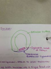
A ball in socket joint. It is made up of:
-the head of the femur -acetabulum of the os coxae |
|
|
What is the function of the acetabular labrum?
|
It increases the surface area of the joint, providing a cushion and helps keep the head of the femur from slipping out of place.
|
|
|
What are the movements of the hip joint?
|
-flexion-extension
-abduction-adduction -medial-lateral rotation -circumduction The range of motion is considerably less than the glenohumeral joint, but that makes it much more stable and less prone to injury. |
|
|
Describe the articular capsule of the hip joint.
|
-Strong and dense, attached to the brim of the acetabulum and the neck of the femur
-Posteriorly, the line of attachment runs across the middle of the neck of the femur -Direction of the fibers are spiral from os coxae to intertrochanteric line, and circular fibers around the femoral neck. |
|
|
What are the intrinsic ligaments of the hip joint?
|
-Iliofemoral
-Ischiofemora -Pubofemoral -Ligament of the head of the femur |
|
|
Describe the iliofemoral ligament of the hip joint.
|
From anterior inferior iliac spine and acetabular margin to the intertrochanteric line. Most important of hip ligaments. It is one of the strongest ligaments in the human body.
|
|
|
Describe the pubofemoral ligament of the hip joint.
|
From acetabular margin (pubis) to superior end of intertrochanteric line. Smallest ligament of the hip joint.
|
|
|
Describe the ischiofemoral ligament of the hip joint.
|
From ace tabular margin (ischim) to superior end of intertrochanteric line.
|
|
|
Describe the ligament of the head of the femor of the hip joint.
|
Transmits blood vessels to the head of the femur.
|
|
|
What bones make up the acetabulum?
|
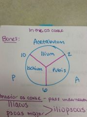
-Ilium
-Ischium -Pubis |
|
|
What muscles cross the hip joint?
|
Anteriorly - iliopsoas
Posteriorly - intrinsic muscles of the hip |
|
|
What makes up the iliopsoas?
|
Two muscles with the same function, the iliacus and psoas major. Both pass under the inguinal ligament and attach to the lesser trochanter.
|
|
|
Iliacus
|
O: iliac fossa
I: lesser trochanter Inv: femoral n. A: flexes hip and thigh |
|
|
Psoas major
|
O: transverse processes T12-L5
I: lesser trochanter of femur Inv: lumbar plexus A: flexes hip and thigh |
|
|
What are the special features of the hip joint?
|
It is a very stable joint, has appropriately shaped bones, strong ligaments, close intrinsic muscles, and powerful extrinsic muscles crossing the joint.
|
|
|
What are some potential problems of the hip joint?
|
-fractures of the neck of the femur
-congenital dislocation -traumatic dislocation -aseptic necrosis |
|
|
What type of joint is the knee joint? What bones make up the knee joint?
|
Hinge joint, but modified with three joints in one capsule (allowing for some rotation). Made up of:
-femur -tibia -patella |
|
|
What are the movements of the knee joint?
|
-flexion-extension
-small amount of rotation -gliding action to lock extension |
|
|
What tibial condyle has a larger surface area?
|
The medial tibial condyle.
|
|
|
What is the intercondylar area?
|
The are b/t the two condyles of the tibial. This is where the cruciate ligaments attach to the ligaments. It is not included in the knee joint because it is outside of the synovial cavity of the joint.
|
|
|
Describe the articular capsule.
|
-Loose and thin posteriorly, reinforced medially, laterally and anteriorly
-fibers mostly vertical from articular margins on both set of condyles (femoral and tibial) |
|
|
What are the ligaments of the knee joint?
|
-Anterior and posterior crucitate ligaments
-Lateral collateral ligament -Medial collateral ligament -oblique popliteal -arcuate popliteal -patellaer ligament |
|
|
Describe the lateral collateral ligament of the knee joint.
|
It is partly overlapped by the biceps femoris tendon. It is smaller and not as strong as the medial collateral ligament.
|
|
|
Describe the medial collateral ligament of the knee joint.
|
It is crossed by the sartorius, gracilis, and semitendinosus muscles. (the pes anserinus)
|
|
|
What is the pes anserinus?
|
Where the tendons of the sartorius, gracilis, and semitendinosus meet on the anteromedial surface of the tibia.
"Goose's feet" |
|
|
What muscles cross the knee joint?
|
-Anterior and posterior actors
-Medial and lateral stabilizers |
|
|
Where is the medial meniscus attached?
|
It is attached to the medial collateral ligament. This makes the medial meniscus more vulnerable to injury.
|
|
|
Where is the lateral mensicus attached?
|
It is NOT attached! The lateral meniscus is separated from the lateral collateral ligament by the tendon of the popliteus.
|
|
|
What are the special features of the knee joint?
|
Largest joint area, synovial capsule and bursae. Structurally unstable, but is functionally strong with the help of:
-cruciate ligaments -lateral and medial menisci cartilages -collateral ligaments (medial and lateral) |
|
|
Are the menisci attached to any bones?
|
NO! They are surround by synovial fluid (which lubricates preventing friction) above and below, they float on top of the tibia.
|
|
|
What are potential problems of the knee joints?
|
-athletic injuries
-infections -post-surgical complications |
|
|
Where does the synovial fluid come from?
|
Microscopic pores of the articular cartilages contain the fluid, and release it whenever there is movement.
|
|
|
Describe the shape of the menisci.
|
The outer periphery is must thicker than the inner surface, there is an incline as you move toward the middle.
|
|
|
How are the fibers of the menisci arranged?
|
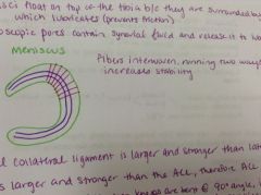
They are made of collagen (very strong). One set of fibers run towards the center, and another set of fibers that run around in a circle. These interwoven fibers allow maximum stability.
|
|
|
Describe the differences between the posterior cruciate ligament and anterior cruciate ligament.
|
The PCL is larger and stronger than the ACL, therefore the ACL is more commonly injured.
|
|
|
What is the function of the posterior cruciate ligament?
|
Supports the body weight when the knees are bent at 90 degree angle. It prevents the anterior displacement of the femur (keep it from sliding forward) and stabilizes and prevents posterior displacement of the tibia (sliding backward).
|
|
|
What is the function of the anterior cruciate ligament?
|
Prevents the femur from sliding backward (posterior displacement) and prevents the tibia from sliding forward (anterior displacement). Prevents hyperextension of the knee.
|
|
|
Where are the bursae of the knee joint?
|
-Above the knee, behind quadriceps tendon
-In front of the patella, provides cushion b/t skin and patella -Behind the patellar tendon, to protect it from the bone. Bursa is a fluid filled sac that protects a bone or tendon from some other structure. |
|
|
What is the blood supply to the menisci of the knee?
|
The meniscus gets more blood supply on the periphery than the inner edge. If there is damage to these vessels during meniscal tears of the outer periphery, damage to occurs d/t lack of blood supply.
|
|
|
When can we have a small amount of rotation of the knee joint?
|
-Locking knees in place
-Unlocking knees |
|
|
What happens when you lock your knees?
|
A small amount of medial rotation of the femur occurs (d/t difference in surface area of the femoral condyles and tibial condyles)
|
|
|
What happens when you unlock your knees?
|
The popliteus muscles performs a slight amount of lateral rotation of the femur.
|
|
|
What type of joint is the ankle joint? What bones make up the joint?
|
A.K.A. talocrural joint. A hinge joint. Made up of the tibia, fibula, and talus.
|
|
|
What kind of movements are possible in the ankle joint?
|
-dorsiflexion
-plantar flexion |
|
|
Describe the articular capsule of the ankle joint.
|
-Loose and thin in the anterior-posterior direction
-Vertical fibers from the edges of the articular surfaces on the inferior tibia and the malleoli |
|
|
What are the medial (deltoid) collateral ligaments of the ankle joint?
|
-tibio-navicular
-tibio-calcaneal -anterior tibio-talar -posterior tibio-talar Are much thicker and stronger, less prone to injury. |
|
|
What are the lateral collateral ligaments of the ankle joints?
|
-Calcaneo-fibular
-anterior talo-fibular -posterior talo-fibular (strongest) Made up of 3 bands, most injuries occur on this side. |
|
|
What muscle tendons cross the ankle joint?
|
Cross anterior ankle:
-Tibialis anterior -extensor digitorum longus -extensor hallucis longus Cross lateral ankle: -peroneus longus -peroneus brevis Cross medial ankle: -tibialis posterior -flexor digitorum longus -flexor hallucis longus |
|
|
What are the special features of the ankle joint?
|
-Stability depends on balance b/t gravity and muscle pull
-Western culture demands unstable ankles for women b/c of elevated shoe heels |
|
|
What are potential problems of the ankle joints?
|
-Torn ligaments
-avulsion of malleoli -response to movement in more distal joints |
|
|
What is the largest joint in the body?
|
The knee joint.
|
|
|
Describe the acetabulum.
|
Cup shaped, has two areas:
-the articular surface with the acetabular notch -the acetabular fossa which is filled with fat The articulating parts are covered with hyaline cartilage. The acetabular labrum (fibrocartilaginous) deepens the socket. |