![]()
![]()
![]()
Use LEFT and RIGHT arrow keys to navigate between flashcards;
Use UP and DOWN arrow keys to flip the card;
H to show hint;
A reads text to speech;
45 Cards in this Set
- Front
- Back
|
What is necrosis? |
- Cell death in a living organism. - May follow from acute cell swelling (if injury exceeds point of no return) - Pathologic process - Inflammation - Often involves multiple cells with loss of membrane integrity |
|
|
What is apoptosis? |
- Programmed cell death - Individual cells shrink and fragment before death - Can be physiological (ie embryogenesis, growth, deletion of redundant immune) - Or can be pathologic (ie viral infections, T cell cytotoxicity, hypoxia, drug induced, glands with blocked ducts, radiation damage) - No inflammation - Often involves single cells or small clusters with intact cell membranes and rapid removal by phagocytosis |
|
|
What is autolysis? |
- Process that involves self digestion or decomposition of tissues by their own enzymes - Occurs in all tissues after death - Tissues appear to fade with no inflammation
Different from putrefaction: putrefaction is tissue degradation that occurs after death and involves bacterial digestion of the tissue.
*Both processes are post-morterm change |
|
|
What processes cause the morphological appearance of necrosis? |
1. Denaturation of proteins 2. Enzymatic digestion of the cell (endogenous enzymes from lysosomes or enzymes from leukocytes) |
|
|
3 nuclear changes in necrosis: |
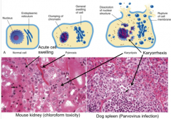
1. Pyknosis: shrunken dark nucleus with clumped chromatin
2. Karyorrhexis: Rupture of the nuclear membrane leads to release of chromatin.
3. Karyolysis: Nuclear remnants begin to dissolve by enzymatic action (appear pale)
4. Nucleus loss |
|
|
4 cytosolic changes in necrosis: |
1. Cytoplasm becomes homogenous and eosinophilic 2. Loss of RNA and digestion of proteins 3. Loss of adhesion to basement membranes 4. Loss of integrity |
|
|
What are the 5 forms of necrosis? |
1. Coagulative necrosis 2. Liquefactive necrosis 3. Caseous necrosis 4. Gangrene 5. Fat necrosis |
|
|
What defines coagulative necrosis? |
- Tissue necrosis with preservation of architecture and cellular outline. - Most common type of necrosis - Characteristic of hypoxic/ischemic death in call tissues except the brain - Protein denaturation predominates over enzymatic digestion.
|
|
|
What does coagulative necrosis look like microscopically? |
- Microscopically, cells are homogenous and opaque. Dead cells = eosinophilic shadow of the original cells. Some cells may be mineralized. - Cytoplasm more eosinophilic due to loss of ribosomes and eosin binding on altered proteins. - Cytoplasm may be hyalinized due to loss of glycogen particles. - Nuclei show pyknosis, karyorrhexis and karyolysis |
|
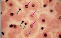
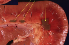
What kind of membrane damage do the green, blue and purple arrows represent? |
Green: pyknosis Blue: karyolysis Purple: karyorrhexis |
|
|
4 etiologies of coagulative necrosis? |
1. Infarction (occluded blood vessels) 2. Bacterial endotoxins 3. Chemical toxins 4. Viral replication |
|
|
What is the gross appearance of coagulative necrosis? |
- Pale tissues due to coagulation of cytoplasmic proteins and decreased blood flow. - Changed texture - Tissues may be swollen or shrunk - May be surrounded by congestion or a thin tan rim of inflammatory cells |
|
|
What is liquefactive necrosis? |
- Type of necrosis with complete destruction of the tissue - Occurs when enzymatic digestion of necrotic cells predominates over protein degeneration - Often seen in bacterial infections due to attraction of neutrophils (which contain proteolytic enzymes) - Seen in hypoxic damage of the brain/spinal cord resulting in rapid enzymatic digestion of the parenchyma = known as malacia |
|
|
What is malacia? |
A form of liquefactive necrosis that occurs in the brain or spinal cord. Rapid enzymatic digestion of the parenchyma with focal areas of dissolution. |
|
|
What is the gross appearance of liquefactive necrosis? |
- Tissue is transforming into a soft viscous to fluid mass - If process initiated by inflammation, then often contains pus (dead white blood cells) |
|
|
What is the microscopic appearance of liquefactive necrosis? |
- Architecture and cell details lost; margins not well defined - A few neutrophils or macrophages may be present - Clusters of amorphous necrotic material |
|
|
3 etiologies of liquefactive necrosis: |
1. Hypoxia in the CNS resulting in rapid enzymatic digestion of the parenchyma 2. Pus producing (pyogenic) bacteria 3. Abscesses filled with fluid pus
Note: Dehydration can transform liquefactive necrosis into something that looks like caseous necrosis. |
|
|
What is caseous necrosis? |
- Variant of coagulative necrosis with loss of tissue architecture and cellular details - Typical of some bacterial diseases such as tuberculosis, caseous lymphadenitis, tularemia - Seen during migration of parasites with secondary bacterial infection of parasitic tracts - Common in birds and reptiles due to presence of heterophils instead of neutrophils (such as from bacterial infections aspergillosis and TB) |
|
|
What is the gross appearance of caseous necrosis? |
- Grey/white, dry (inspissated), friable, pasty areas of necrotic material - Inspissation: process of thickening a liquid by dehydration or evaporation - The friable material results from the nondegradable lipids of bacterial cell walls and dead leukocytes from the host |
|
|
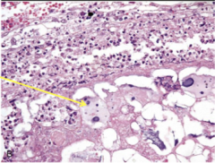
What is the microscopic appearance of caseous necrosis? |
- Tissue architecture and cell details lost (do not retain cellular outline) - Necrotic areas consist of dead cells persisting as amorphous, coarse, granular debris - But do not undergo complete dissolution as in liquefactive necrosis - May see dystrophic mineralization: deposition of calcium as a blue granular material in the centre of a necrotic focus - Caseous necrosis often seen in the center of granulomas (type of chronic inflammation seen in TB)
|
|
|
A classic sheep example of caseous necrosis? |
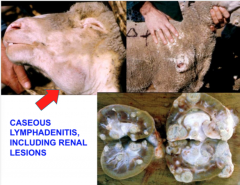
Caseous lymphadenitis |
|
|
What defines gangrenous necrosis? |
Gangrene is a type of necrosis affecting extremities, such as limbs, digits or tips of ears. Usually a result of ischemia. |
|
|
What are the 3 types of gangrene? |
1. Dry gangrene 2. Moist/wet gangrene 3. Gas gangrene |
|
|
What is dry gangrene? |
- Form of coagulation necrosis followed by dehydration and mummification - Often at the distal portion of extremities - Caused by agents that cause ischemia (ie extreme cold, toxins [ie: salmonella endotoxin, ergot, fescue]) - Gross appearance: tissue is dry/shrivelled/brown/gray/black |
|
|
What is moist/wet gangrene? |
- Predominantly coagulative necrosis modified by the liquefactive action of saprophytic bacteria causing putrefaction - Seen on extremities, lung and udder - Caused by ischemia followed by contamination of saprophytic bacteria or aspiration of ingesta/medication (aspiration pneumonia) - Tissue appears soft, moist and reddish brown to black with a putrid odour - Tissue is crepitant: may feel gas bubbles - Affected tissue may slough off |
|
|
What is gas gangrene? |
- Necrosis characterized by production of gas bubbles in the necrotic tissue by invading bacteria. - Caused by anaerobic bacteria such as Clostridium chauvoei (black leg), Clostridium perfringens, Clostridium septicum. - Affected tissue is dark-red/black, contains many gas bubbles |
|
|
What is fat necrosis? |
- Type of necrosis affecting body fat stores - Fat that is free in connective tissue elicits an inflammatory response and phagocytosis - Saponification of fat: fatty acids combine with calcium, precipitating insoluble calcium soaps |
|
|
3 causes of fat necrosis |
1. Trauma: rupture of adipocytes with release of lipase that splits the neutral fat in adipose cells. 2. Enzymatic necrosis of fat, such as in cases of pancreatitis with release of pancreatic lipases from adjacent damaged pancreatic duct system 3. Idiopathic |
|
|
What is the gross appearance of fat necrosis? |
- Firm/hard - White and chalky - Cut surfaces may be gritty
|
|
|
What is the microscopic appearance of fat necrosis? |
- Areas of coagulative necrosis - Cholesterol crystals - Basophilic calcium deposits - Surrounded by inflammatory cells (macrophages, giant cells) |
|
|
Outline of the mechanism of apoptosis |
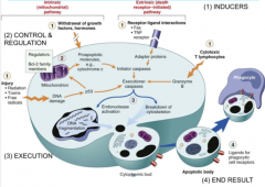
1. Induction 2. Control and regulation 3. Execution 4. End: apoptotic body phagocytosed |
|
|
What are the 2 pathways for inducing apoptosis? |
1. Intrinsic pathway/mitochondrial pathway: - release of proapoptotic molecules (Cyt C and AIF) from mitochondria - initiates caspase activation cascade
2. Extrinsic pathway/death-receptor-initiated pathway: - stimulus triggers a self-programmed, genetically determined, energy-dependent sequence of events - initiated by cell signalling involving death receptors (TNFR1, Fas) |
|
|
What are the regulatory molecules involved with apoptosis? |
- Anti-apoptotic BcI-2 and BcI-X (found in mitochondrial membrane) - When their levels decrease, membrane leaks = release cytochrome C and AIF (initiates caspase activation cascade) |
|
|
What happens in the execution phase of apoptosis? |
- Activated caspase enzymes act as protease and digest the cytoskeleton - Some enzymes also target enzymes involved in DNA transcription, replication and repair - ie caspase 3 is a DNAase |
|
|
What is the histological appearance of apoptosis? |
- Single cells shrunk - Cytoplasm condensation - Chromatin condensation - Nuclear fragmentation - Cytoplasmic buds formed that are phagocytosed by neighboring cells as apoptotic bodies - No inflammatory response |
|

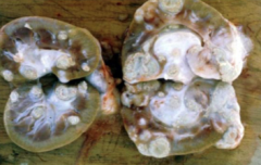
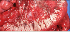
What type of necrosis? |
Coagulative necrosis on a bull testis and a kidney. |
|
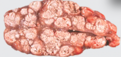
What type of necrosis? |
Renal infarct resulting in coagulative necrosis. Will be wedge shaped. |
|
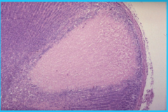
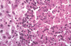
What type of necrosis? |
Microscopic view of coagulation necrosis of an adrenal gland. |
|
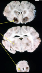
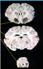
What is this? |
Malacia, a form of liquefactive necrosis. |
|
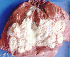
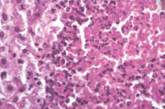
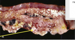
What type of necrosis is this? |
Caseous necrosis of the liver |
|
What is this? |
Renal lesions as a result of caseous lymphadenitis |
|
What caused this caseous necrosis in a bovine lymph node? |
Tuberculosis |
|
What kind of necrosis? |
Caseous necrosis: loss of tissue architecture and cell detail |
|
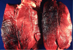
This is the leg of a steer. What type of necrosis? What caused it? |
- Gas gangrene - Black leg - Clostridial myositis |
|
What type of necrosis? |
Traumatic fat necrosis in a dog |