Reading...
![]()
Play button
![]()
Play button
![]()
Use LEFT and RIGHT arrow keys to navigate between flashcards;
Use UP and DOWN arrow keys to flip the card;
H to show hint;
A reads text to speech;
32 Cards in this Set
- Front
- Back
|
Predominant phagocytes
|
Neutrophils & Macrophages
|
|
|
Neutrophils
|
- Neutrophils = 60% of the circulating leukocytes in peripheral blood.
- Neutrophils mature from precursor cells in the bone marrow - 1/2 life ~ 8 hours |
|
|
Macrophages
|
- Derived from monocytes
- 4% of circulating leukocytes - When recruited into tissues - macrophages. - More effective at phagocytosis than monocytes - Have a much longer half-life than neutrophils - Following their activation can serve as antigen presenting cells for CD4+ T cells |
|
|
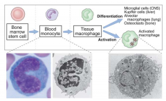
Macrophages in different tissues
|
- Blood - Monocytes
- Bone marrow - Monoblasts - CNS - Microglial cells - Liver - Kupffer - Synovium - Synoviocytes - Lungs - Alveolar macrophages |
|
|
Recognition of Pathogens
|
- 2 Methods: Direct or Opsonin-mediated/Indirect
- Direct = without an intermediary protein - Indirect = intermediary protein bound to an antigen, with a receptor on the phagocyte |
|
|
Direct Recognition
|
- Uses pattern recognition receptors, (PRRs)
- PRRs = membranous or cytosolic receptors - Ligands = PAMPS (pattern activation molecular patterns) |
|
|
NLRs
|
- Group of cytosolic PRRs “nucleotide-binding oligomerization domain–like receptors"
- Nod1 and Nod2 proteins - Mutations in Nod2 associated with Crohn’s |
|
|
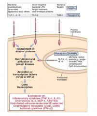
TLRs
|
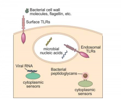
- Group of membranous receptors that exist both on cytosolic vesicles and external cell membranes are the toll-like receptors (TLRs).
- TLRs are so named for their sequence similarity to the Drosphilia protein, Toll - TLRs are expressed on a various cell types - e.g. signaling via TLR2 or TLR4 leads to the activation of NF  B - then activates pro-IL-1 an inflammatory cytokine secreted by activated macrophages |
|
|
Opsonin-mediated Phagocytosis (Indirect Recognition)
|
- Opsonins are the products of:
(i) complement activation (e.g., C3b) (ii) B cell activation (e.g.,IgG ) (iii) cytokine mediated activation of hepatocytes (e.g., C-reactive protein, CRP). - Interaction of any of these pathogen-bound opsonins triggers the process of phagocytosis. |
|
|
Receptor-Opsonin pairing
|
1. Fcγ :FcγR
2. CRP: CRP-BS 3. C3b:CR1 |
|
|
Phagosome
|
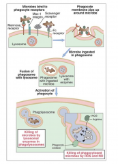
- Vesicles that contain the engulfed pathogen
- Phagocytic vacuole serves as the “battlefield” - Weapons include: (i) lysosomal enzymes (ii) reactive oxygen intermediates (iii) reactive nitrogen intermediates |
|
|
Lysosomes
|
- Fuse with the phagosome to form a fusion product - lysosomal granules are discharged
- Lysosomal granules contain many enzymes (including lactoferrin, lysozyme, and defensins) that are cytostatic/cytotoxic to microorganisms |
|
|
Lactoferrin
|
Binds iron, thereby removing an essential ingredient for microbial growth
|
|
|
Lysozyme
|
Destroys muramic acid in bacterial cell walls
|
|
|
Defensins
|
Permeabilize bacterial and fungal membranes
|
|
|
Myeloperoxidase
|
Generates hypochlorite, a potent antimicrobial agent that mediates its function by halogenating bacterial cell walls
|
|
|
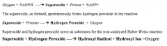
NADPH oxidase
|
Phagocytosis is accompanied by a respiratory burst via NADPH oxidase that uses oxygen, in the presence of cytosolic NADPH
|
|
|
Reactions of NADPH Oxidase
|
|
|
|
Nitric oxide
|
- Lipid and water soluble gas that is cytotoxic/cytostatic to invading microorganisms.
- Many parasites and other intracellular organisms including viruses, intracellular bacteria, parasites, and fungi are susceptible to NO - Even MORE powerful when NO reacts with reactive oxygen intermediates and generates reactive nitrogen intermediates (RNIs) |
|
|
Synthesis of NO
|
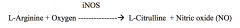
L-Arginine to L-Citrulline and NO in the presence of oxygen.
|
|
|
Roles of Cytokines in Regulation of NO Synthase
|
- Activation occurs by 2 signals: TNF and IFNγ
- Down-regulation occurs: IL-10, IL-1 and TFG β - TFG β = the MOST effective cytokine |
|
|
Activation of Macrophages
|
- MCP-1 (CCL2) = chemoattractant for monocytes and macrophages
- IL-8 (CXCL8) = chemoattractant for neutrophils |
|
|
Cytokines secreted by activates macrophages
|
IL-1, IL-6, IL-12, and tumor necrosis factor (TNF)
|
|
|
TLR Family
|
- PRR subtype is the TLR family ( TLR = Toll-like receptor)
- Extracellular with transmembrane domains |
|
|
NLR Family
|
- Counterpart to the TLR family
- Largest of these families is the NALP/NLRP family of proteins that play a role in inflammation - NALP3 protein, which has a leucine rich (LRR) domain that serves as the recognition portion for various ligands |
|
|
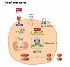
NALP3
|
- NALP3 protein is dormant until it binds a ligand (directly or indirectly) to initiate assembly of the NALP3 inflammasome
- Important for processing of the pro-Caspase-1 zymogen to its active form (Caspase-1/IL-1 converting enzyme) induces proteolytic cleavage of prointerleukin-1b and prointerleukin-18 (pro-IL-18) to their active forms |
|
|
The Inflamasome
|
|
|
|
Macrophage Receptors
|
PRRs, FcγR, CRP-R, CR1
|
|
|
CD200Rs
|
- Expressed primarily on cells of the myeloid lineage and T-cells
- CD4+ T cells express higher amounts than CD8+ T cells - Memory cells express higher amounts of CD200R than naïve or effector cells |
|
|
Eosinophils
|
- Bone marrow derived cells that exist both in the circulation and in tissues
- Only a small percentage of eosinophils released from the bone marrow remain in circulation - 1/2 life ~ 8-10 hours. - Major role: parasites - helminths - FcεR that bind to IgE antibodies themselves bound to epitopes on helminths - Major basic protein (MBP), and eosinophil cationic protein (ECP) = both toxic to helminths other parasites |
|
|
NK Cells
|
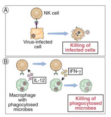
- Arise from bone marrow precursors (lymphoid progenitor cell), and are found predominantly in the blood, spleen, and peritoneal exudate
- NK cells kill infected cells (e.g., viral infections) and some tumors - Express both NK inhibitory receptors and NK activating receptors that interact with specific self-Class I MHC, MHC class I-like molecules and molecules unrelated to MHC - IL-12 and IFN-y |
|
|
Dendritic Cells
|
- Derived from bone marrow progenitor cells
- Most efficient of all antigen-presenting cells, particularly in a primary response to antigen. - Present in all tissues; they express receptors for chemokines that direct them to the appropriate secondary lymphoid tissues following their encounter with antigen - Under the influence of GM-CSF, bone marrow derived myeloid precursors mobilize to the circulation where they differentiate into immature dendritic cells - undergo further differentiation and maturation when they endocyose antigen |