Reading...
![]()
Play button
![]()
Play button
![]()
Use LEFT and RIGHT arrow keys to navigate between flashcards;
Use UP and DOWN arrow keys to flip the card;
H to show hint;
A reads text to speech;
15 Cards in this Set
- Front
- Back
|
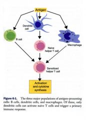
What are antigen presenting cells?
|
Cells that can ingest, process, and present antigen to antigen-sensitive cells in association with MHC class I and class II molecules.
|
|
|
What are MHC molecules?
|
Specialized receptor glycoproteins coded for by genes located in a gene cluster called MHC
Class I MHC present on the surface of most nucleated cells. Class II MHC Present on the surface of the Professional APC |
|
|
What are professional APCs vs nonprofessional APCs?
|
Professional APC (Dendritic cells, Macrophages, and B cells). Pigs have γδ-T cell.
Nonprofessional APC (neutro, eosino, T, NK, endothelial, fibroblasts etc…) |
|
|
What's the difference between the naive helper T-cells and the sensitized helper T-cells?
|
Professional APC (Dendritic cells, Macrophages, and B cells). Pigs have γδ-T cell.
Nonprofessional APC (neutro, eosino, T, NK, endothelial, fibroblasts etc…) |
|
|
What is the function of dendritic cells? Are they more efficient or less efficient than other APCs?
What are their origin, distribution and structure? |
Present antigen to T cells. Only APC that can activate naïve T cells (never previously encountered an antigen)
100 times more efficient than other APCs Origin: Bone marrow Stem cells Myeloid DCs and Lymphoid DC Distribution: all organs except brain, eye and testes Structure: small body with many cytoplasmic processes (“dendrites”=branches of tree) |
|
|
What are the types of dendritic cells, their origin and their functions?
|
Myeloid origin
DC1 Express TLR4, IL-12 Interstitial DC Langerhans DC: Skin immune response and contains Birbeck granules Function: T helper 1 become CMI against intracellular invader Tissue DC Delayed hypersensitivity and allergic contact dermatitis (Poison ivy, I/d antigens like mosq. saliva) – Type IV sensitivity Lymphoid origin DC2: Express TLR9 and IL-1, IL-4, IL-6 Function: T helper 2 is the humoral antibody production against extracellular invader |
|
|
What do immature dendritic cells do?
|
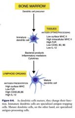
Immature dendritic cells can trap the antigen, but they can’t present it. So what happens to them? They just digest it and keep maturing? Yes – and Pastey doesn’t know why – it doesn’t make much sense why they’d do this.
|
|
|
Which interleukin is a common ingredient in vaccines? Why?
|
IL-12 is a common ingredient in vaccines to stimulate Th1
|
|
|
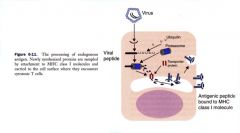
Which MHC mediates the processing of exogenous antigens? What happens if this MHC isn't linked the antigen?
|
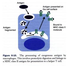
MHCII
If antigen is presented to T cells without linked to MHC II, then T cells die or tolerance results |
|
|
Can an APC present only one or several epitopes?
|
They can present different epitopes simultaneously.
|
|
|
What are the steps in the processing of exogenous antigen?
|
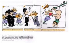
Frogs are MCHII. Charlie Chaplin is invariant chain – these are all complexed in a trimer. Invariant chain is cleaved leaving CLIP in the binding site, until the bacterial enzyme (the big brute) replaces the CLIP with an antigenic peptide for presentation.
1. Antigen Phagocytosed and taken into the phagosome 2. Phagosome fuse with lysosome –phagolysosome 3. Proteases break down antigen into peptide fragments of varying lengths 4. MHC II+peptide invariant chain (Ii) 5. Ii proteolytically cleaved leaving CLIP (class II-associated Ii Peptide) with MHC II 6. CLIP occupies antigen-binding site on the MHC molecule 7. Antigen peptide is exchanged for CLIP 8. Antigen+MHC II presented to T helper on the surface of APC. (1-3 are happening at the same time as 4-6) |
|
|
What is the cellular mechanism of destroying non-functional proteins?
|
the binding of ubiquitin to mark it for destruction. Ubiquitin is recycled. If the protein is foreign, an antigenic peptide will be presented on the surface by MHCI. If it’s a body protein, it will be recycled.
|
|
|
What are the steps in the processing of endogenous antigens?
|
Abnormal proteins linked to ubiquitin (marked for destruction)
Recognized by large enzyme complex called a Proteasome (tubular structure like a cylinder) Ubiquitin is recycled, peptide enters proteasome where it is broken in to 8-15 aa peptides Some peptides bind to TAP (transporter for antigen processing) and carried to ER Aminopeptidase shortens to 9 aa and linked to MHC I CTL recognize MHC I + 9 aa peptide |
|
|
What are the two types of langerhans cell histiocytosis?
What kinds of dogs are they typically found in? |
Cutaneous form and systemic form
Cutaneous form Any dog 3-9 yrs Multiple nodules 30% regress after treatment Systemic form Large breed dogs 4-7 yrs Multiple organs 10% regress after treatment |
|
|
What type of cell growth is a butt cell a cytological characteristic of?
|
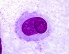
Histiocytoma cell with a nuclear cleft
and typical “butt” cell (nuclear clefts) morphology. Histiocytoma cells have an eccentric, oval to reniform nucleus with cytoplasm that lacks vacuoles and granules. A binucleated cell and mitotic figures are present |