![]()
![]()
![]()
Use LEFT and RIGHT arrow keys to navigate between flashcards;
Use UP and DOWN arrow keys to flip the card;
H to show hint;
A reads text to speech;
41 Cards in this Set
- Front
- Back
|
Immunogenetics |
Study of genetic basis of immunity |
|
|
Immune Response and Immunity |
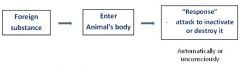
Such response = immune response
|
|
|
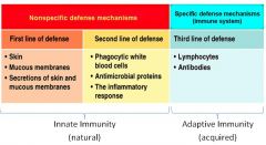
Body defense mechanisms |
|
|
|
Innate immunity |
Anatomical barriers - Skin & mucous membranes, Acidity of stomach |
|
|
Innate immunity - after enter the body |
Phagocytose - kill, and digest whole organisms.
|
|
|
Phagocytosis diagram |
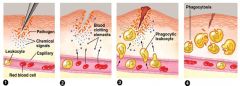
1. Pathogens entering the wound. |
|
|
Phagocytic Cells |
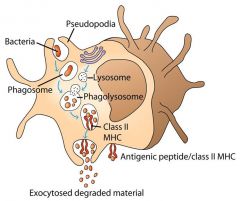
Monocytes, neutrophils (white blood cells) and tissue macrophages (monocytes differentiate into macrophages) - Internalization of whole micoorganism and formation of phagosomes. |
|
|
Phagocytosis process |
|
|
|
difference between pathogens and antigens |
A pathogen is any organism capable of producing disease. An antigen refers to the protein on the surface of a cell (bacterium, fungus or virus) that our bodies recognize as a foreign substance and triggers the immune system into producing antibodies specific to that antigen. This means that if in the future the same antigen is again introduced, our immune system will recognize, remember and produce the right antibodies to "deal" with the intruders.
Pathogens have antigens on their surface. |
|
|
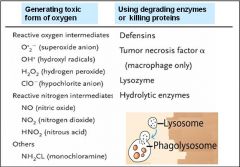
How do lysosomes destroy microbes? |
|
|
|
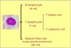
Lymphocytes (specialized white blood cells)
|
originate in the bone marrow stem cells - migrate to Thymus and become T lymphocytes (T cells) |
|
|
Lymphocyte classes |
|
|
|
Lymphocytes show specificity |
T and B lymphocytes recognize and respond to particular microbes and foreign molecules - specificity
|
|
|
immunogen |
A foreign molecule that elicits a specific response by lymphocytes is called an immunogen |
|
|
antigen |
A foreign molecule that binds specifically to an antibody, a B cell or a T cell receptor.
|
|
|
Cell mediated inmmunity |
Development of various T cells after stimulation of antigen - T cells giving rise to lymphokines, soluble factors, enhance destructive action of other white cells e.g. macrophages |
|
|
Humoral Immunity |
B-lymphocytes produce Immunoglobulin (=antibodies). |
|
|
foreign substance initiates |
|
|
|
when the animal is attacked by the same antigen |
- antibody binds with the antigen result :
The protection arising from the production of antibodies is called humoral immunity. |
|
|
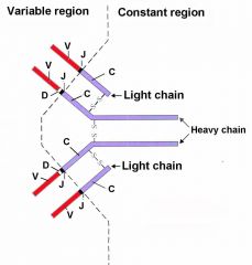
Antibodies |
attach to surface of B-cells or in blood plasma called immunoglobulins (IG) A basic antibody molecule consists of Each chain consists of a variable (V) region and a constant (C) region Variable region differs from one antibody to the next. |
|
|
Antibodies diagram |
|
|
|
Antibodies properties |
Being polypeptides – antibodies are the products of genes. L- and H- chains – produced from different clusters of genes Each different antibody – produced by a different clone of B lymphocytes
Binding of antigens to antibodies triggers immune respone |
|
|
Blood transfusion |
animals do not normally carry antibodies to red-blood-cell antigens (Red-cell antigens), unless they have been specifically challenged with appropriate foreign red blood cells. |
|
|
Blood transfusion cross-match |
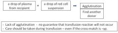
If untyped donors must be used – a simple cross-match should be conducted |
|
|
Transplant rejection |
Organ and tissue transplants and skin grafts
|
|
|
haplotype |
A haplotype is a collection of specific alleles (particular DNA sequences) in a cluster of tightly-linked genes on a chromosome that are likely to be inherited together |
|
|
What is MHC |
group of genes that code for proteins found on the surfaces of cells that help the immune system recognize foreign substances
Both classes – surface antigens |
|
|
What does HTC do? |
Ok, so when there is a pathogen (foreign particle) in your body, the first cells to act are the phagocytic cells (macrophages, neutrpphils...). They engulf the pathogen and partially digest it, then present part of the pathogen on the surface of their cell with the MHCII complex. Another cell, the helper T cell, can now recognize the presentation to activate the cytotoxic T cells. The cytotoxic T cells then destroy any other remaining pathogens. Another way for a pathogen to be recognized is by the B cells, which have antibodies on their surface to recognize the pathogen, then they take the pathogen in and digest it, and present a portion of the antigen on their MHCII complex for the helper T cells to recognize. The helper T cell can then stimulate the B cell to make an increased amount of antibodies to kill the remaining pathogen. All nucleated cells in the body have a MHCI complex on their surface. This is how the body can recognize 'self' from 'nonself' cells. Another cell called the natural killer cell will destroy any cell that has a missing or abnormal MHCI complex |
|
|
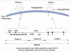
Major Histocompatibility Complex |
Many loci whose gene products play a role in compatibility between tissues (histocompatibility). One group of tightly-linked loci play much more important role than others MHC in mammals |
|
|
MHC Diagram |
|
|
|
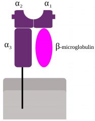
Class I region
MHC Class I |
Contains many genes, each of which codes for a polypeptide which combines with another polypeptide β2-microglobulin (Transport to cell surface) Class I histoglobulin
expressed on almost all nucleated cells Tells immune cells that its not a foreign body (self cells) |
|
|
MHC Class I diagram |
|
|
|
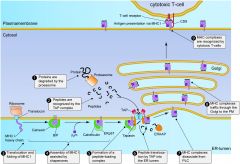
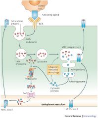
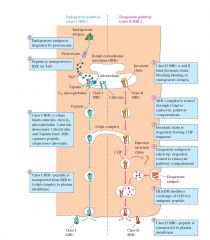
MHC Class I Pathway |
|
|
|
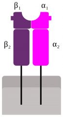
Class II region
MHC Class II |
Contains genes that encode 2 types of polypeptide (α and β chains) which unite to form a class II histoglobulin limited distribution – mainly on lymphocytes, macrophages, and dendritic cells
|
|
|
MHC Class II diagram |
|
|
|
MHC Class I Pathway |
|
|
|
class I and class II MHC Pathways |
|
|
|
Class III region
|
contains a mixture of genes having a wide range of functions only some of which involve in immune response |
|
|
Determining phenotype and genotype at the MHC |
To determine the role of MHC in immunity to disease |
|
|
Determining phenotype and genotype at MHC Methods |
1. Tissue typing 2. Molecular technology |
|
|
MHC polymorphism
|
Many alleles at most loci The chance of choosing any two individuals having the same set of MHC alleles – exceedingly small |