![]()
![]()
![]()
Use LEFT and RIGHT arrow keys to navigate between flashcards;
Use UP and DOWN arrow keys to flip the card;
H to show hint;
A reads text to speech;
100 Cards in this Set
- Front
- Back
- 3rd side (hint)
|
Anterior longitudinal ligament |
Attached superiorly to base of skull - extends anterior to vertebral bodies - anterior surface of sacrum Commonly damaged in whiplash |
|
|
|
Posterior longitudinal ligament |
Attached superiority to base of skull - extends along posterior surface of vertebral bodies - posterior surface of sacrum Part that connects C2 to base of skull is called the tectorial membrane |
|
|
|
Tectorial membrane |
Part of the posterior longitudinal ligament that connects C2 to the base of the skull |
|
|
|
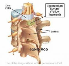
Ligamentum flava |
Between laminae of adjacent vertebrae running superiorly from posterior to anterior Forms the posterior surface of the vertebral canal |
|
|
|
Ligamentum nuchae |
Triangular sheet that forms the upper part of the supraspinous ligament from C7 to skull Broad lateral surface provides muscle attachment sites |
|
|
|
Supraspinous ligament |
Connects tips of the spinous processes from C7 to sacrum (Above c7 gives way to ligamentum nuchae) |
|
|
|
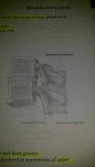
Interspinous ligaments |
Between adjacent vertebral spinous processes Blends with supraspinous ligament posteriorly and ligamentum flava anteriorly |
Definition Anterior and posterior attachments |
|
|
3 groups of back muscles |
Superficial - movement of upper limbs Intermediate - muscles that attach to ribs Deep - muscles innervated by the posterior spinal rami and which are directly related to movement of vertebral column and head |
Name + function |
|
|
Zygapophysial joint |
Between articular processes on adjacent vertebrae Cervical - slope inferiorly a-p to facilitate flexion and extension Thoracic - orientated vertically to limit flexion and extension and facilitate rotation Lumbar - curved and adjecent processes are interlocked to limit movement |
Definition 3 types and why |
|
|
Superficial back muscles |
Trapezius Latissimus dorsi Levator scapulae Rhomboid major Rhomboid minor |
5 |
|
|
Trapezius |
Cervical and thoracic spine Elevate + roate scapula during abduction of humerus |
Origin Function |
|
|
Latissimus dorsi |
T7 - sacrum and iliac crest Extends, adducts and medially rotates humerus |
Origin Function |
|
|
Levator scapulae |
Transverse processes of C1 - C4 Elevate scapula |
Origin Function |
|
|
Rhomboid major |
Spinous processes of T2 - T5 Adducts + elevates scapula |
Origin Functions |
|
|
Rhomboid minor |
Cervical spine Adducts + elevates scapula |
Origin Function |
|
|
Intermediate muscles of the back |
Serratus posterior superior Serratus posterior inferior |
|
|
|
Serratus Posterior superior |
C1 - T3 Elevates ribs 2-5 |
Origin Functions |
|
|
Serratus Posterior inferior |
T11 - L3 Depresses ribs 9 - 12 Rotation and extension of trunk, forced expiration, prevents lower limbs from elevating when the diaphragm contracts |
|
|
|
Deep muscles |
Spinotransversales Erector spinae Transversospinales Interspinales Intertransversarii
|
5 |
|
|
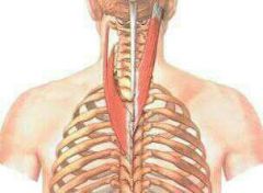
Spinotransversales |
Extensors and rotators of the head and neck Runs from the T6 spinous process to ligamentum nuchae, superiorly and laterally |
Function Origin |
|
|
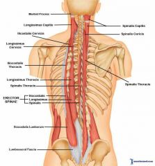
Erector spinae |
Extensor and rotator of vertebral column 3 muscles on each side Posterolateral to vertebral column between spinous processes medially and angle of the ribs laterally Largest group of deep back muscles |
Function Origin |
|
|
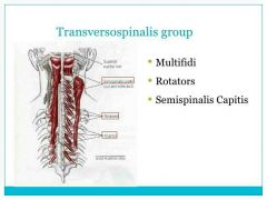
Traversospinales |
Extensor and rotator of vertebral column Runs obliquely upwards and medially from transverse process to spinous process |
|
|
|
Interspinales |
Short segmental muscles that stabilise the vertebral column Between adjacent spinous processes |
Functions Origin |
|
|
Intertransversarii |
Short segmental muscles that stabalise the vertebral column Pass between transverse processes |
Functions Origin |
|
|
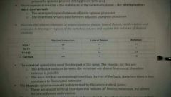
Region of vertebral column with ++ lateral flexion |

|
|
|
|
Region of vertebral column with + lateral flexion |

|
|
|
|
Region of vertebral column with ++ rotation |
|
|
|
|
Region of vertebral column with + rotation |

|
|
|
|
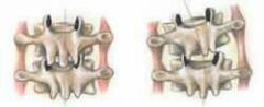
Defining features of the atlas |
Lacks a vertebral body Ring shaped Composed of 2 lateral masses interconnected by an anterior + posterior arch Posterior surface of anterior arch has an articular facet for the dens Dens is held in position by transverse ligaments of atlas Lateral masses articulate superiorly with an occipital condyle and inferiorly with superior articular surfaces of the axis |
Vertebral body Shape Components Articular facet of the anterior arch Superior and inferior articulations |
|
|
Atlanto-occipital joint |
Between atlas and skull Lies behind the mouth Allows for nodding |
Definition Location Function |
|
|
Defining features of the Axis |
Vertebral body extends upwards to form the dens (tooth) process Two superolateral surfaces of the dens have articular impressions that: -serve as attachment sites for alar ligaments - medial occipital condyles -check excessive rotation of the head |
Vertebral body Dens surfaces |
|
|
Surface anatomy of C7 |
Only cervical vertebrae with a prominent spinous process |
|
|
|
Surface anatomy of T3 |
At the level of the medial end of the scapular spine |
|
|
|
Surface anatomy of T7 |
At the level of the inferior angle of the scapula |
|
|
|
Surface anatomy of L2 |
Level of the lowest rib |
|
|
|
Surface anatomy of L2 |
Level of the lowest rib |
|
|
|
Surface anatomy of L4 |
Level of the Iliac crest |
|
|
|
Number of spinal nerves |
31 C - 8 T - 12 L - 5 S - 5 C - 1 |
Number C T L S C |
|
|
Meninges of the spinal cord |
Dura Arachnoid Pia Continuous with the meninges of the brain |
|
|
|
Spinal dura mater |
Outermost meningeal layer Separate from bone therefore forming the epi/extra-dural space which contains connective tissue, fat and internal vertebral venous plexus Superiorly continuous with cranial dura matter at the foramen magnum Inferiorly the dural sac narrows at the level of lower border of S2 and forms an investing sheath for the final part of the Filum terminale that attaches to the posterior surface of vertebral bodies of the coccyx
|
Definition Spaces + what they contain Superior attachment Inferior attachment |
|
|
Spinal arachnoid mater |
Deep but not adherant to the duration mater Seperate from pia material via subarachnoid space Ends at S2
|
Definition Spaces Termination point |
|
|
Arachnoid trabecular |
Interconnect the arachnoid and pia mater Suspend blood vessels within the subarachnoid space |
Functions |
|
|
Subarachnoid space |
Between arachnoid and pia mater Contains CSF Ends at S2 Largest in the region inferior to the end of the spinal cord where it surrounds the cauda equina - CSF sample
|
Definition What does it contain Termination point |
|
|
Spinal pia mater |
Vascular membrane that firmly adheres to the surface of the spinal cord Forms denticulate ligaments |
Definition Protrusions formed |
|
|
Denticulate ligament |
Medially attach to spinal cord Laterally forms a series of triangular extensions that anchor through the arachnoid mater to the dura mater Generally occur between exit points of adjacent posterior and anterior rootlets |
Medial attachments Lateral attachments Positions along spinal cord |
|
|
Difference between spinal and cranial meninges |
The is no cranial extra/epi-dural space since the dura is firmly attached to the skull |
|
|
|
Reasons for carrying out a lumbar puncture |
Sample CSF Spinal anaesthesia |
|
|
|
Describe a lumbar puncture |
Needle passed through skin just lateral to midline at L3/L4 adult or L5/L6 child Passes through ligamentum flavum First give/pop In the epidural space Another give/pop Pierces dura and arachnoid together In the subarachnoid space |
Procedure and observations |
|
|
Dangers of a lumbar puncture without ruling out raise ICP |
Sudden release of CSF can cause brainstem herniation through the foramen magnum - potentially fatal |
|
|
|
Signs of cervical spine injury |
Hypotension Large erection (Custer's last stand) Flaccid paralysis Large bladder with inability to micturate |
4 |
|
|
On scene management of C spine injury |
Assume an unstable fracture Assume neck pain if patient cannot express neck pain Cervical collar + immobilising blocks Only remove once C spine is cleared |
2 assumptions 2 actions |
|
|
In hospital management of C spine injury |
Aims to reduce further damage Lateral + AP C spine CT or MRI Steroids - aim to prevent death of 1cm of spinal cord Treat symptoms |
Aim 3 actions |
|
|
Most common causes of back pain in anatomical terms |
Extending spine from full flexion under a heavy load: -inflame intervertebral joint -place unequal pressure on intervertebral discs — local joint pain or referred pain if pressure is on a spinal nerve Additional attempts at rotation can create extra stress on lumbar joints |
1 cause - 2 things it leads to - 2 possible associated pains 1 cause |
|
|
Most common spinal curvature abnormalities |
Scoliosis Kyphosis Lordosis |
|
|
|
Scoliosis |
Lateral deviation of the vertebral column |
|
|
|
Kyphosis |
Excess thoracic curvature 'hump back' |
|
|
|
Lordosis |
Excess lumbar curvature e.g. due to obesity |
|
|
|
Two divisions the bones of the skull can be divided into |
Cranium Facial |
|
|
|
Bones of the Cranium |
Frontal 2 parietal Occipital 2 Temporal Sphenoid Ethmoid |
6 different bones |
|
|
Facial Bones
|
2 Lacrimal 2 Zygomatic 2 Maxilla Nasal 2 Inferior Conchae Volmer 2 Palatine Mandible |
8 different bones |
|
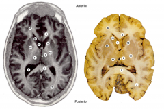
Identify structures in the brain (axial) |
1 - Lateral Ventricle 2 - Third Ventricle 5 - Corpus Callosum 6 - Frontal Lobe 7 - Occipital Lobe 10 - Basal Ganglia - Putamen 11 - Thalamus 12 - Internal capsule |
Occipital Lobe Third Ventrice Lateral Ventricle Internal Capsule Basal ganglia - Putamen Frontal Lobe Thalamus Corpus Callosum |
|
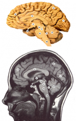
Identify structures in the brain (Saggital) |
2 - Third Ventricle 3 - Fourth Ventricle 4 - Aqueduct 5 - Corpus Callosum 6 - Frontal Lobe 7 - Occipital Lobe 8 - Parietal lobe 13 - Optic Chiasm 14 - Midbrain 15 - Pons 16 - Medulla 17 - Cerebellum |
Aqueduct Parietal Lobe Cerebellum Third Ventricle Optic Chiasm Medulla Corpus Callosum Frontal Lobe Pons Mid brain Fourth Ventricle Occipital Lobe |
|
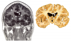
Identify structures in the brain (Coronal) |
1 - Lateral Ventricle 2 - Third Ventricle 5 - Corpus Callosum 6 - Frontal Lobe 9 - Temporal Lobe 10 - Basal Ganglia 11 - Thalamus 12 - Internal Capsule |
Corpus Callosum Lateral Ventricle Basal ganglia Temporal lobe Thalamus Frontal Lobe Internal capsule Third Ventricle |
|
|
Different tissue components of the scalp |
S - Skin C - Connective tissue A - Aponeurosis (epicranial) L - Loose connective tissue - not continuous with pericranium P - Pericranium - continuous with endocranium |
5 |
|
|
Branches of the external carotid artery |
Superior thyroid Ascending pharyngeal Lingual Facial Occipital Posterior auricular Maxillary Superficial temporal |
Some Ancient Lovers Find Old Positions More Stimulating |
|
|
Divisions of the brachial plexus |
Roots Trunks Division Cord |
R T D C |
|
|
Dorsal scapular nerve |
C5 root - Rhomboid major + Rhomboid minor |
|
|
|
Long thoracic nerve |
C5+C6+C7 roots - Serratus anterior |
|
|
|
Suprascapular nerve |
Superior trunk - supraspinatus + infraspinatus |
|
|
|
Subclavius nerve |
Superior trunk - Subclavius |
|
|
|
Terminal branches of the cords of the brachial plexus |
Musculocutaneous - Lateral Axillary - Posterior Radial - Posterior Median - Medial + Lateral Ulnar - Medial |
My Auntie Raped My Uncle |
|
|
Lateral pectoral nerve |
Lateral cord - Pectoralis major |
|
|
|
Medial pectoral nerve |
Medial cord - Pectoralis minor |
|
|
|
Medial cutaneous nerve of arm |
Skin on medial arm - Medial cord |
|
|
|
Medial cutaneous nerve of forearm |
Skin on medial side of arm + forearm - Medial cord |
|
|
|
Branches of the posterior cord of the brachial plexus |
Upper (superior) subscapular Lower (inferior) subscapular Thoracodorsal Radial Axillary |
ULTRA |
|
|
Upper subscapular |
Posterior cord - subscapularis |
|
|
|
Lower subscapular |
Posterior cord - subscapularis + teres major |
|
|
|
Thoracodorsal nerve |
Posterior cord - Latissimus dorsi (adduction, extension, medial rotation) |
|
|
|
Musculocutaneous nerve |
Lateral cord - Flexor muscles of a. compartment of arm - Biceps brachii + brachialis + coracobrachiallis Gives off lateral cutaneous nerve of forearm |
|
|
|
Ulnar nerve |
Medial cord - Flexor digitorum profundus + flexor carpi ulnaris + all intrinsic muscles of hand (EXCEPT thenar muscles + lateral two lumbricals) |
|
|
|
Axillary |
Posterior cord - Deltoid + Long head of triceps brachii + Teres minor Sensory - Gives off superior lateral cutaneous (distal deltoid region) |
|
|
|
Median nerve |
Lateral + Medial cord - Muscles of a. compartment of forearm that the ulnar nerve doesn't innervate + Thenar muscles + Lateral two lumbricals |
|
|
|
Radial nerve |
Posterior cord - Posterior compartment of arm and forearm + dorsal lateral compartment of hand (thumb - lateral half of ring finger) |
|
|
|
Main exit/entry routes of the skull |
Cribiform plate Optic canal Superior orbital fissure Foramen rotundum Foramen ovale Foramen spinosum Carotid canal Internal acoustic meatus Jugular foramen Hypoglossal canal Foramen magnum |
C O S F F F C I J H F |
|
|
Cribiform plate contents |
Olfactory I |
1 |
|
|
Optic canal contents |
Optic II Cantral artery of retina Opthalmic artery |
3 |
|
|
Superior orbital fissure contents |
Oculomotor III Trochlear IV Opthalmic division of trigeminal V Aducens VI Superior opthalmic vein |
5 |
|
|
Foramen rotundum contents |
Maxillary division of trigeminal V |
1 |
|
|
Foramen ovale |
Madibular division of trigeminal V |
2 |
|
|
Foramen spinosum |
Middle meningeal artery + vein |
1 |
|
|
Carotid canal |
Internal carotid artery |
1 |
|
|
Internal acoustic meatus |
Facial VII Vestibulocochlear VIII Labrynthine artery |
3 |
|
|
Jugular foramen |
Glossopharyngeal IX Vagus X Accessory XI Sigmoid sinus - Internal jugular vein |
4 |
|
|
Hypoglossal canal |
Hypoglossal XII |
1 |
|
|
Foramen magnum |
Vertebral arteries Medulla Spinal routes of accessory XI |
3 |
|
|
Boundaries of anterior triangle of neck |
A. border of sternocleidomastoid I. Mandible Midline of neck |
|
|
|
Boundaries of posterior triangle of neck |
A. trapezius P. sternocleidomastoid Middle 1/3 of clavicle |
|
|
|
Contents of anterior triangle of the neck |
Platysma muscle Digastric muscle Strap muscle Mylohyoid muscle Common carotid arteries Internal jugular vein Larynx + Trachea |
P D S M C I L |
|
|
Contents of posterior triangle of the neck |
External jugular Spinal accessory nerve Trunks of brachial plexus Subclavian artery Subclavian vein Phrenic nerve |
E S T S S P |