Reading...
![]()
Play button
![]()
Play button
![]()
Use LEFT and RIGHT arrow keys to navigate between flashcards;
Use UP and DOWN arrow keys to flip the card;
H to show hint;
A reads text to speech;
210 Cards in this Set
- Front
- Back
- 3rd side (hint)
|
What is the study of the structure of body parts and their relationships to one another?
|
Anatomy
|
|
|
|
What concerns the function of the body, how the body parts work and carry out their life sustaining activities?
|
Physiology
|
|
|
|
-Surface Anatomy
-Regional Anatomy -Systemic Anatomy -Developmental Anatomy |
Gross Anatomy
|
Large Structures you can see with your eyes.
|
|
|
-Cytology
-Histology |
Microscopic Anatomy
|
Small Structures you can only see with the use of a microscope.
|
|
|
The study of internal structures as they relate to the overlying skin surface.
|
Surface Anatomy
|
|
|
|
All the structures (muscles, bones, blood vessels, nerves, etc) in a particular region of the body.
|
Regional Anatomy
|
|
|
|
Body structure is studied system by system.
|
Systemic Anatomy
|
|
|
|
Traces structural changes that occur in the body throughout the life span.
|
Developmental Anatomy
|
|
|
|
The study of the cells of the body.
|
Cytology
|
|
|
|
The study of Tissues.
|
Histology
|
|
|
|
Topics of Physiology
|
Integumentary System (Skin)
Nervous System Skeletal System Endocrine System Muscular System Cardiovascular System Lymphatic System Urinary System Respiratory System Digestive System Reproductive System |
11 Topics
|
|
|
Levels of Structural Organization
|
Chemical
Cellular Tissue Organ Organ System Organismal |
6 Levels
|
|
|
Atoms combine together to form molecules.
|
Chemical Level
|
Simplist
|
|
|
Molecules combine together to form cells.
|
Cellular Level
|
|
|
|
Smallest unit of any living thing.
|
Cell
|
|
|
|
Cells combine together for a common function.
|
Tissue Level
|
|
|
|
Tissues combine in different amounts for a common function.
|
Organ Level
|
|
|
|
How many different tissues does it take to form an organ?
|
2 - But most organs have all four.
|
|
|
|
Organs come together for a common function.
|
Organ System Level
|
|
|
|
Sum total of all structural levels working together to promote life.
|
Organismal Level
|
|
|
|
Name the 8 Necessary Life Functions.
|
Maintaining Boundaries
Movement Responsiveness or Irritability Digestion Metabolism Excretion Reproduction Growth |
|
|
|
Every living organism must do this so that its internal environment (inside) remains distinct from the external environment surrounding it (outside).
|
Maintaining Boundaries
|
|
|
|
Includes the activities promoted by the muscular system, such as propelling ourselves from one place to another by running or swimming, and manipulating the external environment with our nimble fingers.
|
Movement
|
|
|
|
The ability to sense changes (stimuli) in the environment and then respond to them.
|
Responsiveness
|
|
|
|
The breaking down of ingested foodstuffs to simple molecules that can be absorbed into the blood.
|
Digestion
|
|
|
|
Includes all chemical reactions that occur within body cells.
|
Metabolism
|
|
|
|
Process of removing excreta (or wastes) from the body.
|
Excretion
|
|
|
|
Cell divides, producing two identical daughter cells that may then be used for body growth or repair.
|
Reproduction
|
|
|
|
An increase in size of a body part or the organism.
|
Growth
|
|
|
|
Name the 5 Survival Needs
|
Nutrients
Oxygen Water Normal Body Temperature Atmospheric Pressure |
|
|
|
Contain the chemical substances used for energy and cell building.
|
Nutrients
|
|
|
|
All chemical reactions that release energy from food require this.
|
Oxygen
|
|
|
|
Accounts for 60-80% of body weight, and is single most abundant chemical substance in the body.
|
Water
|
|
|
|
Chemical Reactions need this to continue at life sustaining rates.
|
Normal Body Tempature
|
|
|
|
Force that air exerts on the surface of the body.
|
Atmospheric Pressure
|
|
|
|
The ability to maintain relatively stable internal conditions even though the outside world changes continuously.
|
Homeostasis
|
|
|
|
3 Components of Homesostasis
|
Receptor
Control Center Effector |
|
|
|
Type of Sensor that monitors the environment and responds to changes, called stimuli, by sending information (input) to the second component, the control center.
|
Receptor
|
How your body knows something has changed.
|
|
|
Determines the set point (the level or range at which a variable is to be maintained).
|
Control Center
|
Holds a set point and contains all the plans to be carried out.
|
|
|
Provides the means for the control center's response.
|
Effector
|
Organs that affect the change.
|
|
|
The output shuts off the original stimulus or reduces its intensity.
|
Negative Feedback Mechanism
|
Causes the variable to change in an Opposite Direction to that of the initial change.
|
|
|
Enhances the original stimulus so that the activity (output) is accelerated.
|
Positive Feedback Mechanism
|
The change that occurs proceeds in the same direction as the initial disturbance.
|
|
|
Example of Negative Feedback System
|
Thermostat is set at 20 C (Control Center). Temperature drops to 18 C outside, and thermometer (receptor) sends input to Thermostat. Control center initiates plan (heater must be turned up). Control center sends plan to heater (effector), and heater turns on until 20 C is reached in the house. Cycle repeats continuously.
|
|
|
|
Why can Positive Feedback be bad?
|
After your body is overworked, (sweating and everything starts to breakdown) Negative feedback that has been trying to maintain Homeostasis will just stop. This will enforce Positive Feedback to start working, and your body will just continue to get hotter and hotter. (Destructive Factors keep happening)
|
|
|
|
Most diseases can be regarded as a result of this.
Negative feedback mechanisms are overwhelmed and destructive positive feedback mechanisms. |
Homeostatic Imbalance
|
|
|
|
Standing upright, head facing forward, feet in normal stance, palms facing anteriorly and thumbs outward.
|
Anatomical Position
|
|
|
|
Toward the head
|
Superior
|
|
|
|
Away from the head
|
Inferior
|
|
|
|
Toward the front of the body
|
Anterior
|
|
|
|
Toward the back of the body
|
Posterior
|
|
|
|
Toward the midline
|
Medial
|
|
|
|
Away from the midline
|
Lateral
|
|
|
|
Between a medial and lateral structure
|
Intermediate
|
Not used very often
|
|
|
Toward the trunk
|
Proximal
|
|
|
|
Away from the trunk
|
Distal
|
|
|
|
Toward the surface of the body
|
Superficial
|
|
|
|
Away from the surface of the body
|
Deep
|
|
|
|
2 Primary regions of the Body
|
Axial Region
Appendicular Region |
|
|
|
Head, Neck, and Trunk
|
Axial Region
|
Does not include shoulders, or anywhere limbs attach.
|
|
|
Limbs
|
Appendicular Region
|
|
|
|
3 Types of Cardinal Planes
|
Frontal
Sagittal Transverse |
Divide Body
|
|
|
Cuts in middle to divide into anterior and posterior halves.
|
Frontal (Coronal) Plane
|
|
|
|
Unequal right and left halves
|
Sagittal Plane
|
|
|
|
Equal right and left halves
|
Median or Midsagittal Plane
|
|
|
|
Cuts body to make top and bottom halves
|
Transverse Plane
|
CT Scan uses this
|
|
|
Internal chambers that hold vital organs.
|
Body Cavities
|
Protect Vital Organs
|
|
|
2 Main Body Cavities
|
Dorsal Body Cavity
Ventral Body Cavity |
|
|
|
Protects the fragile nervous system organs.
|
Dorsal Body Cavity
|
Cranial Cavity and Spinal Cavity; Most Protective; Boney Structures
|
|
|
The more anterior and larger of the closed body cavities.
|
Ventral Body Cavity
|
Thoracic cavity and abdominopelvic cavity.
|
|
|
Surrounded by the ribs and muscles of the chest.
|
Thoracic Cavitiy
|
Subdivision of Ventral Body Cavity
|
|
|
Dome-Shaped muscle important in breathing.
|
Diaphragm
|
|
|
|
Contains the stomach, intestines, spleen, liver, and other organs.
|
Abdominal Cavity
|
Subdivision of Ventral Body Cavity
|
|
|
Contains pelvic area
|
Pelvic Cavity
|
|
|
|
Building block of all matter
|
Atom
|
|
|
|
Subatomic particles of an atom
|
Protons
Neutrons Electrons |
|
|
|
A chemical structure consisting of atoms held together by chemical bonds.
|
Molecule
|
2 or more atoms chemically bonded
|
|
|
A chemical substance composed of two or more different kinds of atoms.
|
Compound
|
8 or more DIFFERENT atoms chemically bonded together.
|
|
|
Substances composed of two or more components physically intermixed.
|
Mixtures
|
Physically mixed, not chemically mixed. (Sand and Water)
No Chemical Reaction needed to separate, and not chemically bonded. |
|
|
Anything that occupies space and has mass.
|
Matter
|
|
|
|
Any chemical reaction in the human body.
|
Metabolism
|
Confused with digestion.
Capture, Storage, and Release of energy. |
|
|
Capacity to perform work.
Movement of an object or change in its physical structure. |
Energy
|
Work
|
|
|
Two Forms of Energy
|
Kinetic Energy
Potential Energy |
|
|
|
Energy Currency of the Body
|
ATP
|
|
|
|
Energy in Action
|
Kinetic Energy
|
Ball Rolling down a hill
|
|
|
Stored energy, that is, inactive energy that has the capability to do work but is not currently doing so.
|
Potential Energy
|
Ball sitting on top of a hill.
|
|
|
Bi product of potential energy changing into kinetic energy.
|
Heat
|
60% is given off as this, 40% is able to perform work.
|
|
|
Storage of energy
|
Fat
|
|
|
|
4 Types of Energy
|
Chemical Energy
Electrical Energy Mechanical Energy Radiant Energy |
|
|
|
The form of energy stored in the bonds of chemical substances.
|
Chemical Energy
|
Potential energy is unleashed, and becomes kinetic energy.
|
|
|
Results from the movement of charged particles.
|
Electrical Energy
|
Sends messages from brain to muscles.
Nervous System |
|
|
Energy directly involved in moving matter
|
Mechanical Energy
|
Movement we use everyday.
(Walking) |
|
|
Energy that travels in waves
|
Radiant Energy
|
Used to expel heat from the body.
(Homeostasis) |
|
|
Reactions of Metabolism
|
Decomposition (AKA Catabolic)
Syntheses (AKA Anabolic) Exchange |
|
|
|
When atoms or molecules combine to form a larger, more complex molecule.
A + B → AB |
Synthesis (AKA Anabolic)
|
Constructive Process
|
|
|
Occurs when a molecule is broken down into smaller molecules.
AB → A + B |
Decomposition (AKA Catabolic)
|
Break large bonds to form small ones.
|
|
|
Involves bonds that are both made and broken.
AB + C → AC + B and AB + CD → AD + CB |
Exchange
|
Rearranging the order; neither losing or gaining anything.
|
|
|
Rules of Reactions
|
All reactions are theoretically reversible.
At equilibrium the rates of two opposing reactions are in balance. Anabolism ↔ Catabolism Being out of Equilibrium can cause problems. |
Technically can be done, but will be difficult.
|
|
|
Factors affecting the rate of chemical reactions.
|
Temperature
Concentration Particle Size Catalysts |
|
|
|
Significance of temperature in relation to chemical reactions
|
Increasing the temperature of a substance increases the kinetic energy of its particles and the force of their collisions. Therefore, chemical reactions proceed quicker at higher temperatures.
|
Increase temp = Increase Reactions
|
|
|
Significance of concentration in relation to chemical reactions.
|
Chemical reactions progress most rapidly when the reacting particles are present in high numbers, because the chance of successful collisions is greater. As the concentration of the reactants declines, chemical equilibrium eventually occurs unless additional reactants are added or products are removed from the reaction site.
|
Higher number of molecules, the faster they will collide.
|
|
|
Significance of particle size in relation to chemical reactions.
|
Smaller particles move faster than larger ones (at the same temperature) and tend to collide more frequently and more forcefully. Therefore, the smaller the reacting particles, the faster a chemical reaction goes at a given temperature and concentration.
|
Smaller they are, the more likely they are going to bump into something.
|
|
|
Significance of catalysts in relation to chemical reactions.
|
Catalysts increase the rate of chemical reactions without themselves becoming chemically changed or part of the product.
|
|
|
|
Cell Theory
|
* The cell is the fundamental unit of structure and function in living things.
* All cells come from pre-existing cells by division. *Energy flow occurs within cells. *Cells contain hereditary information which is passed from cell to cell during cell division * All cells are basically the same in chemical composition. *All known living things are made up of cells. *Some organisms are unicellular, made up of only one cell. Others are multicellular. |
This is why we are able to talk about cells in a generic form, even though there are over 200 types in the human body.
|
|
|
Exceptions to Cell Theory
|
1. Viruses are considered by some to be alive, yet they are not made up of cells.
2. The first cell did not originate from a preexisting cell. |
|
|
|
3 Main Parts of Human Cells
|
1. Plasma Membrane
2. Cytoplasm 3. Nucleus |
|
|
|
The extent of a cell, thereby separating two of the body's major fluid compartments - the intracellular fluid within cells and the extracellular fluid outside cells.
|
Plasma Membrane
|
Thin and Fragile
Picky - picks and chooses what comes in and what goes out. |
|
|
AKA extracellular fluid
|
Interstitial Fluid
|
House Nutrients, among other things.
|
|
|
Depicts the plasma membrane as an exceedingly thin structure composed of a double layer, or bilayer, of lipid molecules with protein molecules dispersed in it.
|
Fluid Mosaic Model
|
|
|
|
The "fabric" of the membrane, which is constructed of phospholipids, with smaller amounts of cholesterol and glycolipids.
|
The lipid bilayer
|
|
|
|
Means "water loving"
|
Hydrophilic
|
|
|
|
Means "water hating"
|
hydrophobic
|
|
|
|
Made up of hydrophilic heads and hydrophobic tails.
|
Phospholipid Molecule
|
|
|
|
phospholipids with attached sugar groups, found only on the outer plasma membrane surface and account for about 5% of the total membrane lipid.
|
Glycolipids
|
|
|
|
Dynamic assemblies of saturated phospholipids associated with unique lipids called sphingolipids and lots of cholesterol.
|
Lipid Rafts
|
|
|
|
2 Distinct Membrane Proteins
|
1. Integral Proteins
2. Peripheral Proteins |
|
|
|
Firmly inserted into the lipid bilayer. Some protrude from one membrane face only, but most are transmembrane proteins that span the entire width of the membrane and protrude on both sides.
|
Integral Proteins
|
Membrane Protein
|
|
|
Not embedded in the lipid. Instead, they attach rather loosely to integral proteins or membrane lipids and are easily removed without disrupting the membrane. Some are enzymes, and others are involved in mechanical functions, such as changing cell shape during cell division and muscle cell contraction, or linking cells together.
|
Peripheral Proteins
|
Membrane Protein
|
|
|
What binds cells together?
|
1. Glycoproteins in the glycocalyx act as an adhesive.
2. Wavy contours of the membranes of adjacent cells fit together in a tongue-and-groove fashion. 3. Special membrane junctions are formed. |
|
|
|
3 Types of Membrane Junctions
|
1. Tight Junctions
2. Desomosomes 3. Gap Junctions |
|
|
|
Series of Integral protein molecules in the plasma membranes of adjacent cells fuse together, forming an impermeable junction that encircles the cell.
|
Tight Junction
|
(Epithelial Tissue)
|
|
|
Anchoring Junctions - mechanical couplings scattered like rivets along the sides of abutting cells that prevent their separation.
|
Desomosomes
|
Plaque (Outside) and Protein filaments bind it to an adjacent cell. (Skin, Muscles, Tendons - Great Mechanical Stress)
|
|
|
A communicating junction between adjacent cells. Plasma membranes are very close, and the cells are connected by hollow cylinders called connexons composed of transmembrane proteins.
|
Gap Junction
|
Fast Communication (Heart)
|
|
|
2 Methods of Membrane Transport
|
1. Passive
2. Active |
|
|
|
Process by which substances cross the membrane without any energy input from the cell.
|
Passive Process
|
Just happens - no energy required
|
|
|
Process by which the cell provides the metabolic energy (ATP) needed to move substances across the membrane.
|
Active Process
|
Energy is Required
|
|
|
Passive Processes
|
1. Diffusion
2. Filtration |
Going along or with the Concentration Gradient.
(No energy required) |
|
|
3 Types of Diffusion
|
1. Simple Diffusion
2. Facilitated Diffusion 3. Osmosis |
|
|
|
Nonpolar and lipid-soluble substances diffuse directly through the lipid bilayer.
|
Simple Diffusion
|
|
|
|
Process in which the transported substance either (1) binds to protein carriers in the membrane and is ferried across or (2) moves through water-filled protein channels.
|
Facilitated Diffusion
|
|
|
|
A transmembrane integral protein that shows specificity for molecules of a certain polar substance or class of substances that are too large to pass through membrane channels.
|
Carrier
|
Sugars and amino acids
|
|
|
Transmembrane proteins that serve to transport substances, usually ions or water, through aqueous channels from one side of the membrane to the other.
|
Channels
|
|
|
|
The diffusion of a solvent, such as water, through a selectively permeable membrane
|
Osmosis
|
|
|
|
When molecules are moving with the concentration gradient, they are flowing from ____ to ____ ground, and eventually they want to _____.
|
High:Low
Equal Out |
|
|
|
The process that forces water and solutes through a membrane or capillary wall by fluid, or hydrostatic pressure.
|
Filtration
|
Not selective; only blood cells and protein molecules too large to pass through membrane pores are held back. (Nutrients and Waste)
|
|
|
The gradient for filtration is a ____ that pushes solute-containing fluid from a higher-pressure area to a lower-pressure area.
|
Pressure Gradient
|
|
|
|
Process in which a cell uses the bond energy (ATP) to move solutes across the membrane.
|
Active Process
|
|
|
|
2 Mechanisms of Active Transport
|
1. Active Transport
2. Vesicular Transport |
|
|
|
Solute Pumps are used in this process to move solutes, most importantly ions (Na⁺, K⁺, and Ca²⁺), "uphill" against a concentration gradient. This requires ATP.
|
Active Transport
|
"Bailing a Boat" Example
Fighting against what wants to happen, but not stopping. |
|
|
Energy comes directly from hydrolysis of ATP.
|
Primary Active Transport
|
Maintain concentration of Na⁺, using energy to achieve the primary goal.
|
|
|
Transport is driven indirectly by energy stored in ionic gradients created by the operation of the active transport pumps.
|
Secondary Active Transport
|
When Na⁺ is pumped, other substances get dragged along with it.
|
|
|
Large particles, macromolecules, and fluids are transported across plasma and intracellular membranes by this process.
|
Vesicular Transport
|
Energized by ATP.
Macromolecules are too large to transport across by themselves. |
|
|
Moving substances from the cell interior to the extracellular space.
|
Exocytosis
|
"out of the cell"
|
|
|
Moving substances across the plasma membrane into the cell from the extracellular environment.
|
Endocytosis
|
"within the cell"
|
|
|
Membrane bound vesicles will bond to the cell, spit out the contents, and vesicle will become attached to the cell.
|
Process of Exocytosis
|
|
|
|
The substance to be taken in by the cell is progressively enclosed by an infolding portion of the plasma membrane, and then enters the cell by a vesicle.
|
Process of Endyocytosis
|
|
|
|
The cellular material between the plasma membrane and the nucleus.
|
Cytoplasm
|
Cell forming material
|
|
|
3 Major Elements of Cytoplasm
|
1. Cytosol
2. Cytoplasmic Organelles 3. Inclusions |
|
|
|
Cytosol
|
The viscous, semi-transparent fluid in which the other cytoplasmic elements are suspended. Thick, egg white substance; Proteins, salts, sugars, and a variety of other solutes are dissolved in this substance.
|
|
|
|
Cytoplasmic Organelles
|
The metabolic machinery of the cell; "little organs" that do all the work.
|
|
|
|
Inclusions
|
Chemical substances that may or may not be present, depending on the cell type. Cells can exist with or without them.
|
|
|
|
Name the 8 Organelles
|
1. Nucleus
2. Endoplasmic Reticulum 3. Ribosomes 4. Golgi Complex 5. Lysosomes 6. Peroxisomes 7. Mitochondria 8. Centrioles |
|
|
|
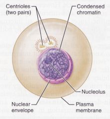
Nucleus
|
1. Control Center
2. Largest Organelle 3. It contains the instructions needed to build nearly all the body's proteins to be synthesized at any one time in response to signals acting on the cell. 4. Can be anuclear, mononuclear, or multinucleated. 5. Contains DNA and RNA 6. Surrounded by the nuclear envelope |
Raw Products cannot function unless in the nucleus.
|
|
|
Nucleoplasm Includes:
|
Chromatin and Nucleoli
|
Contains everything within the nucleus.
|
|
|
Eventually makes Chromosomes
|
Chromatin
|
|
|
|
Nucleoli
|
Where Ribosomes are produced.
|
|
|
|
A double membrane barrier separated by a fluid-filled space. Includes the outer nuclear membrane, inner nuclear membrane, chromatin, and nucleolus.
|
Nuclear envelope
|
|
|
|
Nucleus-Chromatin
|
Composed of about 30% DNA, 60% Globular Histone Proteins, and 10%RNA chains.
Chromosomes. |
|
|
|
Endoplasmic Reticulum
|
1. "Little Network within the Cytoplasm"
2. Interconnected channels called cisternae enclosed by a unit membrane. |
|
|
|
2 Types of Endoplasmic Reticulum
|
1. Rough Endoplasmic Reticulum
2. Smooth Endoplasmic Reticulum. |
|
|
|
Ribosomes are attached to this surface, and they manufacture all proteins secreted from cells. Also produce phospholipids, and synthesizes proteins that are either packaged from others or contained within.
Abundant in Protein Producing Cells. |
Rough Endoplasmic Reticulum
|
AKA "Membrane Factory"
|
|
|
Tubular in shape and no ribosomes attached.
Abundant in detoxifying cells. |
Smooth Endoplasmic Reticulum
|
Tolerance - part is due to growth of this.
|
|
|
Fluid filled cavities (interconnected channels) of Endoplasmic Reticulum
|
Cisternae
|
|
|
|
Ribosomes
|
1. Dark staining granules (sandpaper like)
2. Composed of proteins and RNA called ribosomal RNA (65%, RProteins 35%) 3. Has a top half and a bottom half. 4. Can be attached or free flowing. 5. Function of ribosome depends on if it is membrane bound or not. |
|
|
|
Golgi Complex
|
1. Small System of cisternae
2. Principle traffic director for cellular proteins. 3. Synthesize carbohydrates and finish protein and glycoprotein synthses. 4. Products include: lysosomes, portions of the plasma membrane, or secretory vesicles. 5. "Splice, Dice, and Slice" proteins, and package them into secretory vesicles (or sometimes called golgi vesicles) |
Found in both plant and animal cells.
Shaped like five to eight deflated baloons. Traffic director of the cell. |
|
|
Secretory Vesicles that include Lysosomes
|
Migrate to plasma membrane and others become stored for later use. (Breast milk, etc.)
|
|
|
|
Spherical membranous organelles containing digestive enzymes.
Large and abundant in phagocytes. Hydrolyze proteins, nucleic acids, complex carbohydrates, phospholipids, and other substrates. Autophagy Autolysis |
Lysosmes
|
Bounded by a single unit membrane.
50 have been identified. Produced by Golgi Digest dead or useless cells. |
|
|
Cells that dispose of invading bacteria and cell debris.
|
Phagocytes
|
|
|
|
Lysosomes work best in _____ conditions.
|
Acidic
|
|
|
|
Self digestion of the cell.
The basis for desirable destruction of cells. |
Autolysis
|
|
|
|
Membranous sacs containing a variety of powerful enzymes, the most important of which are oxidases and catalases.
|
Peroxisomes
|
|
|
|
Use Molecular oxygen to detoxify harmful substances, including alcohol and foraldehyde. Also, most important, converts free radicals to hydrogen Peroxide.
|
Oxidases
|
|
|
|
4 Properties of Peroxisomes
|
1. Resemble Lysosomes but do not contain the same enzymes and are not produced by the Golgi complex.
2. General function is to use molecular oxygen to oxidize organic molecules, giving off Hydrogen Peroxide. 3. Neutralize free radicals and detoxify alcohol, other drugs and a variety of blood-borne toxins. 4. They also decompose fatty acids in to two carbon acetyl groups, which is used in the mitochondria for ATP. |
Abundant in kidney and liver cells.
Replicate themselves. |
|
|
Mitochondria
|
Powerhouse of cell, providing most of the ATP supply. They are rod or bean-shaped membranous organelles.
Enclosed by 2 membranes. Uses Aerobic Respiration. (Get 15% more when Oxygen in converted in the mitochondria) Contains mitochondrial DNA which is used to replicate mitochondria. |
We are able to provide more factories for energy if needed
|
|
|
When metabolites are broken down and oxidized, some of the energy released is captured and used to attach phosphate groups to ADP molecules to form ATP.
|
Aerobic Cellular Respiration
|
|
|
|
Centrioles
|
Short cylindrical assembly of microtubles arranged in nine groups of three microtubles.
Play a role in cell division and they form the base for cilla and flagella. |
|
|
|
Cilla
|
Whiplike structures on the surface of the cell that help to move substances in one direction.
|
Move substances across cell membrane.
Do not move the actual cell. Resemble nose hairs. |
|
|
Flagella
|
Single projection that is substantially longer than the cilla and are used for propulsion.
|
Sperm Cells
Not as numerous as Cilla. Propels and moves the cell. |
|
|
Cytoskeleton
|
Collection of protein filaments and cylinders that:
1. Determine the shape of the cell 2. Lend it structural support 3. Organize its contents 4. Move substances through the cell 5. Contribute to movements of the cell as a whole. |
Like skeleton
|
|
|
Inclusions
|
2 Types
1. Stored Cellular products (Glycogen, pigment granules, fat particles) 2. Foreign bodies (Dust, viruses, intercellular bacteria. Not enclosed in a unit membrane Not essential to cell survival Storage for nutrients wastes, and cell products. |
|
|
|
2 major phases of the life cycle of a cell
|
Interphase and Cell Division
|
|
|
|
3 Phases of Interphase
|
G₁ Phase (First Gap Phase)
S Phase (Synthesis Phase) G₂ Phase (Second Gap Phase) |
|
|
|
The series of changes a cell goes through from the time it is formed until it reproduces.
Divides into 2 Daughter Cells |
The cell life cycle
|
|
|
|
G₁ Phase (First Gap Phase)
|
Cell spends most of its functional life in this phase.
Cell synthesizes proteins, grows and carries out its preordained tasks for the body. Cells accumulate the materials needed to replicate their DNA in the next phase. Stimuli trigger cell division event. |
|
|
|
Triggers for Synthesis Phase of Cell
|
1. To replace dead or dying cells
2. To produce more cells to enlarge the organism (growth and development) 3. Reproduction (increase number of unicellular organisms) 4. To reduce the surface volume ratio. (Want more surface available to volume of nutrients.) |
|
|
|
S Phase (Synthesis Phase)
|
Cell makes duplicate copies of its centrioles and all of its DNA. Often referred to as DNA replication.
|
Allows 2 sets to be available to each daughter cell.
Without this phase, cell will not properly divide, and eventually will die. |
|
|
G₂ Phase (Second Gap Phase)
|
Brief interval between DNA replication and cell division.
Cell finishes replicating its centrioles and synthesizes enzymes that control cell division. |
Final Touches, Final Preparation before dividing.
|
|
|
2 Events of Mitotic Phase
|
1. Mitosis
2. Cytokinesis |
|
|
|
Properties of Mitotic Phase
|
1. Cell replicates nucleus and then pinches in two.
2. Forms 2 Daughter Cells |
|
|
|
4 Phases of Mitosis
|
1. Prophase
2. Metaphase 3. Anaphase 4. Telephase |
|
|
|
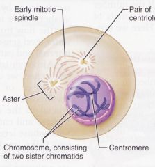
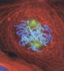
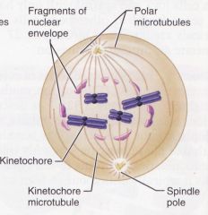
Prophase
|
Chromosomes shorten and thicken.
Nuclear envelope disintegrates and releases chromosomes into the cytosol. Centrioles begin to sprout spindle fibers. Spindle fibers grow toward the chromosomes and attach at the kinetochore, which is the center of the chromosome. |
Chromatins form chromosomes (little squiggles)
Spindle fibers tug at chromosomes, making them line up in center. |
|
|
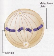
Metaphase
|
Chromosomes are aligned on the cells equator awaiting a signal that stimulates them to split.
Spindle fibers for the mitotic spindle Long micro tubules attach to chromosomes while shorter micro tubules form the aster. |
Chromosomes are in the center.
Asters are anchors for spindle fibers. (Prepare to split chromosomes) |
|
|
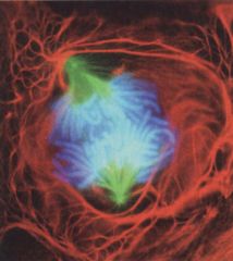
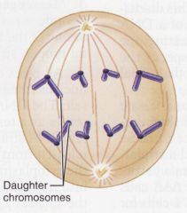
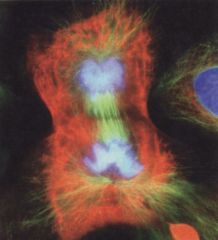
Anaphase
|
An enzyme splits the chromosomes into two sister chromatids.
Spindle fibers shorten and the sister chromatids are drawn to opposite poles of the cell. |
Split Chromosomes
Draw into the ends of the cell. Exact Division of genetic info to each daughter cell. |
|
|
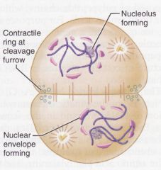
Telephase
|
Chromatids cluster on each side of the cell.
Rough ER produces a new nuclear envelope that encircles the chromatids. Chromatids uncoil and return to their original chromatin form. Mitotic spindle breaks up Each new nucleus forms nucleoi |
Chromatids split up
|
|
|
Cytokinesis
|
Division of the cytoplasm into two cells.
Sometimes overlaps with the telephase. |
Actual Dividing, continues until 2 separate cells are formed.
|
|
|
Free Flowing Ribosomes
|
Makes proteins
|
|
|
|
Membrane Bound Ribosomes
|
Synthesizes proteins destined to be incorporated inside the membrane or exported to be used in extracellular fluid.
|
|
|
|
Cell Mortality
|
There is a natural limit to how many times cells can divide
Biochemical errors accumulate during cell division and reproduction which eventually result in cell death. Immortality of cancer cells shows there is a way to keep cells alive. Cells can become immortal if transformed by infecting them with a virus (Cancer, HPV virus) Cell death is part of normal development, and it is also important for regenerating tissues or bones after injury. |
50 to 100 times before cell dies.
Important for cell division, repair and mending. |
|
|
Cells Divide When:
|
They grow large enough to have enough cytoplasm to distribute their own 2 daughter cells.
They have replicated their DNA so they can give each daughter cell a duplicate set of genes. They receive adequate supply of nutrients. They are stimulated by growth factors. Neighboring cells die, leaving space in the tissue to be occupied. |
Cells cannot die without having enough DNA to replicate.
Growth Factors (major contributor) helps us to continue to grow taller and bigger from birth to mid twenties. |
|
|
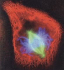
Anaphase
|
|
|
|
Anaphase
|
|
|
|
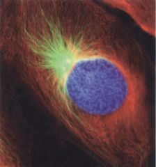
Early Prophase
|
|
|
|
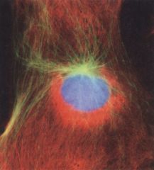
Interphase
|
|
|
|
Interphase
|
|
|
|
Late Prophase
|
|
|
|
Telophase and cytokinesis
|
|
|
|
Telophase and cytokinesis
|
|
|
|
Early Prophase
|
|
|
|
Metaphase
|
|
|
|
Metaphase
|
|
|
|
Late Prophase
|
|