![]()
![]()
![]()
Use LEFT and RIGHT arrow keys to navigate between flashcards;
Use UP and DOWN arrow keys to flip the card;
H to show hint;
A reads text to speech;
243 Cards in this Set
- Front
- Back
|
Gross Anatomy |
Seen without magnification |
|
|
Histology |
Microscopic anatomy of tissues |
|
|
Frontal/Coronal Plane |
Section of body cut to separate front from back |
|
|
Sagittal Plane |
Section of body cut to separate left from right |
|
|
Transverse Plane |
Section of body cut to separate top from bottom |
|
|
Anterior |
Towards front of body |
|
|
Posterior |
Towards back, reverse of anterior |
|
|
Dorsal |
At the back side of body, can refer to actual back |
|
|
Ventral |
At belly side of body |
|
|
Dorsal cavity contains |
Contains: Cranial Cavity (Head) & Vertebral Cavity (spinal cord) |
|
|
Ventral cavity contains |
Contains: - Thoracic: Pleural Cavity (lungs), Pericardial Cavity (heart) & Mediastinum - Abdominal - Pelvic |
|
|
Thoracic Cavity contains |
Contains: - Pleural cavity (lungs) - Pericardial Cavity (heart) - Mediastinum |
|
|
Plasma Membrane
|
Outer layer of the cell |
|
|
Plasma Membrane is made of |
A double layer (bilayer) of lipid molecules (50%) with protein molecules (45%) embedded in it. 5% carbohydrates. |
|
|
Cytoplasm |
The cellular material between the membrane and the nucleus |
|
|
Cytosol |
The fluid in which cytoplasmic elements are suspended |
|
|
Organelles |
- Endoplasmic Reticulum (ER) - Rough ER - Golgi Apparatus - Mitochondria |
|
|
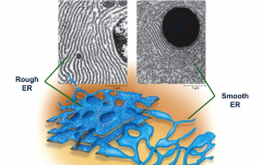
Rough Endoplasmic Reticulum |
- Studded with ribosomes. - Synthesises protein, phospholipids, and cholesterol molecules. |
|
|
Ribosomes |
- Sites of protein synthesis - Found on RER. |
|
|
Smooth Endoplasmic Reticulum |
- Lacks Ribosomes - Synthesises lipids, steroid hormones and carbohydrates - Plays role in carbohydrate, fat and drug metabolism |
|
|
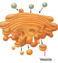
Golgi Apparatus |
- Receives proteins from RER - Packages protein secretions for export/delivery |
|
|
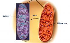
Mitochondria |
- Generates ATP (adenosine triphosphate) |
|
|
Cristae |
- Folds in a mitochondrion - Enzymes responsible for production of ATP are concentrated here |
|
|
Smooth and Rough Endoplasmic Reticulum |
|
|
Golgi Apparatus |
|
|
Mitochondrion |
|
|
Nucleus |
- Controls cell function and reproduction - Contains Chromatin (DNA & Histone protein) |
|
|
Supportive Connective Tissues |
- Cartilage - Bone Tissue |
|
|
Cartilage contains |
Contains: - cells called chondrocytes (found in spaces called lacunae) - rubber like matrix (many collagenous fibres and ground substance composed of proteoglycans) |
|
|
Types of Cartilage |
- Hyaline Cartilage - Fibrocartilage - Elastic Cartilage |
|
|
Hyaline Cartilage |
- Found over ends of bones at moveable joints - Acts as shock absorber or binding agent - Strong and flexible - Glass-like matrix |
|
|
Fibrocartilage |
- Strongest cartilage - Found in pad-like structures |
|
|
Cells in Bone Tissue |
- Osteoblast (builds bone) - Osteocyte (maintains bone) - Osteoclast (breaks down bone) |
|
|
Matrix in Bone Tissue |
- 1/3 collagenous fibres - 2/3 inorganic matter (salts) |
|
|
Classifications of Bone Tissue |
- Compact (cortical/dense) bone - Spongy (cancellous/trabecular) bone |
|
|
Compact bone |
- Found in areas with lots of pressure or weight - Main structural unit of this bone type is an osteon |
|
|
Spongy bone |
- Porous lattice of bony trabeculae which enclose cavities that contain red bone marrow where blood cells are produced |
|
|
Osteon |
- A group of layers called lamellae surrounding central (Haversian) canal - Blood vessels run through these canals |
|
|
Osteon Canals |
- Haversian canal (parallel) - Volkman canal (right angle) |
|
|
Bone Membranes |
- Periosteum - Endosteum |
|
|
Periosteum |
- Membrane lining external surface of bones - Allows tendons, ligaments and joints to attach - Contains osteoblasts and osteoclasts - Anchored to the bone with Sharpey's fibres |
|
|
Endosteum |
- Membrane on other side of bone - Delicate and thin - Contains lots of cells capable of breaking down or building up bone |
|
|
Axial Skeleton contains |
- Skull - Vertebral Column - Bony Thorax |
|
|
Skull Bones |
- Cranial bones (8) - Facial bones (14) |
|
|
Cranial bones |
- Frontal bone - Parietal bones (x2) - Temporal bones (x2) - Occipital bone - Sphenoid bone - Ethmoid bone |
|
|
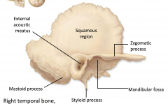
Temporal bone |
|
|
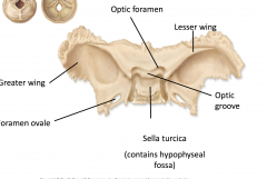
Sphenoid bone |
|
|
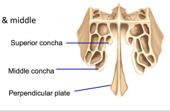
Ethmoid bone |
|
|
Facial Bones |
- Mandible - Maxillae (x2) - Zygomatic bones (x2) - Nasal bones (x2) - Lacrimal bones (x2) - Vomer - Palatine bones (x2) - Inferior Nasal Conchae (x2) |
|
|
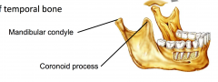
Mandible |
|
|
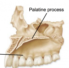
Maxillae |
|
|
Cranial Fossa |
1. Anterior fossa - supports frontal lobe 2. Middle fossa - supports temporal lobes 3. Posterior fossa - supports cerebellum |
|
|
Nasal cavity |
- Where air goes before it reaches the lungs - Separated into left and right by septum |
|
|
Paranasal sinuses |
- Linked to the nasal cavity via ducts - Holes in various skull bones - Humidify/clean air |
|
|
Orbits |
- Bony cavity that encases the eyeballs |
|
|
Vertebral Column |
- Provides vertical support for body - Houses and protects spinal cord |
|
|
Vertebrae |
- Cervical (7) - Thoracic (12) - Lumbar (5) - Sacral (sacrum and coccyx) |
|
|
Vertebral Structure |
- Body - Vertebral foramen (hole for spinal cord) - 7 processes: Spinous process, Transverse x2, Inferior Articular x2, Superior Articular x2 - Vertebral arch (surrounds vertebral foramen) |
|
|
Atlas (C1) |
- No distinct body - No spinous process - Superior articular facets articulate with occipital condyles of skull, allowing nodding movement |
|
|
Axis (C2) |
- Knoblike odontoid process (dens) - Provides pivot for rotation of atlas |
|
|
Thoracic Vertebrae |
- Heart shaped body - Long spinous processes angled inferiorly - Costal facets for articulation with ribs |
|
|
Lumbar Vertebrae |
- Kidney shaped body - Relatively short processes - Bear the most weight |
|
|
Sacrum |
- Formed by 5 fused vertebrae - Articulates with coxal bones of pelvis |
|
|
Coccyx |
- Formed by 4 fused vertebrae |
|
|
Bony Thorax contains |
- Thoracic Vertebrae - Ribs - Sternum |
|
|
Ribs |
- 12 pairs - First 10 pairs attach to the sternum |
|
|
True Ribs |
- First 7 pairs of ribs attach directly to sternum via independent strips of hyaline cartilage |
|
|
False Ribs |
- Ribs 8-10 attach indirectly to sternum via common cartilage strip - Ribs 11 & 12 don't attach to sternum (floating ribs) |
|
|
Sternum Elements |
1. Manubrium Sterni 2. Body/Corpus 3. Xiphisternum (Xiphoid process) |
|
|
Appendicular Skeleton contains |
1. Pectoral girdle 2. Pelvic girdle 3. Bones of the limbs |
|
|
Pectoral Girdle contains |
- Clavicle - Scapula |
|
|
Clavicle |
- Medial (sternal) end articulates with axial skeleton at manubrium sternum bone - Lateral (acromial) end articulates with acromion of scapula |
|
|
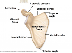
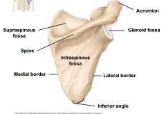
Scapula borders |
- Superior border - Medial (vertebral) border - Lateral border |
|
|
Scapula angles |
- Superior angle - Inferior angle - Anterior angle |
|
|
Scapula features |
- Coracoid Process - Acromion - Glenoid Fossa/Cavity - Spine |
|
|
Coracoid Process |
- Arises from lateral superior border of the scapula - Muscle attachment point |
|
|
Acromion |
Attaches to clavicle |
|
|
Glenoid Fossa/Cavity |
Concave surface that articulates with humerus |
|
|
Scapula Spine |
Thin shelf of bone that projects from the posterior surface of scapula |
|
|
Scapula Fossas |
- Subscapular Fossa (formed by entire ventral surface) - Supraspinous Fossa (on dorsal surface, superior to the spine) - Infraspinous Fossa (on dorsal surface, inferior to the spine) |
|
|
Scapula (anterior view) |
|
|
Scapula (posterior view) |
|
|
Bones of the Upper Limb |
- Humerus - Ulna - Radius - Carpals - Metacarpals - Phalanges |
|
|
Humerus |
Only bone in brachium (arm) |
|
|
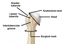
Humerus features (proximal) |
- Head (hemispherical projection that forms articulation with glenoid fossa) - Greater and Lesser Tubercles (sites of muscle attachment) - Intertubercular Sulcus (deep groove running between tubercles, where biceps brachii muscle tendon fits) |
|
|
Proximal end of Humerus |
|
|
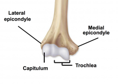
Humerus features (distal) |
- Medial and Lateral Epicondyles (origin point for several flexor and extensor muscles of antebrachium) - Capitulum (articulates with head of radius) - Trochlea (articulates with trochlear notch of ulna) - Olecranon Fossa (accommodates olecranon process of ulna bone) |
|
|
Distal end of Humerus |
|
|
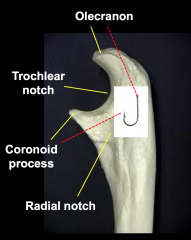
Ulna |
Medial bone in antebrachium (forearm) |
|
|
Ulna features |
- Trochlear Notch (deep C-shaped indentation that articulates with humerus) - Olecranon Process (posterior prominence of trochlear notch, projects into olecranon fossa of humerus) - Coronoid Process (anterior prominence of trochlear notch) - Radial Notch (articulates with head of radius) |
|
|
Proximal end of Ulna |
|
|
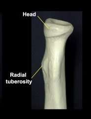
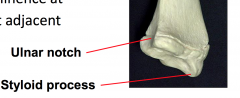
Radius |
Lateral bone in antebrachium (forearm) |
|
|
Radius features (proximal) |
- Head (circular disk that articulates with capitulum of humerus and radial notch of ulna) - Radial Tuberosity (roughened area located on medial side, attachment point for biceps brachii muscle) |
|
|
Proximal end of Radius |
|
|
Radius features (distal) |
- Ulna Notch (articulates with distal aspect of ulna) - Styloid Process (bony prominence that can be felt proximal to thumb) |
|
|
Distal end of Radius |
|
|
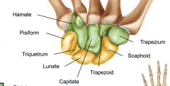
Carpal Bones |
- 8 short bones in carpus (wrist) - Organised into 2 rows of 4 bones |
|
|
Proximal row of Carpal bones (lateral to medial) |
- Scaphoid (articulates with radius) - Lunate (articulates with radius) - Triquetrum - Pisiform (embedded in tendon of flexor carpi ulnaris muscle) |
|
|
Distal row of Carpal bones (lateral to medial) |
- Trapezium - Trapezoid - Capitate - Hamate |
|
|
Carpals |
|
|
Metacarpals |
- 5 small long bones in palm that radiate from wrist - Assigned Roman Numerals (I-V) lateral to medial - I starts at thumb - Proximal ends articulate with carpals - Distal ends (knuckles) articulate with phalanges |
|
|
Phalanges |
- 14 individual phalanx bones in fingers - Proximal phalanx (closest to metacarpals) - Middle phalanx - Distal phalanx (tips of fingers) - Assigned Roman Numerals (I-V) lateral to medial - I starts at thumb - Thumb only has proximal and distal phalanx |
|
|
Pelvis |
- All the bones that surround the pelvic girdle |
|
|
Bones in Pelvis |
- Sacrum of vertebral column - Coccyx of vertebral column - Left and right Ossa Coxae (hip bones) |
|
|
Pelvic Girdle |
- Attaches the lower limb bones to the axial skeleton - Protects visceral organs in pelvic cavity - Refers to left and right Ossa Coxae only |
|
|
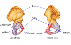
Major Features of Os Coxa bone |
- Regions: 1. Ilium (superior aspect, articulates with sacrum) 2. Ischium (posterior inferior aspect) 3. Pubis - Acetabulum - Obturator Foramen |
|
|
Major features of Os Coxa bone |
|
|
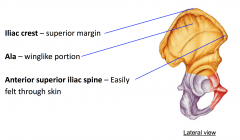
Ilium features |
- Iliac Crest (superior margin) - Ala (winglike portion) - Anterior Superior Iliac Spine (lateral to sacrum) |
|
|
Ilium features |
|
|
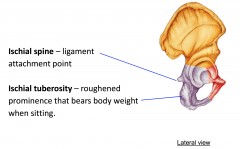
Ischium features |
- Ischial Spine (ligament attachment point - Ischial Tuberosity (roughened prominence that bears body weight when sitting and is an attachment point for hamstring) |
|
|
Ischium features |
|
|
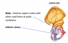
Pubis features |
- Body (anterior aspect unites with other coxal bone at pubic symphysis) - Inferior Ramus |
|
|
Pubis features |
|
|
Acetabulum |
Deep socket in pelvic girdle that articulates with head of femur |
|
|
Obturator Foramen |
Large hole inferior to acetabulum through which blood vessels and nerves pass |
|
|
Bones of the Lower Limbs |
- Femur - Patella - Tibia - Fibula - Tarsals - Metatarsals - Phalanges |
|
|
Femur |
- Only bone in the femoral (thigh) - Longest, strongest bone in the whole body |
|
|
Femur features (proximal) |
- Head (rounded projection that articulates with acetabulum) - Neck (connects head to shaft) - Greater Trochanter (projects laterally from junction of neck and shaft, muscle attachment point) - Lesser Trochanter (projects posteromedial from surface of neck) |
|
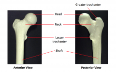
|
Proximal end of Femur |
|
|
Femur features (distal) |
- Lateral and Medial Condyles (large rounded projections that articulate with tibia) - Lateral and Medial Epicondyles (projections extending from the condyles) - Intercondylar Fossa (deep recess on posterior surface that separates the two condyles) - Patellar Surface (medial depression on anterior surface that articulates with patella) |
|
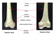
|
Distal end of Femur |
|
|
Patella |
- Forms knee cap - Enclosed in quadriceps femoris tendon - Articulates with patellar surface of femur |
|
|
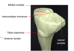
Tibia |
- Medial bone of the crural (leg) - Receives the weight of the body from the femur and transmits it to the foot |
|
|
Tibia features (proximal) |
- Medial and Lateral Condyles (articulate with condyles of femur and form a hinge joint) - Intercondylar Eminence (an irregular projection between the condyles, ligament attachment point) - Tibial Tuberosity (triangular shaped prominence on the anterior surfaces of the tibial condyles, site of insertion for anterior thigh muscles) |
|
|
Proximal end of Tibia |
|
|
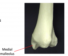
Tibia features (distal) |
- Medial Malleolus (a prominent process that forms the medial bulge of ankle) |
|
|
Distal end of Tibia |
|
|
Fibula |
- Lateral bone of the crural (leg) - Does not bare any weight - Provides sites for muscle attachment |
|
|
Fibula features |
- Head (attaches to superior aspect of tibia) - Lateral Malleolus (articulates with talus and forms lateral ankle bulge) |
|
|
Tarsal Bones |
- 7 short bones in tarsus - Split into 2 rows, 3 proximal and 4 distal |
|
|
Proximal row of Tarsal bones (lateral to medial) |
- Calcaneus - Talus - Navicular |
|
|
Distal row of Tarsal bones (lateral to medial) |
- Cuboid - Lateral Cuneiform - Intermediate Cuneiform - Medial Cuneiform |
|
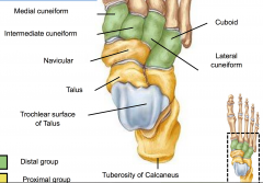
|
Tarpals |
|
|
Metatarsals |
- 5 small long bones in palm that radiate from tarsus - Assigned Roman Numerals (I-V) lateral to medial - I starts at big toe - Distal ends articulate with phalanges |
|
|
Phalanges |
- 14 individual phalanx bones in toes - Proximal phalanx (closest to metatarsals) - Middle phalanx - Distal phalanx (tips of toes) - Assigned Roman Numerals (I-V) lateral to medial - I starts at big toe - Big toe only has proximal and distal phalanx |
|
|
Joints |
- Structures where bones meet - Allow movement or provide stability |
|
|
Functional classification of Joints |
- Synarthrosis (immobile) - Amphiarthrosis (slightly mobile) - Diarthrosis (freely mobile) |
|
|
Structural classification of Joints |
- Fibrous (held together with dense connective tissue) - Cartilaginous (held together with hyaline or fibrocartilage) - Synovial (no direct connection between bones) |
|
|
Synovial Joints |
- Most common type of joint - Typically allow movement - Because there is no direct link between bones, there is a cavity enclosed by a capsule |
|
|
Materials in Synovial Cavity |
- Articular Cartilage (lines bones) - Fibrous Connective Tissue (forms outer layer/capsule) - Synovial Membrane (creates lubricating fluid) - Synovial Fluid (from synovial membrane) - Ligaments or Discs (reinforce joint) |
|
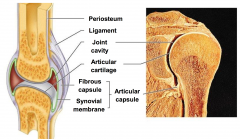
|
Synovial Joint |
|
|
Axes of Movement - Synovial Joints |
- Nonaxial (no movement) - Uniaxial (one axis only) - Biaxial (two axis) - Multiaxial (multiple axis) |
|
|
Structural Shape - Synovial Joints |
- Plane/gliding (nonaxial) - Hinge (uniaxial) - Pivot (uniaxial) - Condyloid/Circular (biaxial) - Saddle (biaxial) - Ball and socket (multiaxial) |
|
|
Joint Movement |
- Angular Movement - Circular Movement |
|
|
Angular Movements of Joints |
- Flexion (reduces angle) - Extension (increases angle) - Abduction (lateral, away from axis of body) - Adduction (medial, towards axis of body) |
|
|
Circular Movements of Joints |
- Conical (cone shape) - Rotation (circular movement along longitudinal axis of a bone) |
|
|
Meninsci |
- Two crescent-shaped pads of fibrocartilage - Lateral and medial - Absorb shock and prevent femur rocking from side to side on tibia |
|
|
Intracapsular Cruciate Ligaments in Knee Joint |
- Prevent forward and backward displacement of tibia - Cross each other to form an X 1. Anterior Cruciate Ligament (ACL) 2. Posterior Cruciate Ligament (PCL) |
|
|
Extracapsular Ligaments in Knee Joint |
- Limit rotation of extended knee 1. Fibular Collateral Ligament (lateral) 2. Tibial Collateral Ligament (medial) |
|
|
Muscle Tissue |
- Responsible for movement in the body - Exhibits contractibility (shortens forcefully) and excitability (responds to stimuli) |
|
|
Types of Muscle Tissue in the body |
1. Skeletal 2. Cardiac 3. Smooth |
|
|
Skeletal Muscle |
- Attached to the skeleton - Controlled voluntarily (somatic motor division) - Striated (striped) - Fibres are very long with multiple nuclei |
|
|
Cardiac Muscle |
- Found in the heart - Controlled involuntarily (autonomic motor division) - Striated (striped) - Fibres are branched cylinders with 1-2 nuclei per fibre |
|
|
Smooth Muscle |
- Found in the walls of many hollow visceral organs - Controlled involuntarily (autonomic motor division) - Non-striated (no stripes) - Spindle-shaped fibres with only 1 nucleus |
|
|
Fascicles |
- Bundles of 10-100 skeletal muscle fibres that are wrapped by connective tissue |
|
|
Structures in Skeletal Muscle Fibres (cells) |
- Sarcolemma (muscle fibre plasma membrane) - Sarcoplasm (muscle fibre cytoplasm) - Myofibrils (long rod-like structures) |
|
|
Myofibrils |
- Many in each skeletal muscle fibre - Contain contractile units called sarcomeres which facilitate muscle movement |
|
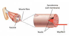
|
Skeletal muscle fibre structure |
|
|
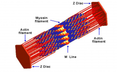
Sarcomeres |
- Contain interlocking actin (thin) and myosin (thick) filament proteins - Contract when stimulated by a signal from a neurone - The arrangement of the contractile proteins give skeletal muscle fibres their striated appearance |
|
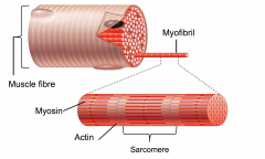
|
Sarcomere |
|
|
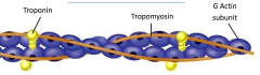
Actin |
- Composed of Globular (G) Actin Subunits - Stiffened by Tropomyosin - Myosin heads bind to these with the help of the molecule Troponin - Anchored to the Z Disc |
|
|
Actin |
|
|
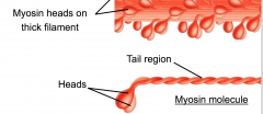
Myosin |
- Molecules have two heads and a tail - The heads contain sites that bind to the actin when contracting - Anchored to the M Line |
|
|
Myosin |
|
|
Structure of a Sarcomere |
|
|
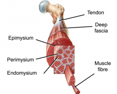
Connective Tissues in Skeletal Muscle Organs |
1. Deep Fascia (lines the body wall and separates muscles into functional groups) 2. Epimysium (outermost of layers 2-4, surrounds an individual muscle and blends with fascia) 3. Perimysium (bundles together individual muscle fibres into fascicles) 4. Endomysium (connective tissue wrapping an individual muscle fibre) |
|
|
Connective Tissues in Skeletal Muscle Organs |
|
|
Naming Muscles |
- Location (eg. costal = ribs) - Shape (eg. deltoid = triangle) - Size (eg. maximus = largest) - Fibre Direction (eg. rectus = straight) - Origin Number (eg. biceps = 2 origins) - Location of Attachments (eg. sterno = sternum) - Action (eg. flexor = flexion) |
|
|
Muscle Actions |
- Prime Movers (produce the major force for a specific movement) - Antagonist (oppose or reverse a particular movement. Help return back to original position) - Synergist (add force to a movement or reduce unnecessary movements) |
|
|
Anatomical Features of a Skeletal Muscle |
- Origin (attachment point of muscle, does not move during muscle contraction) - Belly (thicker middle region of muscle) - Insertion (point where muscle attaches to location which will be moved) |
|
|
Sternocleidomastoid |
- Major head flexors - Can allow lateral and rotational movements of head O = manubrium and clavicle I = temporal and occipital bones |
|
|
Trapezius |
- Stabilises, raises and rotates scapula - Can extend head with scapula fixed O = occipital bone, C7 and all thoracic verterbrae I = clavicle and scapula |
|
|
Rhomboid Major |
- Acts to retract, rotate and stabilise scapula O = vertebrae T2-T5 I = scapula |
|
|
Pectoralis Major |
- Prime mover of arm flexion - Can rotate and adduct shoulder joint O = clavicle, sternum and rib cage I = humerus |
|
|
Deltoid |
- Prime mover of arm abduction - Can flex and extend arm O = clavicle and scapula I = humerus |
|
|
Latissimus Dorsi |
- Prime mover of arm extension - Can adduce and rotate arm O = spine, pelvis and ribs I = humerus |
|
|
Rotator Cuff |
- Group of 4 muscles with reinforce shoulder joint - SITS: Supraspinatus, Infraspinatus, Teres Minor, Subscapularis O = scapula I = rotator cuff muscle tendons blend with the fibrous capsule of the shoulder joint |
|
|
Biceps Brachii |
- Flexes the elbow joint - Supinates forearm O = scapula I = radius |
|
|
Triceps Brachii |
- Powerful forearm extensor O = scapula and humerus I = ulna |
|
|
External Intercostals |
- Elevates ribs during inspiration O = superior rib I = inferior rib |
|
|
Internal Intercostals |
- Depresses rib cage during expiration O = inferior rib I = superior rib |
|
|
Rectus Abdominis |
- Flexes and rotates lumbar region of vertebral column - Stabilises pelvis when walking O = pubic crest and symphysis of pelvis I = sternum and cartilage linking anterior aspects of ribs to sternum |
|
|
External Oblique |
- Rotation of vertebral column - Assist action of rectus abdominis O = inferior 8 ribs I = sternum and aponeurosis abdominal wall |
|
|
Gluteus Maximus |
- Powerful extensor of the thigh - Can laterally rotate and abduct hip joint O = pelvis and lower vertebral column I = femur |
|
|
Quadriceps |
- Powerful knee joint extensors - 4 separate heads which have a common tendon inserting into the patella and tibia: - Vastus Lateralis, Vastus Medialis, Vastus Intermedius O = femur - Rectus Femoris O = pelvis |
|
|
Biceps Femoris |
- Extends the thigh and flexes the knee - Can laterally rotate knee joint O = pelvis (long head) = femur (short head) I = fibula and tibia |
|
|
Gastrocnemius |
- Plantar flexion of the foot (standing on toes) O = femur/knee capsule I = calcaneus bone in foot |
|
|
Cells in Nervous Tissue |
1. Neurones 2. Glial Cells (neuroglia) |
|
|
Neurones |
- Allow rapid cellular communication (transmit information) - Branching, excitable cells - Generate action potentials (movement of ions) - Relatively large cells |
|
|
Glial Cells |
- Provide structural and/or metabolic support to neurones - Outnumber neurones 10:1 |
|
|
Examples of Glial Cells |
- Astrocytes (form links between neurones and blood vessels in CNS) - Oligodendrocytes (provide insulating myelin sheath for CNS neurones) - Microglia (provide immunity from disease and remove debris in CNS) - Schwann Cell (provide insulating myelin sheath for PNS neurones) |
|
|
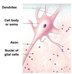
Cellular Components of Neurones |
1. Cell Body (Soma) 2. Dendrites 3. Axons |
|
|
Structure of Neurones |
|
|
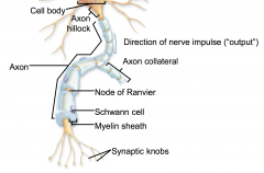
Axons |
- Transmit signals away from cell body - Longest part of the neurone - Lacks Nissl bodies (rough endoplasmic reticulum) |
|
|
Types of Axons |
1. Myelinated (axon is covered by a myelin sheath. These axons are usually quite long and needs to deliver signal very quickly) 2. Non-Myelinated (axon is enveloped by a single fold of a sheath cell |
|
|
Myelinated Axon |
|
|
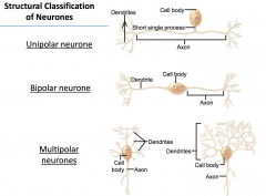
Structural Classification of Neurones |
- Classified according to the number of processes rising from the cell body - Unipolar = 1 process - Bipolar = 2 processes - Multipolar = many processes |
|
|
Structural Classification of Neurones |
|
|
Functional Classification of Neurones |
- Classified by the direction of information flow - Sensory (afferent) = transmit signals to CNS - Motor (efferent) = transmit signals to muscles or glands |
|
|
How Neurones Work |
- The ability to generate a signal involves the movement of charged ions (Na+, K+) across the neurone cell membrane - Ions move across the neurone via active transport (pumped) or diffusion |
|
|
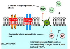
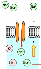
Resting potential |
- At rest, the sodium-potassium pump actively pumps sodium (Na+) and potassium (K+) ions across the cell membrane - In one cycle of the pump: - 3 sodium ions pumped out of cell - 2 potassium ions pumped into cell - As a result the inner surface becomes more negatively charged (-70mv) |
|
|
Resting potential |
|
|
Action potential |
- When stimulated, ion channels in the cell membrane open and close rapidly - This allows ions to quickly enter or exit the cell - When ions move they take their charge with them |
|
|
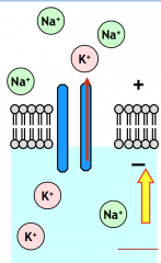
First Phase of Action Potential - Depolarisation |
- Na+ channels open rapidly - Na+ diffuses into the neurone - As a result, the inner membrane surface becomes more positively charged (+30mv) |
|
|
Depolarisation |
|
|
Second Phase of Action Potential - Repolarisation |
- After Depolarisation, Na+ channels close - K+ channels open rapidly - To help reset resting conditions, K+ quickly diffuses out of the neurone - As a result, the inner membrane surface becomes more negatively charged |
|
|
Repolarisation |
|
|
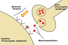
The Synapse |
- A point of communication between a neurone and another cell - Communication is in one direction only - No direct contact between neurone and responsive cell - Communication facilitated by release and reception of chemicals called neurotransmitters |
|
|
Neurotransmitters |
- Chemicals released by the synapse - Contained within vesicles in synaptic bulbs at axon terminal |
|
|
The Synapse and Neurotransmitters |
|
|
Central Nervous System |
- Brain - Spinal Cord |
|
|
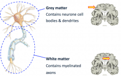
General Tissue Structure |
- Grey Matter (contains many neurone cell bodies and dendrites) - White Matter (contains myelinated axon tracts) |
|
|
Grey and White Matter |
|
|
The Cerebrum |
- The largest part of the brain (80%) - Responsible for high level, conscious mental function - Separated into 2 hemispheres and 5 lobes |
|
|
2 hemispheres of Cerebrum |
1. An outer layer of grey matter (2-4mm thick) 2. A layer of white matter deep into the cortext |
|
|
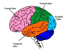
5 lobes of Cerebral Cortex |
1. Frontal (clearly visible on surface) 2. Parietal (clearly visible on surface) 3. Occipital (clearly visible on surface) 4. Temporal (clearly visible on surface) 5. Insula (not visible, under temporal lobe) |
|
|
Visible lobes in Cerebral Cortex |
|
|
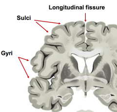
Folds of the Cerebral Cortex |
- Gyri (elevated ridges) - Sulci (shallow grooves) - Fissures (deeper grooves) |
|
|
Folds of the Cerebral Cortex |
|
|
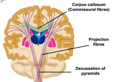
Tracts in Cerebral White Matter |
- Axons in White Matter are organised into pathways called tracts 1. Commissural Tracts (link corresponding areas of grey matter in cerebral hemispheres, the largest is the Corpus Callosum) 2. Projection Tracts (link cerebral hemispheres to inferior CNS regions) |
|
|
Tracts in Cerebral White Matter |
|
|
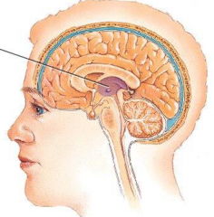
The Thalamus |
- In the central core of the forebrain - Is a major relay station for sensory data to specific areas of the cerebral cortex - Consists of 2 lobes with a midline commissure *imagine the Thalamus is like a computer server relaying data from the internet to a specific PC* |
|
|
The Thalamus |
|
|
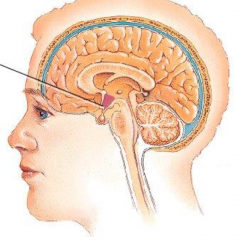
The Hypothalamus |
- Is an autonomic control centre (no thought required) - Regulates many mechanisms of homeostasis: - Body temperature - Water balance - Food intake - Endocrine (hormones) |
|
|
The Hypothalamus |
|
|
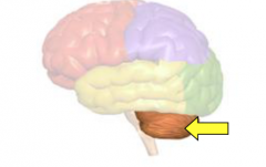
The Cerebellum |
- Makes up 11% of total brain mass - Receives input from cerebral motor cortex, brainstem nuclei, and sensory receptors - Develops the plans needed for smooth coordinated movements - Functions subconsciously - Sits on the posterior cranial fossa, dorsal to the pons and medulla |
|
|
The Cerebellum |
|
|
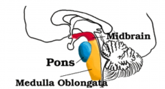
The Brain Stem |
- Composed of Midbrain, Pons and Medulla Oblongata - Controls many basic life functions such as breathing, heart rate and blood pressure - Provides exit points for cranial nerves - Provides connection for higher brain to spinal cord |
|
|
The Brain Stem |
|
|
Protection of the Brain |
1. Skull 2. Meninges (connective tissue membranes) 3. Cerebrospinal Fluid CSF (fluid cushion) |
|
|
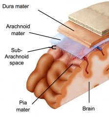
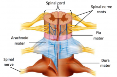
Meninges - 3 Major Layers (PAD) |
1. Pia Mater (inner layer) 2. Arachnoid Mater 3. Dura Mater (outer layer) |
|
|
Meninges |
|
|
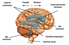
Ventricles of the Brain |
- A series of interconnected cavities that contain cerebrospinal fluid: - Lateral Ventricles (x2) - Third and Fourth Ventricles - Choroids Plexuses (vascular structures on the ventricle roof responsible for CSF production) |
|
|
Ventricles of the Brain |
|
|
Cerebrospinal Fluid (CSF) |
- Floats, nourishes and protects the brain - Flows through the central canal of the spinal cord and subarachnoid space around the brain and spinal cord - A healthy body produces 500mL per day |
|
|
Spinal Cord |
- Connects the brain to the spinal nerves - Extends from medulla to upper lumbar vertebrae - Enclosed within vertebral column - 31 pairs of spinal nerves arise from it - Enlarged in the cervical and lumbar regions where nerves that serve the limbs arise |
|
|
Meninges on the Spinal Cord |
|
|
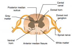
Anatomy of the Spinal Cord |
- Contains a central H-shaped (butterfly) mass of grey matter surrounded by white matter - Ventral Horns (anterior) - Doral Horns (posterior) |
|
|
Spinal Cord Anatomy |
|
|
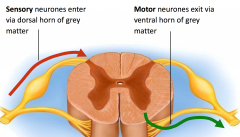
Ventral Horns of Spinal Cord |
- 2 rounded projections of grey matter - Anterior - Contains cell bodies of motor neurones - Motor neurones exit via these horns |
|
|
Dorsal Horns of Spinal Cord |
- 2 pointed projections of grey matter - Posterior - Contains cell bodies of neurones that synapse with sensory neurones - Sensory neurones enter via these horns |
|
|
Spinal Nerve Neurones in Spinal Cord |
|
|
Functions of Motor Nerves |
- Somatic: targets skeletal muscles - Autonomic: targets glands, cardiac & smooth muscle - Sympathetic: promotes responsesrequired for emergencies - Parasympathetic: promotes rest &digest activities |