Reading...
![]()
Play button
![]()
Play button
![]()
Use LEFT and RIGHT arrow keys to navigate between flashcards;
Use UP and DOWN arrow keys to flip the card;
H to show hint;
A reads text to speech;
58 Cards in this Set
- Front
- Back
|
Describe the five main function of bone
|
1. support
2. movement 3. Protection 4. Store bone marrow 5. Reservoir of Ca, PO4, and other ions |
|
|
What are osteoprogenitor cells?
|
Function- repair and growth, can differentiate to osteoblasts
Structure- resemble fibroblasts found in periosteal surfaces |
|
|
What is structure function and location of osteoblasts?
|
function-
1. bone forming cells 2. makes organic components of bone 3. eventually become osteocytes Structure- simple epithelium structure, location- side by side on surfaces of bone |
|
|
What is structure, function and location of osteocytes?
|
Function-
1. maintain bone matrix Structure- 1. flat, almond shaped Location- matrix within lacuna |
|
Identify each and describe roles and structure in bone..
|
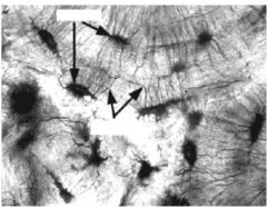
Lacunae are black holes- "cavity" in bone matrix between lamellae
they contain osteocytes and is forced after osteocyte forms Canaliculi- connects with each other and lacunae lead to HC, function to carry nutrients from blood vessels to rest of cell |
|
label left to right
|
Haversion Canal, Canaliculi, lacuna, cement
|
|

What is picture? Role and structure?
|
osteoclasts-
Origin- come from bone marrow-derived cells, Structure - have many nuclei, located at periphery, many organelles (mitochondria) Function- resorption and remodeling |
|
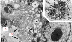
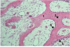
Label appropriate A and B
|
A is clear zone surrounds ruffled border
B is ruffled border involving osteoclasts |
|
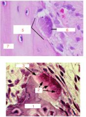
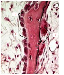
Label 1-6
|
1. bone matrix
2. nuclei 3. osteoclasts 5. resorption bay 6. osteoclast 7. bone |
|
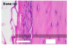
what is pictured?
|
osteocyte
|
|

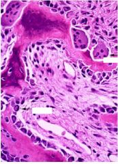
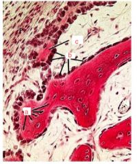
what are the arrows pointing to including the red one...
|
top to bottom... Osteoclasts, howship's lacuna, same
|
|
|
What are the organic (2) and inorganic (2) components of bone matrix
|
Organic-
1. Fibers 2. ground substance- Inorganic- 1. Ions 2. hydration shell for ion exchange |
|
|
What controls osteoclasts?
|
Horomones-calcitonin, PTH- simulate
Cytokines |
|
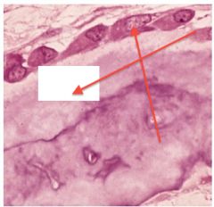
What are arrows pointing to?
|
osteoclast and blast on bottom
|
|

Marble bones?
|
osteopetrosis
|
|
What is 1-3?
|
1. osteoblasts
2. osteoid 3. osteoclasts |
|
|
What disease shows a mosaic patter of disorganized matrix?
|
a. paget's disease- due to remodeling problems
|
|
|
What is one of the components of bone matrix... (Fibers)... made of and role?
|
Structure-
mostly Type I collagen Primary- woven Secondary- lamellar Role- needed for bone growth and structure |
|
|
What are the four main components of ground substance? State a brief fact about the roles of each one and what is ground substance made by?
|
Ground substance is secreted by blasts and clasts
4 main proteins- 1. proteoglycans- strength of bone and inhibit mineralization 2. glycoproteins- promote calcification of matrix 3. vitamin K dependent- Ca capture 4. growth factors and cytokines- |
|
|
What is result of the following...
a. vitamin C deficiency b. Vitamin D deficiency |
a. scurvy
b. rickets- osteomalacia |
|
λαός, ὁ
|
people; nation
|
|
|
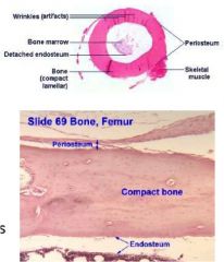
Compare endosteum and
periosteum in terms of location and structure and function |
Have same function both bone associated connective tissue that line bones that provide nutrition and growth/repair
endosteum- internal surface of bone structure- thinner than peri. made of single layer of osteoprogenitor cells periosteum- around/ surrounding bone Structure- dense connective tissue with two layers: outer: collagen and fibroblasts inner: osteoprogenitor cells |
|
|
What are shin splints?
|
inflammation of periosteum?
|
|
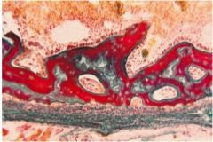
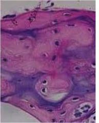
Red in picture is?
|
calcification
|
|
Fill in blanks
|

|
|
Label
|
|
|
|
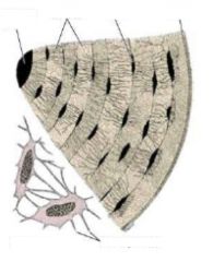
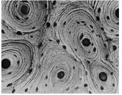
What is the osteon's location and function...
|
Secondary Bone type cell
"haversion system" functionl unit of most compact bones |
|
What type of microscopic bone?
|
primary "woven"
|
|
what type of bone cell?
Where in a adult might you find it permanently? |
primary- immature bone
or sutures of skull, tooth sockets, insertions of some tendons |
|
What are we seeing
|
primary "primitive" bone
a. osteocytes b. osteoblasts c. osteoid Top left is = periosteum |
|
what type of microscopic bone does this show?
|
secondary mature bone
|
|

What is arrow pointing to and role?
|
volkmann's canal- used by haversion canals as means of communication
|
|
|
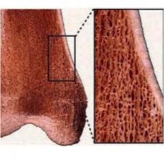
What are the two types of macroscopic bones?
Briefly describe each... |
1. compact (cortical)- no cavities, outer cortex of solid bone (osteons), encloses inner compartment
Located- beneath periosteum, 2. Cancellous (spongy/trabecular)- many interconnecting cavities (woven or lamellar) |
|
What type of macroscopic bone is this?
|
spongy/trabecular
|
|
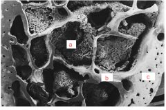
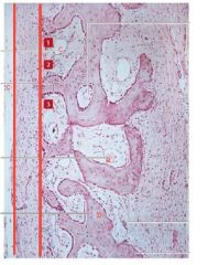
What are the labels of?
|
a. marrow
b. Cancellous or spongy bone c. compact bone |
|
|
What are the 4 types of bones (gross)?
|
1. long
2. flat 3. irregular 4.short |
|
|
Describe and give example of long bones...
|
three parts with "physis"- area bone that lengthens
1. epiphysis- top part, has spongy covered by thin compact 2. diaphysis- growing between- has medullary cavity 3. metaphysis- region of growing, area of growth plate |
|
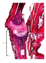
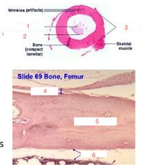
Where is markers pointing to?
|
1. diaphysis
2. metaphysis 3. epiphysis |
|
|
Describe and give example of flat and short bones...
|
flat- have 2 layers of compact bone called plates seperated by dipole (spongy bone)
ex. bones of skull short- spongy core surrounded by compact - metacarpals |
|
|
what are the two types of bone formation?
|
1. intramembranous- mesenchymal replaced by bone
2. endochondral- skeletal cartilage templates replaced by bone |
|
|
Describe the steps in intramembranous bone formation...where does it take place and controlling factors?
Which bones undergo this type of formation process? |
Takes place in condensations of mesenchyme tissue
Controlled by GDF - flat bones, contributes to growth of short, thickening of long 1. mesenchymal cells aggregate 2. Form osteoblasts, bone blastema formed and syncytium formed 3. Matrix (osteoid) deposited by osteoblasts and Ca is taken into for minerals and bone tissue is formed |
|
Label parts
|
|
|
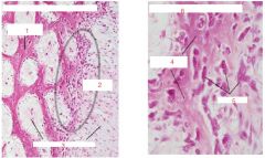
Label the picture... what is going on?
|
a. osteoblasts
b. osteocyte c. osteoid d. primary or woven bone e. mesenchyme |
|
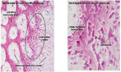
Find the a-e labeling what is it?
|
intra membranous ossification
a. osteoblasts b. osteoprogenitor c. bone spicules- form circles around vessels d. blood vessels e. mesenchyme |
|
What is going on here?
|
intramembranous ossification
a. bone spicule b. periosteum c. osteoblast d. mesenchyme |
|
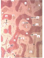
What is going on between the two lines?
What are labels and 1-3? |
1-3 are recap of steps
1. well-vascularized primitive connective tissue 2. not preceded by cartilage formation 3. agreget of cells differentiates a blood vessel b. trabecula c. blood vessel lines are where periosteum is formed |
|
|
Whats the difference between spongy and compact bone ossification?
|
Compact- deposition of bone on trabecular surfaces
Spongy- thickening of trabecular surfaces do not occur and primary spongiosa turns into cancellous bone |
|
|
Describe the location of endochondral bone formation, when does it occur? What bones are formed?
|
occurs within cartilage on piece of hyaline cartilage, both bone and cartilage are present (fetus and child growth)
short/long bones (extremities) |
|
|
What are the 6 stages of endochondrial ossification?
|
1. development of hyaline cartilage model
2. growth of cartilage model 3. formation of primary ossification center 4. formation of marrow cavity 5. formation of secondary ossification center 6. formation of epiphyseal plate and articular cartilage |
|
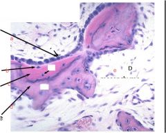
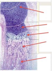
What is shown?
|
periosteal bone collar
a. hyaline cartilage b. hypertrophic cartilage c. periosteol bone collar d. perichondrium |
|
|
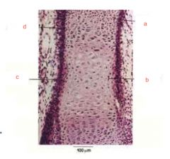
Describe the cartilaginous structure and function of an epiphyseal plate.
|
Structure- thin plate of cartilage left connects two epiphyses to diaphysis
Role- responsible for area of bone growth, will contain specific histologic zones during growth |
|
|
What are the five zones of epiphyseal plate?
|
1. zone of resting (closest to articular, hyaline surface)
2. Zone of proliferation- columns of flattened chondrocytes 3. zone of hypertrophy- columns of enlarged cells 4. zone of calcification- dying chondrocytes 5. zone of ossification- osteoid deposited by osteoblasts |
|
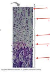
What is going on here?
|
epiphyseal zones
1. zone of resting 2. zone proliferation 3 Hypertrophic zone 4. calcification zone 5. zone of ossification |
|
Where is zone of proliferation? hypertrophic zone?
|
prolif- second from top
hypertrophic zone- third from top |
|
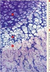
which zones are which?
|
1. proliferative zone
2. hypertrophic zone 3. vascularization zone |
|
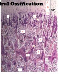
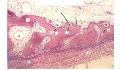
What are the labels?
|
this is endochondrial ossification...
a. resting b. proliferation c. hypertrophic d. calcification e. marrow f. bone (ossification |
|
|
Describe how fractured bone get repaired...6-steps
|
1. blood clots on fracture
2. vessels around undergo shutdown 3. loss of blood supply kills haversian cannals 4. macrophages come clean up 5. intense proliferation of osteoprogenitor cells by periosteum and endosteum to form collar 6. bone callus forms, which is remodeled with stress placed on it |
|
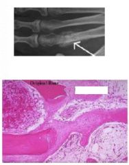
What is arrow on x-ray and what is blank spot?
|
both are fracture calluses
|