Reading...
![]()
Play button
![]()
Play button
![]()
Use LEFT and RIGHT arrow keys to navigate between flashcards;
Use UP and DOWN arrow keys to flip the card;
H to show hint;
A reads text to speech;
90 Cards in this Set
- Front
- Back
|
Function of the Respiratory system:
|
provide oxygen to the tissues of the body in exchange for carbon dioxide
|
|
|
2 major divisions of the respiratory system:
|
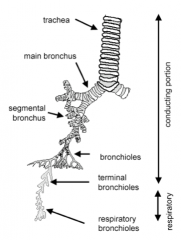
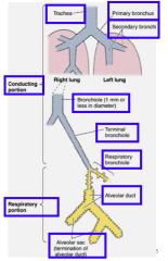
1. Conducting portion: airways that deliver air to the lungs
2. Respiratory portion: structures within the lung where gaseous exchange occurs |
|
|
Conducting Portion of respiratory system:
|
- DELIVERs air to the respiratory tissue
- warm, moisten and filter the air before it reaches the respiratory tissue |
|
|
What does the conduction portion of the respiratory system include:
|
nose
pharynx larynx trachea bronchi bronchioles down to and including the terminal bronchioles |
|
|
Respiratory portion of respiratory system:
|
*where EXCHANGE of gases take place
|
|
|
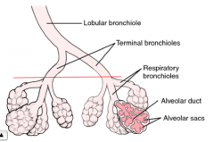
What does the respiratory portion include:
|
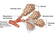
respiratory bronchioles
alveolar ducts alveolar sacs alveoli *these parts of the system are INTRApulmonary |
|
|
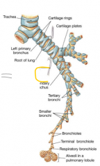
The main divisions of the respiratory tract include:
|
|
|
|
What are nares:
|
= nostrils --> whose outermost portions are lined by extensions of skin
|
|
|
Epithelium of the nares:
|
Keratinized stratified squamous epithelium
- contains sweat glands, hair follicles, and sebaceous glands |
|
|
Nasal cavity include:
|
Vestibule
Respiratory Olfactory |
|
|
Vestibule is:
|
the first portion of the nasal cavity
|
|
|
Skeleton of the vestibule:
|
Hyaline cartilage
|
|
|
What type of epithelium does the vestibule have?
|
Stratified squamous keratinized
- Posteriorly, the lining changes to respiratory epithelium (pseudostratified ciliated columnar epithelium with goblet cells). *NO cilia * No Goblet cells |
|
|
What type of glands does the vestibule contain?
|
Sebaceous
sweat glands |
|
|
Feature(s) of the Vestibule:
|
contains Vibrissae (thick short hairs) that filter out large particles from the inspired air
|
|
|
Lamina propria layer of the vestibule:
|
Vascular (contains many venous plexuses)
has a number of seromucous glands |
|
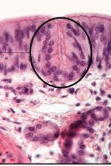
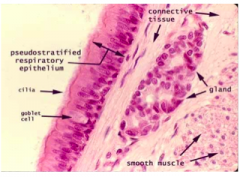
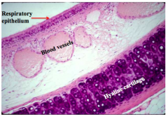
What is seen in this slide?
|
Nasal cavity : intraepithelial gland
|
|
|
Skeletal structure of the Respiratory region of the nasal cavity:
|
Bone & hyaline cartilage
|
|
|
What type of epithelium does respiratory region have?
|
Pseudostratified ciliated columnar
*HAS cilia *HAS goblet cells |
|
|
What type of glands does the respiratory region contain?
|
Seromucous
|
|
|
Feature(s) of Respiratory region:
|
Large venous plexus
|
|
|
Skeletal structure of the olfactory region:
|
Nasal conchae (bone)
|
|
|
What type of glands can you find within the olfactory region?
|
Bowman's glands
|
|
|
Feature(s) of the olfactory region:
|
Basal cells
sustentacular cells olfactory cells nerve fibers |
|
|
Olfactory epithelium is located at the:
|
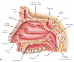
Roof of nasal cavity, on either side of the nasal septum and onto the superior nasal conchae
|
|
|
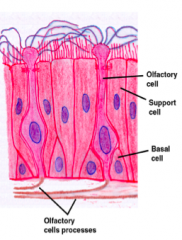
Epithelial layer of the olfactory region consists of:
|
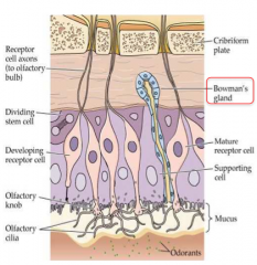
Pseudostratified ciliated columnar
*HAS cilia *NO Goblet cells |
|
|
What are the types of cells within the Olfactory mucosa?
|
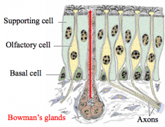
Pseudostratified ciliated columnar with 3 types of cells:
a. olfactory b. supporting c. basal |
|
|
What are olfactory cells?
|
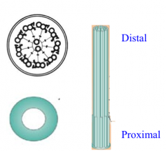
- bipolar nerve cells characterized by a bulbous projection (olfactory vesicle) from which several modified cilia extend
- olfactory cilia --> acts as receptors (nonmotile & very long) - proximal 1/3 contains a typical axoneme but their distal 2/3s is composed of 9 peripheral singlets surrounding 2 central singlets |
|
|
What are the structures of the supporting (sustentacular) cells?
|
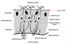
- Possess nuclei that are more apically located than those in the other two cell types.
- Have many microvilli and a prominent terminal web |
|
|
Where are the basal cells located?
|
Rest on the basal lamina but do not extend to the surface and form an incomplete layer of cells
|
|
|
What are basal cells?
|
The are believed to be regenerative for all three-cell types
|
|
|
Lamina Propria of the Basal cells consists of:
|
many veins
unmyelinated nerves Bowman’s glands |
|
|
Pharynx includes which 2 regions?
|
Nasal
Oral |
|
|
What are the supporting structures for the Nasal & Oral region?
|
Muscle
|
|
|
What types of gland does the nasal region have?
|
Seromucous glands
|
|
|
Epithelial layer of the nasal region?
|
Pseudostratified ciliated columnar
*HAS cilia *HAS goblet cells |
|
|
Features of the nasal region:
|
Pharyngeal tonsil
eustachian tube |
|
|
What type of glands are located within the oral region of the pharynx?
|
Seromucous glands
|
|
|
Epithelial layer of the oral region:
|
Stratified squamous non-keratinized
*NO cilia *NO goblet cells |
|
|
Feature(s) of the oral region of the pharynx?
|
Palatine tonsils
|
|
|
The larynx connects....
|
pharynx with trachea
|
|
|
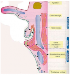
Skeletal structure of the larynx (division):
|
Wall is supported by:
a. Hyaline cartilage (thyroid, cricoid, lower part of arytenoids) b. Elastic cartilages (epiglottis, corniculate and tips of arytenoids) Remainder of the wall contains striated muscle and CT with glands |
|
|
What type of glands located in the larynx?
|
Mucous & seromucous glands
|
|
|
Epithelial layer of the Larynx:
|
Stratified squamous non-keratinized
Pseudostratified ciliated columnar *HAS cilia *HAS goblet cells |
|
|
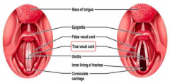
Features of the Larynx:
|
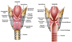
Vocal cords
epiglottis some taste buds |
|
|
How do the vocal cords work?
|
Muscles within the larynx contract and change the size of the opening between the vocal cords, which provide the means for sounds of different frequencies to be produced
|
|
|
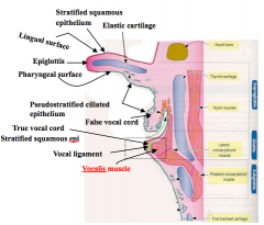
What does the true vocal cords consists of?
|
- skeletal muscle (the vocalis)
- the vocal ligament (formed by a band of elastic fibers) - a covering of stratified squamous nonkeratinized epithelium |
|
|
Where does the change in lining epithelium occur?
|
- It changes to respiratory epithelium at base of epiglottis, inferior to the vocal cords
- Respiratory epithelium lines air passages down through trachea and primary bronchi |
|
|
What is the vestibular fold?
|
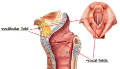
= false vocal cord
= a fold of loose connective tissue containing glands and lymphoid aggregations - covered by respiratory epithelium |
|
|
Where are the Vestibular fold (false vocal cord) located?
|
Lies superior to the true vocal cord
|
|
locate different structures:
|
|
|
|
Trachea & extrapulmonary (primary) bronchi are supported by:
|
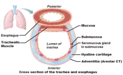
C-rings of hyaline cartilage (with open ends facing posteriorly)
- smooth muscle (trachealis) extends between open ends of the cartilage - Dense fibroelastic connective tissue superior and inferior to each cartilage --> facilitates the elongation of the trachea during inhalation |
|
|
Types of gland you find within the trachea & extrapulmonary bronchi:
|
Mucous & seromucous glands
|
|
|
Epithelial layer of the trachea & extrapulmonary (primary) Bronchi is composed of:
|
Pseudostratified ciliated columnar
*HAS cilia *HAS goblet cells |
|
|
Features of the trachea & primary bronchi:
|
Trachealis muscle
elastic lamina |
|
|
The lumen of the trachea and extrapulmonary bronchi are lined by:
|
respiratory epithelium
|
|
|
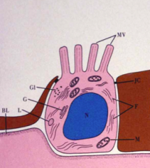
In human, the respiratory epithelium consists of different cell types:
|
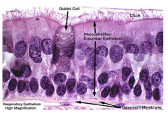
- ciliated cells
- mucous cells - mature goblet cell - enteroendocrine cells |
|
|
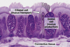
Importance of the Ciliated cells:
|
- protect delicate lung tissue from possible damage by inhaled particulate matter
- have long actively motile extensions that beat in the direction of the pharynx * Ciliated cells also contain microvilli |
|
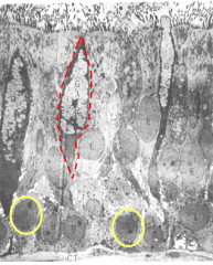
What type of epithelium is this? what stain is used?
|
Respiratory epithelium (pseudostratified ciliated columnar)
Pararosaniline—toluidine blue (PT) stain |
|
|
What is an axoneme?
|
the central core of a cilium
|
|
|
Cilia are anchored to the cells by:
|
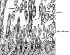
U-shaped basal bodies
|
|
|
The two types of mucous cells are:
|
1. small mucous granule "brush" cell
2. mature goblet cells |
|
|
What are small mucous granule "brush" cells?
|
- called "brush" because of the numerous microvillil
- Actively divides and thus might be able to replace desquamated cells; might also be a goblet cell that has secreted its mucus - contains varying numbers of small mucous granules |
|
|
What are mature goblet cells?
|
- best known because of its shape
- filled with large mucous droplets that are secreted to trap inhaled particles - have short basal, cells "STEM" - are able to divide |
|
|
Where are the mature goblet cells located?
|
Rest on the basal lamina but do not extend to the lumen, making epithelium pseudostratified
|
|
|
What are enteroendocrine cells?
|
- also "APUD": Amine Precursor Uptake Decarboxylase
- small granule cells also from part of the epithelium - contain many small granules concentrated in their basal cytoplasm |
|
|
Where are the functions of the enteroendocrine cells?
|
- These cells exert a local affect on nearby structures and cell types (paracrine regulation)
- Various types of enteroendocrine cells synthesize different polypeptide hormones |
|
|
What is a basement membrane?
|

a very thick layer that underlies the epithelium
|
|
|
Trachea lamina propria:
|
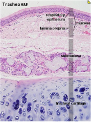
- A thin layer of connective tissue that lies beneath the basement membrane.
- Elastic fibers run longitudinally and separate the lamina propria from the submucosa *distinct line between lamina propria and submucosa! |
|
|
Trachea Submucosa contains what type of glands?
|
seromucous glands
|
|
|
Outer layer of the trachea is formed by:
|
- Adventitia
- Contains C-shaped cartilages |
|
|
cross section of the wall of the trachea
Thick cartilage within the adventitia layer |
|
|
Origin of the intrapulmonary bronchi (secondary bronchi):
|
Arise from subdivision of the primary bronchi and divide many times
|
|
|
Feature(s) of the Intrapulmonary Bronchi (secondary bronchi)
|
Have irregular cartilage plates in their walls
- respiratory epithelium lines the lumina of the intrapulmonary bronchi |
|
|
Intrapulmonary Bronchi (secondary bronchi) can be divided into which 3 regions?
|
Secondary bronchi
Bronchioles Terminal bronchiole |
|
|
Secondary bronchi is supported by what type of skeletal structure?
|
Plates of hyaline cartilage
|
|
|
What type of glands does the secondary bronchi have?
|
Seromucous glands
|
|
|
What is the epithelial layer of the secondary bronchi?
|
Pseudostratified ciliated columnar
*HAS cilia *HAS goblet cells |
|
|
What are the distinctive features of the secondary bronchi?
|
Two helically oriented ribbons of smooth muscle
|
|
|
What are the structures that support the bronchioles?
|
Smooth muscle
|
|
|
What kind of glands can you find within the bronchioles?
|
NONE!
|
|
|
What lines the bronchioles?
|
Simple columnar to simple cuboidal
*HAS cilia *HAS goblet cells BUT ONLY IN larger bronchiole |
|
|
Special features of the bronchioles:
|
Clara cells
= dome-shaped cells with short microvilli found in the small airways (bronchioles) of the lungs |
|
|
The terminal bronchiole is supported by...
|
smooth muscle
|
|
|
Are there any glands within the terminal bronchiole?
|
NO!
|
|
|
What lines the surface of the terminal bronchiole?
|
Simple cuboidal
*HAS some cilia *NO goblet cells |
|
|
Features/structures of the terminal bronchiole:
|
Less than 0.5 mm in diameter
Clara cells |
|
|
Lamina Propria of the Intrapulmonary Bronchi (secondary bronchi) is separated from the submucosa by...
|
layers of spiraling smooth muscle
|
|
|
Glands (seromucous) of the intrapulmonary are present in which layer?
|
Submucosa
|
|
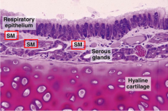
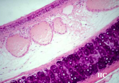
Identify all the layers of the secondary bronchus.
|
- respiratory epithelium with goblet cells and columnar ciliated cells
- The connective tissue of the lamina propria contains serous glands and smooth muscle (SM) - In the lower half of the photomicrograph is a large piece of hyaline cartilage *Smooth muscles are not present in the primary bronchi and trachea |