![]()
![]()
![]()
Use LEFT and RIGHT arrow keys to navigate between flashcards;
Use UP and DOWN arrow keys to flip the card;
H to show hint;
A reads text to speech;
66 Cards in this Set
- Front
- Back
|
Functions of blood vessels
|
Distribution of blood (arteries) Exchange at the tissues (capillaries) Return of blood to the heart(veins) |
|
|
Starling law of the heart (also known as Starling's law or the Frank–Starling mechanism or Maestrini heart's law) states that
|
the stroke volume of the heart increases in response to an increase in the volume of blood filling the heart (the end diastolic volume) when all other factors remain constant.
|
|
|
diostole |
relaxation |
|
|
systole |
contraction |
|
|
structure of blood vessels |
Structure:
Most have the same basic structure: – 3 layers surrounding a hollow lumen |
|
|
arteries and veins are composed of three tunics
|
tunica interna/intima
tunica media tunica externa |
|
|
Capillaries are composed of |
endothelium resting on a sparse basal lamina. |
|
|
Tunica interna (intima):
|
Single endothelial layer that lines the lumen of all vessels.
In vessels larger than 1 mm, a subendothelial connective tissue basement membrane is present |
|
|
Tunica media:
|
Smooth muscle and elastic fiber layer, regulated by sympathetic nervous system Circumferentially arranged
Controls vasoconstriction/vasodilation of vessels |
|
|
Tunica externa (adventitia):
|
•Collagen fibers that protect and reinforce vessels
Longitudinally arranged Larger vessels contain vasa vasorum More pronounced in veins than in arteries |
|
|
The vasa vasorum
|
a network of small blood vessels that supply the walls of large blood vessels, such as elastic arteries (aorta) and large veins (vena cava).
|
|
|
Tunica Intima
|
Innermost smooth layer
Simple squamous epithelium Continuous with the endocardium Present in all vessels |
|
|
Tunica Media
|
layer of smooth muscle
- circular arrangement – contains elastin Supplied by sympathetic division of the ANS ̈Depending on demands– lumen is narrowed (vasoconstriction) or widened (vasodilation) |
|
|
Tunica Externa (Adventitia)
|
Thin layer of Connective Tissue
Elastic & collagen fibres |
|
|
The Vessels Types of Vessels
|
Arteries– carry blood away from the heart
Veins – carry blood towards the heart Capillaries – the most important part of the vascular system; site of exchange of materials |
|
|
Arteries
|
blood from heart
¤Strong & Elastic ¤Conduct blood to capillaries ¤Sphincters ̈ |
|
|
Capillaries:
|
exchange with cells ̈
|
|
|
Veins
|
¤Return blood to heart
¤Valves |
|
|
Make Up of Blood Vessels: Arteries and Arterioles
|
̈Endothelium
̈Elastic tissue -(Recoil in diastole ,Evens flow) ̈Smooth muscle Fibrous tissue (Tough and Resists stretch ) |
|
|
functions of Endothelial cells |
Critical to blood homeostasis Rod-like inclusions – Wiebel Palade Bodies-----Contain vWF (Factor VIII) ̈ Maintain selective permeability A non-thrombogenic barrier that maintains blood fluidity Alter vascular resistance Immune response Secrete growth factors Convert Ang I to Ang II Inactivate norepinephrine, prostaglandins, serotonin Oxidise LDL |
|
|
healty endothelium |
antithrombic anti inflammatory anticoagulant protrombolytic antihypertrophic endothelium-dependent vasodilation |
|
|
the tunica media of the aorta is made up of |
multiple elastic layers |
|
|
elastic arteries |
Thick-walled arteries near the heart; the aorta and its major branches.
Large lumen allows low-resistance conduction of blood. Contain abundant elastin in all three tunics. ̈walls stretch and recoil to propel blood ̈Withstand and regulate large blood pressure fluctuations. Allow blood to flow fairly continuously and uniformly |
|
|
Smooth Muscle,
|
Spindle shaped
elongate nucleus No fibroblasts Smooth muscle cells have synthetic activity and can proliferate and migrate to intima in repair or atherosclerosis |
|
|
atherosclerosisˌ
|
a disease of the arteries characterized by the deposition of fatty material on their inner walls.
|
|
|
Tunica media of elastic arteries is most pronounced and contains
|
ELASTIN – fenestrated sheets/lamellae between muscle layer, concentrically arranged.
Smooth Muscle Collagen and proteoglycan |
|
|
fenestrated
|
having perforations, apertures,
|
|
|
Muscular arteries –
|
distal to elastic arteries; deliver blood to body organs
Have thick tunica media with more smooth muscle and less elastic tissue Active in vasoconstriction |
|
|
Arterioles
|
– smallest arteries; lead to capillary beds Control flow into capillary beds via vasodilation and constriction.
close to capillaries - single layer of muscle spiralling around the endothelial lining
Smooth muscle before a capillary bed – forms a precapillary sphincter |
|
|
tunica intima in elastic arteries |
endothelium smooth muscle connective tissue |
|
|
tunica media of elastic arteries |
alternating between elastic fibres and smooth muscle |
|
|
Adventitia/tunica externa of elastic arteries |
thin layer of collagen,elastic fibers and fibroblasts |
|
|
tunica intima of mascular arteries |
endothelium ,internal elastic membrane |
|
|
tunica media of mascular arteries |
smooth muscle and collagen fibers |
|
|
tunica externa of muscular arteries |
external elastic membrane fibroblasts collagen fibers |
|
|
Tunics of veins
|
less well defined
|
|
|
veins have
|
Thinner walls and larger lumens, may be collapsed
Semilunar valves- that convey blood against gravitational pull |
|
|
Pericytes are
|
contractile cells that wrap around the endothelial cells of capillaries and venules throughout the body.
|
|
|
Postcapillary venules–
|
smallest venules, composed of endothelium and a few pericytes
Site of WBC extravasation |
|
|
Muscular venules
|
have one or two layers of smooth muscle (tunica media)
Distal to post capillary venules |
|
|
high endothelial venules |
site of ; fluid absorbtion(via -aquaporin -1 channels) which cause lymph flow
exit of lymphocytes from blood stream trough diapedesis.
|
|
|
diapedesis
|
The outward passage of blood cells through intact vessel walls.
|
|
|
medium and large veins |
Most have valves in lower body
Three layers Tunica media contains smooth muscle as a thin layer In large veins, the tunica adventita is larger and pronounced Capacitance vessels (blood reservoirs) that contain 65% of the blood supply |
|
|
Veins have _____________ blood pressure and ____________walls than arteries
|
much lower
thinner |
|
|
special adaptations that veins have to return blood to the heart, |
Large-diameter lumens, which offer little resistance to flow Valves (resembling semilunar heart valves), which prevent backflow of blood Venous sinuses – specialized, flattened veins with extremely thin walls (e.g., coronary sinus of the heart and dural sinuses of the brain) |
|
|
Veins and Venules (Contrasted to Arteries)
|
Thinner walls
Larger diameter Closer to skin Less muscle Less elastic |
|
|
Anastomoses
|
Merging blood vessels, more common in veins than arteries
|
|
|
Arterial anastomoses provide
|
alternate pathways (collateral channels) for blood to reach a given body region
If one branch is blocked, the collateral channel can supply the area with adequate blood supply |
|
|
Thoroughfare channels are examples of
|
arteriovenous anastomoses
|
|
|
Capillaries
|
Smallest vessels, with diameters less than erythrocytes
Sites of exchange with tissues A single layer of endothelial cells resting on a basal lamina |
|
|
Types
|
¤Continuous
¤Fenestrated ¤Discontinuous |
|
|
Continuous capillaries are abundant in
|
the skin
muscles CNS lung |
|
|
continuous capillaries have: |
Endothelial cells that provide an uninterrupted lining
Adjacent cells that are held together with tight junctions Intercellular clefts of unjoined membranes that allow the passage of fluids Pericytes (Rouget cells) may surround capillaries |
|
|
Continuous capillaries of the brain:
|
Have tight junctions completely around the endothelium .
Constitute the blood-brain barrier. |
|
|
fenestrated capillaries are abundant in
|
small intestines endocrine glands and kidneys Found wherever active capillary absorption or filtrate formation occurs |
|
|
fenestrated capillaries are characterized by: |
An endothelium riddled with pores (fenestrations) Greater permeability to solutes and fluids than other capillaries
|
|
|
Sinusoids / Discontinuous Capillaries
|
Highly modified, leaky, fenestrated capillaries with large lumens
Allow large molecules (proteins and blood cells) to pass between the blood and surrounding tissuesBlood flows sluggishly, allowing for modification in various ways |
|
|
discontinuous cappillaries are mainly found in |
the liver
bone marrow lymphoid tissue some endocrine organs |
|
|
Capillary Beds
|
A microcirculation of interwoven networks of capillaries, consisting of:
True capillaries metarterioles |
|
|
Metarteriole
|
a short vessel that links arterioles and venules. Instead of a continuous tunica media, they have individual smooth muscle cells placed a short distance apart
|
|
|
True capillaries
|
10 to 100 per capillary bed, capillaries branch off the metarteriole and return to the thoroughfare channel at the distal end of the bed
|
|
|
Precapillary sphincter
|
¤smooth muscle that surrounds each true capillary
Regulates blood flow into the capillary Blood flow is regulated by vasomotor nerves and local chemical conditions, so it can either bypass or flood the capillary bed |
|
|
corrugated |
shaped into a series of parallel ridges and grooves so as to give added rigidity and strength. |
|
|
histology of aorta |
in H and E staining elastic lamelli not well defined Resorcin-Fuschin stain - fenestrated lamella easily seen |
|
|
Endothelium
|
a type of epithelium that lines the interior surface of blood vessels and lymphatic vessels
|
|
|
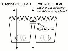
transcellular vs paracellular paths,
|
|