Reading...
![]()
Play button
![]()
Play button
![]()
Use LEFT and RIGHT arrow keys to navigate between flashcards;
Use UP and DOWN arrow keys to flip the card;
H to show hint;
A reads text to speech;
127 Cards in this Set
- Front
- Back
|
Extrinsic glands of the digestive system are located...
|
outside the wall of the alimentary canal
- deliver secretion into lumen via a system of Ducts |
|
|
Function of extrinsic glands:
|
-Provide enzyme, buffers, emulsifiers, and lubricants for the digestive tract
-Provide hormones, proteins, globulins ...etc. to the remainder of the body |
|
|
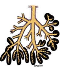
Extrinsic glands include:
|
Salivary glands (parotid, sublingual, submandibular)
Pancreas Liver |
|
|
Salivary glands are:
|
=exocrine glands in the mouth
-produce saliva (function in digestive, lubricating, protective) -pH: 6.5-6.9 |
|
|
Saliva is important in:
|
buffering
evaporative cooling (in nonhuman species) |
|
|
3 pairs of the large salivary glands are:
|
1. Parotid
2. Submandibular 3. Sublingual -in addition to minor glands in mucosa and submucosa throughout the oral cavity --> secrete 10% of the total vol. of saliva |
|
|
Reduced function of the major salivary glands (due to diseases/radiotherapy) is associated with:
|
caries - atrophy of the oral mucosa and speech difficulties (destroy bone structures i.e. teeth)
|
|
|
Each major salivary gland is surrounded by:
|
A capsule of connective tissue
|
|
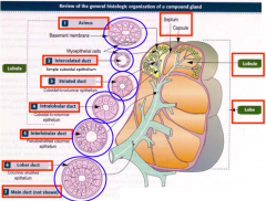
Organization of a compound gland:
|
Main duct < Lobar duct < INTERlobular duct < INTRAlobular duct < Striated duct < Intercalated duct < Acinus
|
|
|
What lines intralobular duct?
|
Cuboidal to columnar
|
|
|
What lines striated duct?
|
Cuboidal to columnar
|
|
|
What lines interlobular duct?
|
Pseudostratified columnar
|
|
|
What lines lobar duct?
|
Columnar stratified
|
|
|
What lines intercalated duct?
|
Simple cuboidal
|
|
|
Secretion of each gland can be:
|
Serous (fluid)
Seromucous Mucous (viscous) *depending on its glycoprotein mucin content |
|
|
Saliva from the Parotids is:
|
Serous & watery
|
|
|
Submandibular and sublingual glands produce what type of secretion?
|
Seromucous secretion
with mostly mucus from minor glands |
|
|
Saliva is modified in which way?
|
Much Na+ & Cl- reabsorbed
Certain growth factors and digestive enzymes are added |
|
|
What are serous cells?
|
Polarized protein-secreting cells
usually pyramidal in shape broad base resting on basal lamina narrow apical surface facing lumen |
|
|
Ajacent serous cells are joined by...
|
Junctional complexes --> form a spherical mass of cells = Acinus
|
|
|
Shape of acini and their duct system?
|
like grapes attached to a stem
|
|
|
Serous acinar cells produce:
|
Digestive enzymes and other proteins
|
|
|
Shape of the mucous cells:
|
~cuboidal / columnar with nuclei pressed toward the bases of the cells
|
|
|
What do the mucous cells contain?
|
hydrophilic glycoprotein mucins
*important for the moistening and lubricating functions of the saliva |
|
|
Mucous cells are organized in what shape?
|
most often tubules rather than acini
-produce mostly mucins |
|
|
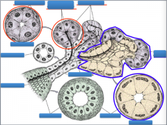
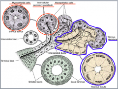
Where are myoepithelial cells found?
|
Inside the basal lamina of the secretory units
and (less in) the initial part of the duct system |
|
|
Myoepithelial cells are sometimes called...
|
Basket cells
|
|
|
Shape of the myoepithelial cells?
|
1. those surrounding the secretory portion --> Well developed and branched
2. those associated with initial ducts --> spindle-shaped & lie parallel to the duct's length |
|
|
Function of the myoepithelial cells:
|
- prevent distention of the endpiece when lumen fills with saliva
- contraction to accelerate secretion of the product |
|
|
Course of salivary glands:
|
Secretory endpieces (serous acinus/mucous tubule) --> intercalated ducts --> striated ducts --> intralobular ducts --> interlobular ducts --> lobar ducts --> main duct
|
|
|
Structure of the Columnar cells of striated ducts:
|
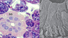
Radial striations extending from the cell bases to the level of the nuclei
- striations consist of infoldings of the basal plasma membrane *the folds increases cell surface area --> facilitate ion absorption & ion transport |
|
|
Columnar cells of striated ducts contain:
|
numerous mitochondria aligned parallel to the infolded membranes which contain ion transporters
|
|
|
Striated ducts converge and drain into ducts located in the CT septa and becomes...
|
Interlobular ducts
|
|
|
Epithelium of the ducts:
|
Initiallly: pseudostratified/stratified cuboidal
Distal part: stratified columnar with a few mucous cells |
|
|
The main duct of each large salivary gland empties into...
|
Oral cavity
|
|
|
Epithelium of the oral cavity:
|
Nonkeratinized-stratified squamous epithelium
|
|
|
Vasculization of the salivary glands:
|
- vessels and nerves enter the large salivary glands at a hilum
- a rich vascular and nerve plexus surrounds the secretory and ductal components of each lobule - capillaries surrounding secretory endpieces --> imp for secretion of saliva (stimulated by autonomic n.s.) |
|
|
Parasympathetic stimulation of the salivary glands:
|
- elicited by smell/taste of food
- provokes copious (=plenty) watery secretion with relatively little organic content |
|
|
Sympathetic stimulation of the salivary glands:
|
- inhibits secretion --> dry mouth (often associated with anxiety)
|
|
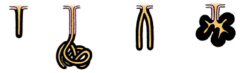
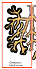
Name the different structures of ducts
|
1. simple tubular
2. simple coiled tubular 3. simple branched tubular 4. simple branched acina |
|
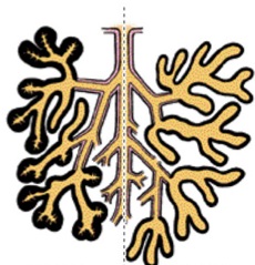
Structure of this duct?
|
left: compound tubuloacinar
right: compound tubular |
|
Structure of this duct:
|
Compound acinar
|
|
|
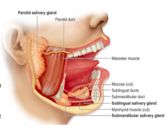
Know the location of the major salivary glands
|
|
|
|
Classification of parotid gland:
|
Purely serous
branched acinar |
|
|
What forms the capsule of the parotid gland?
|
by the continuation of superficial cervical fascia - mostly collagenous
--> forms broad bands of trabeculae (septa) that subdivide the gland into lobes and lobules |
|
|
Trabeculae of the parotid glands:
|
convey blood and lymph vessels, ducts, and nerves into the substance of the gland
- contain fat cells in older individuals |
|
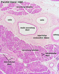
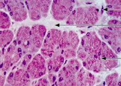
What type of gland is this?
|
Parotid gland
|
|
|
Acini of parotid glands:
|
Composed of seromucous instead of purely serous cells (serous cells predominate)
-surrounded by myoepithelial cells -contains lumen in the center |
|
|
Secretory portion of the parotid gland consists of:
|
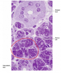
-Serous amylase-producing cells that store this enzyme in secretory granules
-intralobular (intercalated & striated) ducts *slide stained with toluidine blue (PT) |
|
|
Structure of the Acinar cells:
|
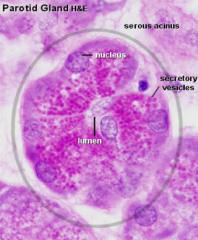
-Pyramidal cells
-apical aspects usually contain secretory granules -nucleus is round and basally located -cytoplasm contains extensive RER, well-developed golgi apparatus, numerous mitochondria |
|
|
Structure of the Myoepithelial cells:
|
-Stellate-shaped
-cytoplasm is difficult to distinguish with LM -with EM, they resemble smooth muscle cells and are contractile |
|
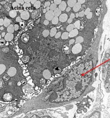
Red arrow is pointing to:
|
nucleus of a myoepithelial cell
|
|
|
Stensen’s Duct:
|
- largest (main parotid duct)
- opens into the oral vestibule at the parotid papilla |
|
|
What kind of epithelium lines the Stensen's duct?
|
Simple columnar or pseudostratified epithelium
|
|
|
Mode of secretion is classified into:
|
1. Merocrine (process of exocytosis)
2. Apocrine 3. Holocrine |
|
|
What are the smallest ducts
|
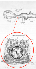
Intercalated ducts
- lie interposed (between) among the acini (initial part of the duct system) |
|
|
What lines the intercalated ducts?
|
Simple cuboidal epithelium
|
|
|
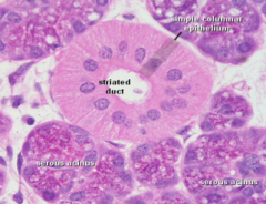
Striated ducts are lined by:
|
High cuboidal cells
|
|
|
Structure of the striated ducts:
|
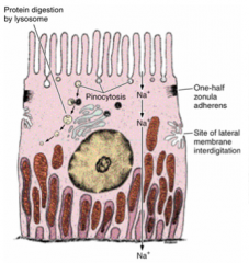
- basal striations that parallel the longitudinal axis of the cell
- striations are due to deep infoldings of the basal plasma membrane (compartmentalize mitochondria) |
|
What type of gland is this?
Arrow are pointing to ... |
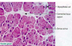
Parotid gland
|
|
|
What do the granules of the parotid glands contain?
|
-rich in proteins (proline-rich proteins)
-enzymes (amylase, peroxidase, lysozyme) -proteins with antimicrobial activity (cystatins & hystatins) |
|
|
Structure of the parotid gland:
|
-has the longest intercalated ducts
-myoepithelial cells at the periphery of each acinus -CT & vessels surround the serous acini |
|
|
Classification of sublingual gland:
|
-Contains both pure mucous & mixed secretory units
-Mixed, compound-branched tubuloacinar gland *mucous cells predominate |
|
|
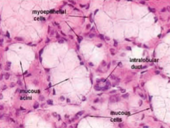
What type of cells does sublingual gland contain:
|
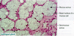
Mucous cells that predominate with serous cells only present in demilunes on mucous tubules
-mucous cells resemble goblet cells of the intestinal epithelium |
|
|
Major salivary product of the sublingual gland is:
|
mucus
-cells of the serous demilunes secrete amylase and lysozyme |
|
|
Shape/structure of the sublingual mucous cells:
|
-Pyramidal
-flattened nuclei against the basal plasma membrane -LM display pale blue cells with a "frothy" appearance (with hematoxylin & eosin) -EM display apical region of cell that contains large # of mucinogen granules -organelles of the cell (Golgi, RER, mitochondria) are located between the granules and nucleus |
|
|
Serous Demilune cells of the sublingual gland are located :
|
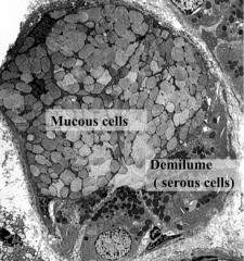
Peripheral to the mucous acini
-deliver their secretions into their spaces between the neighboring mucus-producing cells |
|
|
Ducts of the sublingual gland:
|
short intercalated and striated ducts are present, but are in number
|
|
What gland is this?
|
Sublingual gland
stained with H&E |
|
|
Classification of Submandibular (=Submaxillary) gland:
|
Mixed, compound (branched) tubuloacinar gland
*Serous cells predominant |
|
|
What are the differences between submandibular and sublingual glands?
|
The submandibular gland is mostly serous.
The sublingual glands are mostly mucous. |
|
|
Acini of submandibular gland is composed of:
|
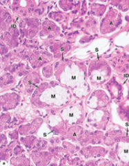
-mostly serous acini
-has groups of mucous acini with serous demilunes (10%) -secretory portions contain both mucous and serous cells |
|
Identify each type of ducts
|
|
|
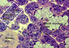
What type of gland is this?
|
Submandibular/submaxillary gland
|
|
|
Ducts of the submandibular glands:
|
-intercalated ducts are short
-striated ducts are long and clearly evident *striated ducts lined by cuboid cells with basal infoldings, containing mitochondria |
|
|
Major salivary products of the sublingual gland are:
|
mucus
serous demilunes secrete amylase & lysozyme |
|
|
Salivary products of the submandibular gland are:
|
alpha-amylase
proline-rich proteins serous cells secrete enzymes (lysozyme) *lysozyme imp. in hydrolyzing the walls in many types of bacteria |
|
|
Salivary products of the parotid gland are:
|
serous cells contain secretory granules with abundant alpha amylase and proline-rich proteins
|
|
|
Average daily production of saliva is:
|
1.0 - 1.2 liters
|
|
|
Function of the saliva include:
|
1. protects the oral cavity by lubrication, control of bacterial flora (by presence of lysozyme, lactoferrin, and IgA), and its cleansing action
2. assists in taste sensation 3. initiates digestion of carbohydrates via action of its enzyme, salivary amylase |
|
|
Primary secretion of the saliva is by:
|
Acini
-resembles extracellular fluid prior to modification by the system of ducts |
|
|
Final secretion of the saliva is by:
|
a. Intercalated ducts --> may deliver bicarbonate ions into the saliva
b. Striated ducts (via their sodium pump) --> remove Na+ and Cl- ions from luminal fluid and actively pump potassium ions into it |
|
|
Saliva is a (hypertonic/hypotonic/isotonic) solution?
|
HYPOtonic!
|
|
|
Exocrine function of the pancreas is served by:
|
acinar cells --> secrete digestive enzymes
centroacinar cells intercalated ducts |
|
|
Endocrine portion of the Pancreas is composed of:
|
Islets of Langerhans
|
|
|
What type of gland is Pancreas ?
|
Retroperitoneal gland
- produces both exocrine & endocrine secretions a. exocrine - serous acinus b. endocrine - islet of Langerhans |
|
|
Pancreas is subdivided into what regions?
|
head
uncinate process neck body tail |
|
|
Which region of the pancreas contains the highest concentration of enocrine tissue (islets of Langerhans)?
|
Tail region
|
|
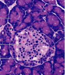
What type of gland is this?
|
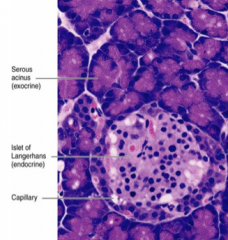
Pancreas
*acini contain secretory cells with basophilic cytoplasm - different types of endocrine cells are seen in the islet |
|
|
Capsule of the pancreas is composed of:
|
- Delicate connective tissue --> subdivides the gland into lobules by forming numerous septa
- Septa convey blood and lymph vessels, nerves, and ducts in and out of the gland |
|
|
Classification of the exocrine pancreas:
|
Pure serous, compound tubuloalveolar gland
|
|
|
Acini of the exocrine pancreas contain:
|
Only serous
centers contain centroacinar cells --> form the beginning of the duct system in the pancreas |
|
|
Shape & structure of the Acinar cells:
|
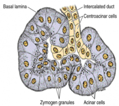
-Pyramidal in shape
-round nucleus that is basally located -Apical region densely packed with secretory granules (zymogen granules) -RER at their base --> strongly basophilic basal region -intercalated duct cells known as centroacinar cells partly penetrates the acini -NO myoepithelial cells ! |
|
|
Intercellular canaliculi...
|
lead from between acinar cells to the lumen of the acinus
|
|
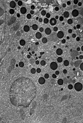
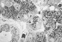
What type of cell is this?
|
Acinar cell of the pancreas
(N) - nucleus (G) - golgi (C) - condensing vacuoles (S) - mature secretory (zymogen) granules (L) - lumen --> contains proteins recently released from the cell by exocytosis *Acinar cells are pyramidal in shape with round nucleus that is basally located |
|
|
What type of organelles can you find within in Acinar cell under EM?
|
-abundant RER
-extensive golgi apparatus -numerous mitochondria -many free ribosomes *highly active cell |
|
|
Structure and appearance of the secretory granules?
|
- All membrane bound
- occasionally in the process of being released in "chains" that extend to the lumen of the acinus |
|
|
Are there any myoepithelial cells in the pancreas?
|
NO!
(difference between parotid glands and pancreas) |
|
|
Ducts of the acini (in pancreas):
|
Extend from within the acini
No striated duct! |
|
|
Shape of the centroacinar cells:
|
Low cuboidal
- constitute the initial part of the intercalated duct |
|
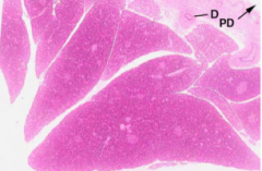
What is this slide of?
|
This slide is showing an excurrent duct (D), and the main pancreatic duct (PD)
- Septums dividing the glands |
|
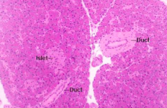
What type of cells/duct is seen here?
|
Pancreas (*Islet)
|
|
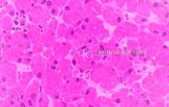
What is this slide of?
|
Pancreas
-cells are packed with dense granules pale area = centroacinar cells surrounding the acinar cells *note: intercalated ducts |
|
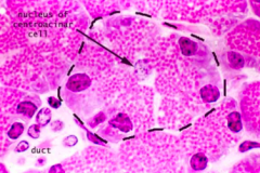
Identify centroacinar cell.
|
Pale-stained nucleus (arrow) = centroacinar cell
Darkly stained Round nucleus = acinar cells packed with zymogen granules (pink tiny dots) |
|
|
note: intertubular ducts on lower left corner
abundant amount of zymogen granules within the acinar cells |
|
|
Intercalated ducts lead into ...
|
intercalated ducts lead into small number of inTRAlobular ducts, which then empty into the large inTERlobular ducts
* No striated ducts in pancreatic glands (difference from parotid gland) |
|
|
What type of acini are present in the exocrine pancreas?
|
ONLY serous acini
|
|
|
A unique feature of the pancreatic acinus is the presence of...
|
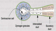
Centroacinar cells (squamous-to-cuboidal epithelial) = starting point of exocrine pancreas
*Striated ducts and Myoepithelial cells are NOT present in exocrine pancreatic glands |
|
|
Where are the interlobular ducts located?
|
between lobules in extensive CT and empty into the main (or accessory) pancreatic duct
|
|
|
Pancreatic duct:
|
the main pancreatic duct delivers pancreatic secretions into the duodenum at the papilla of Vater
*Papilla of Vater = junction between the embryological foregut & midgut |
|
|
What controls the release of pancreatic secretions?
|
By 2 hormones that are released by APUD (Amine Precursor Uptake Decarboxylase) cells of the digestive tract
*hormones are secreted by the DUODENUM: 1. Cholecystokinin (pancreozymin) 2. Secretin |
|
|
Cholecystokinin (pancreozymin):
|
induces the acinar cells to release pancreatic juices rich in digestive enzymes
|
|
|
Digestive enzymes of the pancreatic juice released by the acinar cells are:
|
Trypsin
Chymotrypsin Peptidase Pancreatic amylase Lipase Ribo-nuclease Deoxyribonuclease |
|
|
Secretin:
|
induces the intercalated ducts to secrete large quantities of an enzyme-poor, alkaline fluid that probably functions in neutralizing the acidic chyme that enters the duodenum
|
|
|
Endocrine Pancreas:
|
= Islets of Langerhans
|
|
|
Shape & structure of Islets of Langerhans:
|
-spherical clumps of richly vascularized endocrine cells
-lie scattered in random fashion among acini of the exocrine pancreas *more numerous in the tail region -composed of several cell types with different functions |
|
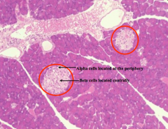
What are the structures circled in red?
|
Islets of Langerhans (endocrine area in Pancreas)
-cells are indistinguishable in regular stain but can be differentiated by the use of special stains |
|
|
Structure and location of the Alpha cells:
|
-preferentially positioned at the islet's periphery
-under EM: spherical, membrane-bound, electron-dense granules |
|
|
What type of cells constitute the majority and occupy mostly the center of each islet?
Importance of this type of cells? |
Beta cells
*produce hormone insulin --> lower blood glucose level |
|
|
Characteristics of the granules of the Beta cells:
|
-membrane bound
-smaller than those of alpha cells -characterized by presence of an irregular dense core, surrounded by an electron-lucent periphery |
|
|
Delta1 Cells:
|
-less common than alpha- / beta- cells
-recognized in the EM by their relatively large, electron-lucent granules -membranes of granules are not well defined |
|
|
Delta1 cells produce:
|
Somatostatin
-depresses all the actions -hormone that controls the release of insulin and glucagon --> reduces vol. of alkaline-rich pancreatic juices |
|
|
Other cell types include:
|
C,E,G,PP cells
|
|
|
Each cell type produces a specific hormone, Alpha and Beta cells produce...
|
Alpha cells --> glucagon: ELEVATES blood glucose levels
Beta cells --> insulin: LOWERS blood glucose levels |
|
|
C cells are:
|
represent immature / degranulated islet cells
|
|
|
PP cells release:
|
Pancreatic polypeptide
- controls both exocrine and edocrine cells - hormone that may act to reduce the release of both enzyme-rich and alkaline-rich pancreatic secretions |
|
|
G cells release:
|
Gastrin
- hormone that functions in modulating the release of HCL by the parietal cells of the stomach |