Reading...
![]()
Play button
![]()
Play button
![]()
Use LEFT and RIGHT arrow keys to navigate between flashcards;
Use UP and DOWN arrow keys to flip the card;
H to show hint;
A reads text to speech;
52 Cards in this Set
- Front
- Back
|
Liver:
|
= 2nd largest organ
- composed of a single type of parenchymal cell = hepatocyte *IGF-I *Angiotensionogen *Thrombopoietin |
|
|
Hepatocyte..
|
possess a myriad of both endocrine and exocrine functions
|
|
|
What is a Glisson's Capsule?
|
composed of thin CT that subdivides the liver into lobes and lobules
|
|
|
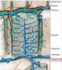
Blood supply to the liver:
|
Derived from 2 sources:
1. Abdominal aorta, via the hepatic artery (=branch of celiac trunk) 2. Portal vein --> brings nutrient-laden blood from the alimentary tract and the spleen |
|
|
Porta Hepatis:
|
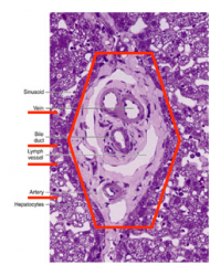
the region where:
- the hepatic artery and portal vein ENTER liver - the hepatic ducts LEAVE the liver |
|
|
Drainage of blood of the liver is via:
|
Hepatic vein
- hepatic vein is formed by the union of numerous sublobular veins - sublobular veins collect blood from the central vein of each classical liver lobule |
|
|
Transportation of bile:
|
- leaves the liver via hepatic ducts
- delivered to the gallbladder |
|
|
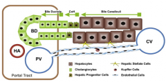
Portal triad consists of:
|
Hepatic artery
Bile duct Hepatic vein |
|
|
3 types of liver lobules are:
|
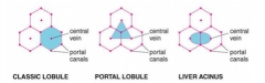
1. classical (hexagonal)
2. portal lobule (triangular) 3. liver acinus of Rappaport (liver acinus - diamond-shaped) *nutrient-rich blood towards central vein --> out of liver *bile towards peripheral to portal canals --> intestine |
|
|
Features of the classical lobule:
|
- based on the pig's liver, where CT elements clearly delineate it
- portal area (portal canal; triad) is present at each corner of the lobule - portal area contains branches of the portal vein, hepatic artery, bile duct, lymph vessel* |
|
|
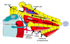
Structure of the Liver:
|
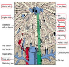
Kupffer cells (=marophages) within the sinusoid
-hepatocytes (plate-like) arranged in columns separated by sinusoides |
|
|
The liver lobule is surrounded by:
|
Portal space
|
|
|
What occupies the portal spaces?
|
Arteries
Veins bile ducts Nerves CT lymphatic vessels |
|
|
What forms the radial disposition of the plates in the lobule?
|
Hepatocytes
- plates are separated by the Sinusoidal capillaries - Bile canaliculi seen between the hepatocytes |
|
|
What drains the blood from the live lobules?
|
Sublobular (intercalated) veins
|
|
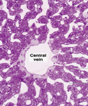
What type of tissue is this?
|
Liver
- purple: hepatocytes - white spaces: sinusoids |
|
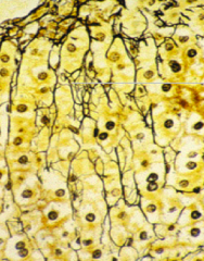
What type of tissue is seen in this slide?
|
Collagen type 3 reticular fibers in the lobule --> forming a scaffold for the hepatic tissue
Stain: silver impregnation |
|
|
Plates of Hepatocytes:
|
- compose the bulk of the lobule
- arranged in a radial fashion, radiating from the region of a central vein - blood flows from the periphery of the lobule forward the central vein |
|
|
Portal triad is composed of:
|
Portal vein
Hepatic artery Bile duct *lymphatics |
|
|
Bile Canaliculi:
|
= slender intercellular spaces between neighboring hepatocytes
- convey bile to canals of Herring - canals of Herring deliver bile ducts in the portal area at the periphery of the classical lobule |
|
|
Flow of bile and blood is...
|
in Opposite direction
|
|
|
Hepatocytes are jointed by...
|
Tight (occluding) junctions
where bile cannot leak |
|
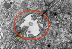
What is show in this micrograph?
|
Bile canaliculus (rat liver)
-microvilli in lumen -junctional complexes (yellow arrows) =[tight junction] |
|
|
The junction of bile and canaliculi and bile ductules are lined by...
|
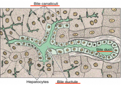
Cuboidal epithelium
- the ductules merge with bile ducts in the portal spaces |
|
|
What are sinusoids?
|
= endothelial-lined spaces between neighboring plates of hepatocytes
- receive blood from the vessels in the portal area and deliver it to the central vein |
|
|
Endothelial cells lining the sinusoids are:
|
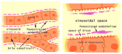
NOT continuous, have large fenestrations
- display discontinuous between neighboring cells |
|
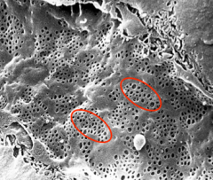
What type of tissue is this?
|
Liver
sinusoidal capillary with large fenestrations (red circle) |
|
|
What are Kupffer cells?
|
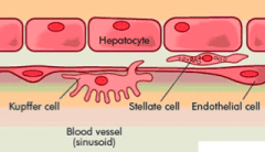
= phagocytic cells that are derived from monocytes
- they are also present outside/within the endothelial lining |
|
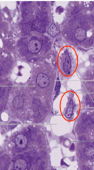
What are the structures circled in red?
|
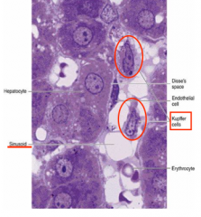
Kupffer cells (present towards the sinusoid)
- sinusoid capillaries close to hepatocytes - Disse = thin slit between the hepatocytes and the endothelium |
|
|
Space of Disse are located at:
|
visualized by EM as a subendothelial space between the liver cells (hepatocyts) and the lining cells of the sinusoids
|
|
|
Space of Disse contains:
|
-stellate-shaped fat storing cells (preferentially store vit. A)
- reticular fibers (maintains the architecture of the sinusoids) - NON-myelinated nerve fibers - short, blunt microvilli of hepatocytes (increase S.A.) |
|
|
Functions of the Space of Disse?
|
exchange of material between the bloodstream and the hepatocytes (which do not contact the
bloodstream) |
|
|
Is Basal Lamina present in the Space of Disse?
|
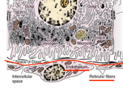
NO!
|
|
|
Where is the portal lobule located?
|
-it is based on the liver's exocrine function; in many exocrine glands, the ducts is in the center of a lobule
|
|
|
Portal Lobule:
|
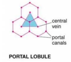
a triangular region whose 3 apices are neighboring central veins
-Has a portal canal in a central position and central veins at the edges of the cross-sectioned lobule |
|
|
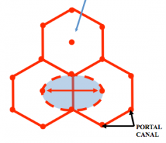
Liver Acinus (of Rappaport):
|
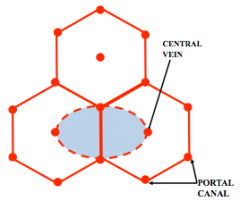
= another interpretation of lobulation in the liver
- based on blood flow - diamond-shaped structure - distributing vessels in the center & central veins at each pole - portal area shared by the 2 neighboring classical lobules |
|
|
What is Zonation?
|
blood enters the sinusoids from vessels located in the interface between the two neighboring classical lobules (bases of the equilateral triangles)
|
|
|
Hepatocytes:
|
- first to be "exposed" to the entering blood
- near the central vein, they are the last to be "exposed" |
|
|
Three zones within each acins of Rapport:
|
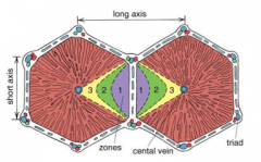
1: in the immediate vicinity of the blood supply
3: in the area of the central vein 2: in between zone 1 & 3 |
|
|
Structure of Hepatocytes:
|
- large, polyhedral cells that stain light pink with Hematoxylin & eosin (H&E)
- usually possess 1 or 2, centrally placed round nucleus (polyploid) - frequently >2 nuclei may be present in a single cell - bile canaliculi are between neighboring hepatocytes |
|
|
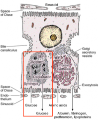
Organelles within the liver cells:
|
Under EM
- rich in RER & SER - rich in mitochondira - several golgi regions - lysosomes - peroxisomes - lots of lipid droplets - lots of glycogen |
|
|
Carbohydrate stored in the liver in the form of:
|
glycogen
- usually associated with SER, when glucose is needed, glycogen is degraded |
|
|
Proteins produced by hepatocytes are synthesized in...
|
RER
|
|
|
Hepatocyte lesions or starvation lead to ...
|
decrease in amounts of albumin, fibrinogen, prothrombin in a pt's blood
--> imp for blood's osmotic pressure and for coagulation |
|
|
10% of bile acids are made in the liver, how are they made?
|
by conjugation of cholic acid with the amino acids glycine and taurine
- this process occurs in the SER |
|
|
Secretion of bilirubin:
|
the Water-insoluble form of bilirubin is derived from the metabolism of hemoglobin in macrophages
*Bilirubin can be "conjugated" with a molecule of glucuronic acid which makes it soluble in water *Bilirubin diglucuronide is a conjugated form of bilirubin |
|
|
2 types of the surfaces (liver cells):
|
1. those that border the space of Disse
2. those that are adjacent neighboring hepatocytes |
|
|
What takes place in the adjacent space of Disse?
|
Microvilli assist in the transfer of materials to and from hepatocytes. It is here that the endocrine secretion of the liver also take place.
*microvilli extend into the bile canaliculus from each hepatocyte |
|
|
What do the adjacent neighboring hepatocytes form?
|
- Form small, tunnel-like bile canaliculi that represent intercellular spaces
- Form occluding junctions at each surface of the bile canaliculus |
|
|
Bile Canaliculi:
|
Receive the exocrine secretion of the liver (bile)
--> representing the beginning of the duct system |
|
|
Neighboring hepatocytes contact one another via?
|
Gap Junctions
|
|
|
Intrahepatic Bile ducts consists of:
|
1. bile canaliculi
2. cholangioles 3. canals of Herring (bile ductules), lined by a layer of low cuboidal cells 4. bile ducts, lined by a single layer of cuboidal cells |