Reading...
![]()
Play button
![]()
Play button
![]()
Use LEFT and RIGHT arrow keys to navigate between flashcards;
Use UP and DOWN arrow keys to flip the card;
H to show hint;
A reads text to speech;
46 Cards in this Set
- Front
- Back
|
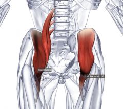
Gluteus maximus
origin: ilium, sacrum, coccyx insertion: posterior, proximal femur and iliotibial tract group: superficial gluteus muscle location: superficial posterior hip ("butt") function: extends thigh |
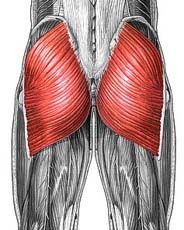
Name the muscle
|
|
|
Gluteus medius
origin: ilium insertion: greater trochanter group: superficial gluteus muscle location: under maximus, origin at ilium close to iliac crest function: abduct and medially rotate thigh, stabilise pelvis |

Name the muscle
|
|
|
Guteus Minimus
origin: ilium insertion: greater trochanter group: superficial gluteus muscle location: inferior to medius (origin half way on fossa "wing" of ilium) function: abduct and medially rotate thigh, stabilise pelvis |
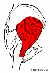
Name the muscle
|
|
|
Tensor fasciae latae
origin: anterior ilium insertion: iliotibial tract group: superficial gluteus muscle function: tense fascia lata, abduct and medially rotate thigh, stabilise pelvis |
Name the muscle
|
|
|
Piriformis
Group: deep gluteus group Function: lateral hip rotation |
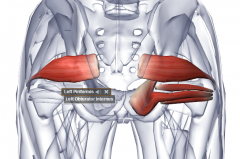
Name this muscle
|
|
|
obturator internus
Group: deep gluteus group Function: lateral hip rotation |
Name this muscle
|
|
|
gemelli superior
group: deep gluteus group Function: lateral hip rotation |
Name this muscle
|
|
|
Gemelli inferior
group: deep gluteus group Function: lateral hip rotation |
Name this muscle
|
|
|
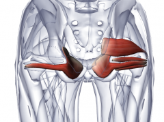
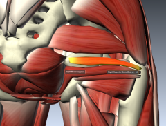
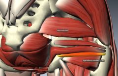
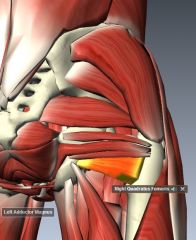
Quadratus Femoris
group: deep gluteus muscle function: lateral hip rotation |
Name this muscle
|
|
|
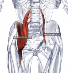
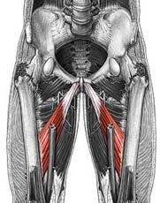
Iliacus
origin: ilium (close to iliac crest) insertion: lesser trochanter function: flex thigh at hip joint |
Name the muscle
|
|
|
Psoas Major
origin: inferior most thoracic vertebrae and lumbar vertebrae insertion: lesser trochanter function: flex thigh at hip joint |
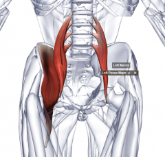
Name the muscle
|
|
|
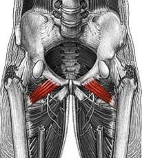
Psoas minor: superior and anterior to major
origin: vertebrae, superior lumbar region insertion: pelvic bone Not everyone has it Note: does not affect hip joint Function: flex thigh at hip joint |
Name the muscle
|
|
|
Sartorius
origin: anterior superior iliac spine insertion: medial surface of proximal tibia (after winding behind the medial aspect of the lower thigh) Function: flex, abducts, lateral rotate thigh at hip joint; flex knee joint |
Name the muscle
|
|
|
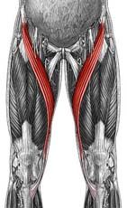
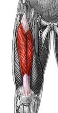
Rectus femoris (Quadraceps femoris)
origin: anterior ilium insertion: patella and tibial tuberosity Function: extend knee |
Name the muscle
|
|
|
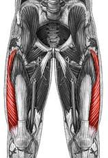
Quadriceps femoris group: Vastus lateralis
origin: greater trochanter and linea aspera of femur insertion: patella and tibial tuberosity Function: extend knee |
Name the muscle
|
|
|
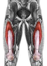
Quadriceps femoris group: Vastus medialis
origin: linea aspera insertion: patella and tibial tuberosity Function: extend knee |
Name the muscle
|
|
|
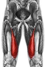
Quadriceps femoris group: Vastus intermedius
origin: anterior and lateral surfaces of proximal shaft of femur insertion: patella and tibial tuberosity Function: extend knee |
Name the muscle
|
|
|
Adductor longus
origin: pubis insertion: linea aspera group: medial thigh innervation: obturator nerve blood supply: femoral artery drain into: femoral vein location: inferior to pectineus, pectineus + adductor longus = most superficial anterior view of medial thigh (under Quadratus femoris from anterior view) Function: adduct thigh |
Name the muscle
|
|
|
Pectineus
origin: pubis insertion: linea aspera group: medial thigh innervation: obturator nerve blood supply: femoral artery drain into: femoral vein location: superior to adductor longus, pectineus + adductor longus = most superficial anterior view of medial thigh (under Quadratus femoris from anterior view) Function: adduct and flex thigh |
Name the muscle
|
|
|
Adductor magnus
femoral --> adductor hiatus --> popliteal origin: Ischium and pubis insertion: linea aspera group: medial thigh innervation: obturator nerve blood supply: femoral artery drain: femoral vein location: anterior view - under adductor longus (but can see medial edge slightly), deep posterior - whole muscle, very big (magnus), hole = adductor hiatus, inferior to adductor brevis Function: adduct thigh at hip |
Name the muscle
|
|
|
Adductor Brevis
origin: pubis insertion: linea aspera group: medial thigh innervation: obturator nerve blood supply: femoral artery drain: femoral vein location: anterior view - under adductor longus, superior to adductor magnus, longer and skinnier and deeper (anterior) than pectineus Function: adduct thigh at hip |
Name the muscle
|
|
|
Gracilis
origin: pubis insertion: medial, proximal tibia group: medial thigh innervation: obturator nerve blood supply: femoral vein: femoral location: most medial muscle, more likely directly medial to adductor magnus Function: adduct thigh at hip, flex leg, medial leg rotation |
Name the muscle
|
|
|
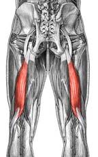
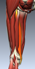
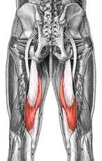
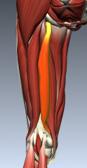
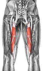
Biceps femoris: 2 head
origin: long - ischium, short - linea aspera insertion: head of fibula and lateral condyle of tibia group: posterior thigh innervation: tibial nerve (branch sciatic) blood supply: femoral artery vein: femoral vein location: most superficial lateral posterior thigh, long head superficial to short head, common insertion Function: extend thigh (long head only) , flex and lateral rotate leg |
Name the muscle
|
|
Semimembranosus
origin: ischium insertion: medial, proximal tibial shaft group: posterior thigh innervation: tibial nerve (sciatic branch) blood vessel: femoral artery vein: femoral vein location: posterior view - deep to semitendonosus Function: extend hip, flex knee |
Name the muscle
|
|
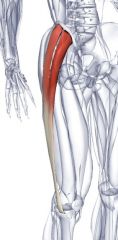
Semitendinosus: tendons between
origin: ischium insertion; medial, proximal tibia group: posterior thigh innervation: tibial nerve (sciatic branch) blood supply: femoral artery vein: femoral vein location: posterior view - superficial to semimembranosus, medial posterior thigh Function: extend hip, flex knee |
Name the muscle
|
|
|
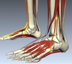
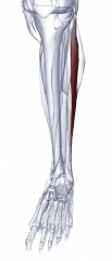
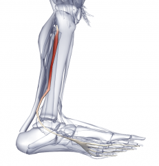
Tibialis anterior
origin: lateral, proximal tibia insertion: medial, dorsal foot Group: anterior leg innervation: fibular nerve (sciatic branch) blood supply: drain into: location: most superficial anterior muscle of leg, most medial of anterior leg group, tendon ends base of first metacarpal |
Name the muscle
|
|
|
Extensor digitorum longus
origin: proximal tibia and fibula insertion: superior surfaces of phalanges 2-5 group: anterior leg innervation: fibular nerve (branch sciatic) blood supply: drain into: location: lateral to tibialis anterior, superficial anterior leg, tendon to base of distal phalanges Function: |
Name the muscle
|
|
|
Extensor hallucis longus
origin: anterior surface of tibia insertion: dorsal, lateral foot group: anterior leg innervation: fibular nerve (branch sciatic) blood supply: drain into: location: under extensor digitorum longus, medial to fibularis brevis, lateral to tibialis anterior, proximal and medial to fibularis tertius Function: |
Name the muscle
|
|
|
Fibularis tertius
Origin: distal anterior surface of fibula Insertion: dorsal lateral foot group: anterior leg innervation: fibular nerve (branch sciatic) blood supply: drain into: location: under extensor digitorum longus, most lateral anterior leg muscle, end lateral superficial to extensor digitorum brevis Function: |
Name the muscle
|
|
|
Fibularis longus
origin: proximal fibula insertion: tendon crosses plantar foot to insert in medial, plantar foot (1st metatarsal) group: lateral leg innervation: fibular nerve (branch sciatic) blood supply: drain into: location: superficial lateral leg, superficial to fibularis brevis, lateral to extensor digitorum longus and fibularis brevis, closest posterior muscle: soleus. Tendon wrapped by superior and inferior fibular retinaculum Function: everts foot + weak plantar flexion |
Name the muscle
|
|
|
Fibularis brevis
origin: lateral, distal fibula insertion: base of 5th metatarsal group: lateral leg innervation: fibular nerve (branch sciatic) blood vessel: drain into: location: under fibularis longus, lateral to extensor hallucis longus & fibularis tertius (under EHL) & extensor digitorum longus (superficial to EHL, FT). Tendon wrapped by superior and inferior retinaculum Function: everts foot + weak plantar flexion |
Name the muscle
|
|
|
TIbialis posterior
origin: interosseous membrane and adjacent shafts of tibia and fibula insertion: tarsal and metatarsal bones group: deep posterior leg innervation: tibial nerve blood supply: drain: location: chunky tendon on tarsal bones, medial to flexor hallucis longus and lateral to flexor digitorum longus, tendon crosses with FDL tendon - tendon then medial to FDL tendon Function: plantar flex ankle, inverts foot (supinate) |
name the muscle
|
|
|
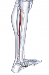
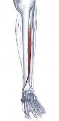
Flexor digitorum longus
origin: posteromedial surface of tibia insertion:inferior surfaces of distal phalanges 2-5 group: deep posterior leg innervation: tibial nerve blood supply: drain: location: medial leg, tendon crosses tendon of TP - tendon end lateral to TP tendon Function: plantar flexion, flex 4 digits, support longitudinal arches of foot |
Name the muscle
|
|
|
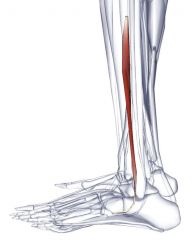
flexor hallucis longus
origin: posterior surface of fibula insertion: inferior surface of distal phalanx of great toe group: deep posterior leg innervation: tibial nerve (branch sciatic) blood supply: drain: location: under and lateral to tibialis posterior Function: assist plantar flexion, flex big toe, support medial longitudinal arch of foot |
Name the muscle
|
|
|
popliteus: divergent
origin: lateral, distal femur insertion: posterior proximal tibial shaft group: deep posterior innervation: tibial nerve blood supply: drain into: location: under plantaris (& entire superficial group) function: weak knee flexion, medially rotates tibia (of unplanted limb) |
name the muscle
|
|
|
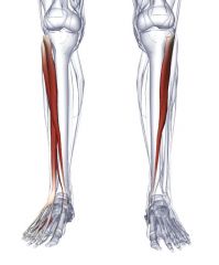
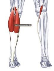
Gastrocnemius: 2 head structure
origin: posterior, distal femur insertion: calcaneus (calcaneal tendon) group: superficial posterior leg innervatioin: tibial nerve (branch sciatic) blood supply: drain into: location: most superficial two headed posterior muscle, inserts into calcaneous tendon Function: plantar flexion when knee extended + knee flexion + raise heel during walking |
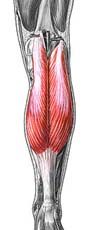
Name the muscle, structure
|
|
|
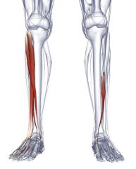
Soleus
origin: posterior, proximal tibia and fibula insertion: calcaneous (calcaneous tendon) group: superficial posterior leg innervation: tibial nerve (branch sciatic) blood supply: drain into: location: under gastrocnemius, "sole" shaped Function: plantar flexion when knee extended |
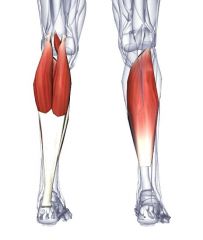
Name the muscle
|
|
|
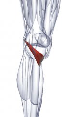
Plantaris
origin: lateral, distal femus insertion: calcaneus & sometimes plantar aponeurosis group: superficial posterior leg innervation: tibial nerve blood supply drain: location: very long tendon from lateral to medial, most proximal and deep of superficial posterior leg Function: weak gastrocnemius plantarflexing ankle |
Name the muscle
|
|
Extensor hallucis brevis (big toe)
Extensor digitorum brevis No hand equivalent |
Name the two muscles
|
|
|
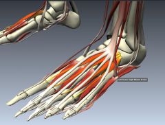
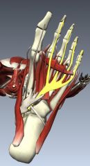
Abductor hallucis
Abductor digiti minimi Layer 1 |
Name the two muscles
|
|
|
Flexor digitorum brevis
No hand equivalent Layer 1 |
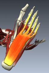
Name the muscle
|
|
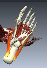
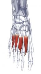
Lumbricals
deep to tendon of flexor digitorum longus Layer 2 |
Name the muscle
|
|
|
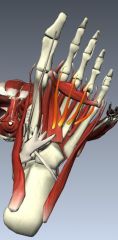
Quadratus Plantae
No hand equivalent Layer 2 |
Name the muscle
|
|
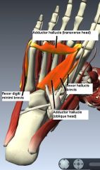
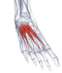
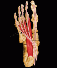
Flexor hallucis brevis
Flexor digiti minimi brevis Adductor hallucis (transverse and oblique head) layer 3 |
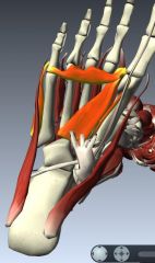
Name layer and muscles
|
|
|
Plantar interossei
|
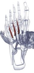
Name the muscle
|
|
|
Dorsal interossei
|
Name muscle
|