Reading...
![]()
Play button
![]()
Play button
![]()
Use LEFT and RIGHT arrow keys to navigate between flashcards;
Use UP and DOWN arrow keys to flip the card;
H to show hint;
A reads text to speech;
31 Cards in this Set
- Front
- Back

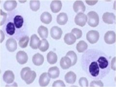
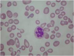
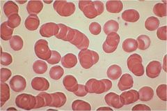
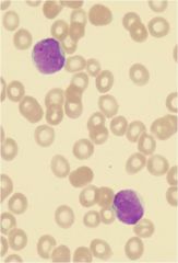
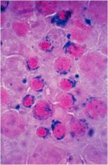
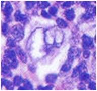
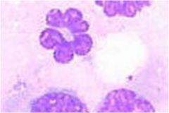
What is wrong with the neutrophils seen here?
|
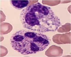
Toxic granulations. The azurophilic granules are indicative of immaturity. Patient could have a massive infection or another reason to be putting out immature cells
|
|
What autosomal dominant congenital abnormality has basophilic inclusions that may resemble Dohle bodies? What other findings are characteristic on blood smear?
|

May-Hegglin anomaly, but also GIANT platelets. Usually benign, sometimes thrombocytopenia
|
|
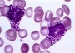
What is a neutrophil anomaly associated with mucopolysaccharidoses, and what are the findings on blood smear?
|
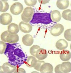
Alder-Reilly anomaly. Blood smear will show many azurophilic granules in granulocytes, lymphocytes and monocytes.
|
|

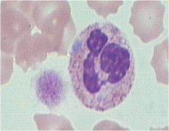
What are the two possible causes of the pince-nez appearance of these polys?
|
Congenital (innocuous)
or acquired (pseudo Pelger-Huet - myelodysplasia) |
|
If this cell was on a peripheral smear of a child with oculocutaneous albinism, what else would you expect on history?
|

Frequent infections from Chediak-Higashi syndrome, a defect in fusion/trafficking of vesicles (including melanosomes)
|
|

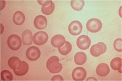
What type of anemia is seen here?
|
Iron deficiency anemia - hypochromatic with increased central pallor and pencil cells
|
|
What type of anemia is seen here?
|
Thallassemia - increased central pallor, target cells
|
|
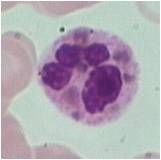
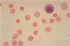
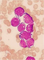
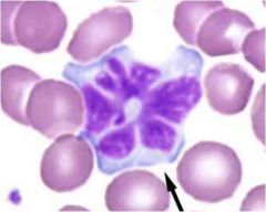
What causes this type of anemia?
|

B12 deficiency - multilobulated PMN, irregular shapes
|
|

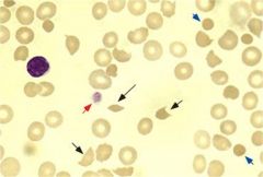
What can cause the appearance of these cells?
|
Thalassemia, Liver disease, severe Iron deficiency, Heme C disease, asplenia
|
|
What causes spherocytosis?
|

Hereditary spherocytosis caused by mutations in cytoskeleton proteins
Autoimmune hemolysis |
|

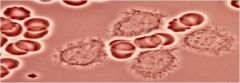
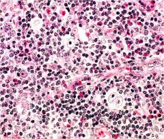
What conditions can produce schistocytes?
|
Shearing stress caused by microangiopathies (DIC), aortic stenosis, or a mechanical heart valve
|
|

What disorder causes Rouleux, and what is the pathophysiology of it?
|
Multiple Myeloma produced paraprotein, which coats RBCs and carries a negative charge which causes them to stack
|
|

What infections, and which immunoglobulin is associated with this slide's appearance?
|
IgM, Mononucleosis, mycoplasm
|
|

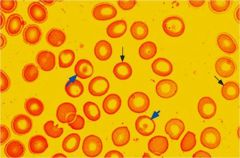
What is seen here?
|
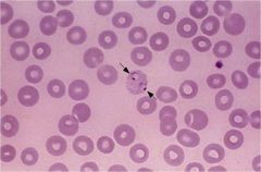
RBC infected with malaria
|
|
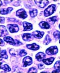
What type of lymphoma is seen here?
|
Burkitt's - note large vacuoles
|
|
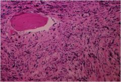
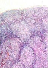
What process is shown in this bone marrow biopsy?
|

Idiopathic myelofibrosis - collagen III fiber deposition
|
|

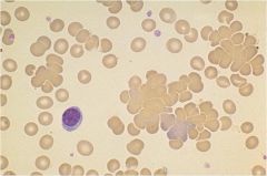
What type of ALL is seen here, and in what population is it most common
|
L1 - Small cell ALL, most common in pediatric cases. Note lymphocytes are similar in size to RBCs
|
|
What type of ALL is seen in this peripheral smear, and in what population is it most common?
|

Large cell ALL - most common in adults. Note size of lymphocytes compared to RBCs
|
|
What are the multinucleate RBCs here indicative of?
|
myelodysplasia
|
|
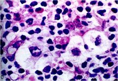
What are the ringed sideroblasts here indicative of?
|
myelodysplasia - iron not leaving with RBCs stuck in marrow
|
|
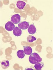
What feature of myelodysplastic syndrome is seen here?
|

Micromegakaryocyte, washed out appearance
|
|

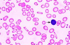
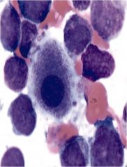
What are these cells' appearance typical of?
|
Hairy cell leukemia - rare CLL, normal life expectancy
|
|
What is the appearance of these cells suggestive of in a patient with cervical adenopathy?
|
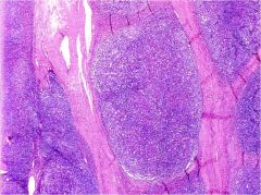
Lacunar cells, indicative of nodular sclerosis (here in lower power), most common type of HL
|
|
What are these popcorn appearing cells indicative of?
|

Lymphocyte-predominant (non-classic) HL. Very rare. CD 15/30 negative
|
|
If you didn't have a good heme pathologist, what might you mistake this biopsy from a retroperitoneal node for?
|

A sarcoma. It is lymphocyte depleted HL
|
|
How does this 2nd most common HL subtype differ from nodular sclerosis, the most common HL?
|

Mixed cellularity HL - has R-S cells (nod sclerosis does not) and lacks collagen bands.
|
|
What would these small, cleaved cells in a patient with diffuse rubbery adenopathy and CD19, 20 positive cell infiltrate suggest?
|
Follicular lymphoma (NHL)
|
|

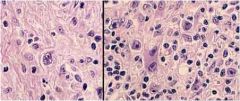
How might a patient with this cell on peripheral smear present?
|
With raised cutaneous plaques of malignant T cells - Mycosis fungoides/Sezary syndrome
|
|
A patient presents with lymphocytosis, hypercalcemia, elevated LDH, and splenomegaly. You see this clover-leaf cell on peripheral smear. He is from Japan. What do you suspect, and what is prognosis?
|
Suspect Human T-cell leukemia/lymphoma, and prior infection by HTLV.
Poor prognosis: <1 year |
|
What do these cells suggest in a child with a quickly growing sub-mandibular mass?
|
Burkitt's Lymphoma
|
|
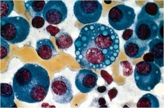
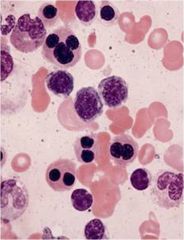
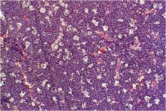
A patient with M-protein in urine has this bone marrow appearance. What do you expect from their clinical presentation?
|

Multiple myeloma: CRAB:
HyperCalcemia Renal impairment Anemia Bone lesions |