Reading...
![]()
Play button
![]()
Play button
![]()
Use LEFT and RIGHT arrow keys to navigate between flashcards;
Use UP and DOWN arrow keys to flip the card;
H to show hint;
A reads text to speech;
24 Cards in this Set
- Front
- Back
- 3rd side (hint)
Splenic resection. Which statement is true about this entitiy?
A. It expresses CD31 but not CD34 B. Metastatsis is common C. It is the only small B-cell leukemia to affect the red pulp. D. It expresses HHV8 |
A. This is a littoral cell angioma. Micro: anastomosing monotonous vascular channels resembling splenic sinuses, but lined by tall endothelial cells with variable hemophagocytosis; channels have irregular lumina, often papillary projections and cystic spaces; endothelial cells frequently detach into vascular spaces; no sclerosis, no atypia. Stains: CD31, CD68 and CD21 are positive. CD34 is usually negative. Re B: Angiosarc is rare in the spleen and shows much atypia. C. Blood lakes are a feature of HCL however no infiltrate is seen. D. Kaposi is spindled with small vessels
|
|
|
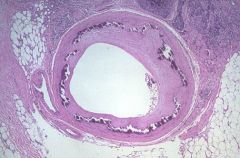
Which statement is true regarding this entity?
A. Significant luminal narrowing occurs over time. B. These changes are usually limited to the coronary arteries. C. It is usually not symptomatic unless severe. D. apolipo proteins are an important part of the pathogenesis. |
C. This is Monckeberg's arteriosclerosis, AKA medial calcific sclerosis. It is usually more benign than other forms of arteriosclerosis because it does not cause narrowing of the lumen. It is most commonly found in the radial or ulnar arteries but can occur anywhere. It is age related and associated with diabetes.
|
|
|
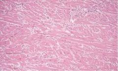
The gross findings of this histology most likely are...
A. Asymmetric thickening of the ventricular septum, 1.5 times that of the posterior left ventricular wall. B. Myocardial thinning and four chamber dilatation. C. A lesion with central, yellow-tan softening with surrounding hyperemia. D. No gross changes would be recognizable in most cases. |
A. This is myofiber disarray and fibrosis seen in hypertrophic cardiomyopathy. Usually it cannot be diagnosed by endomyocardial biopsy because abnormal tissue is within the septum (B) describes dilated cardiomyopathy. (C) describes an MI at 3-7 days.
|
|
|
|
Which of the following statements is not true regarding Anthracycline toxicity?
A. It is graded; grade 1, 1.5, 2, 2.5 or 3. B. Endomyocardial biopsy is the gold standard for assessing toxicity. C. Grading is based on cytoplasmic vacuolization and myofibrillar loss. D. Doxorubicin causes more cardiac toxicity than daunorubicin (4.4% vs. 1.7%) |
D is not true. Daunorubicin is more toxic. The other statements are true. Grade 1 is when <5% of cells are damaged; grade 1.5 is 6-15% of cells; 2= 16-25%; 2.5= 26-35%, 3= >35% of cells. Grade 2.5 allows for one more dose to be given. Grade 3 means that no more drug should be given. Toluidine blue can be used to highlight the cytoplasmic vacuolization. EM is also used.
|
|
|
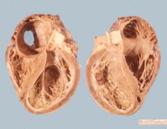
Which of the following are is NOT a cause of dilated cardiomyopathy?
A. Fabry's disease B. Loffler's endomyocarditis C. Hemochromatosis D. Doxorubicin toxicity |
B. Loffler's endomyocarditis is characterized by a restrictive cardiomyopathy with fibrosis and an intense infiltrate of eosinophils, necrosis and arteriolitis. Fabry's disease is X-linked recessive with angiokeratomas, renal insufficiency, eye keratopathy and dilated cardiomyopathy. It is a lysosomal storage disease characterized by a deficiency in alpha-galactosidase A with the accumulation of ceramide trihexoside. Hemochromatosis usually causes dilated cardiomyopathy but it can rarely be restrictive. Doxorubicin causes dilated cardiomyopathy in 1.7% of patients.
|
|
|
The image demonstrates what disease process?
A. Sarcoidosis B. Fungal myocarditis C. Whipple's disease D. Rheumatic heart disease E. Giant cell myocarditis |
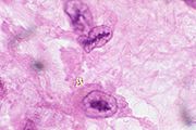
D. The image is an Aschoff nodule which is seen in rheumatic heart disease. The above picture shows Anichkow cells which are pathognomonic of rheumatic heart disease.
Followup question: Which valve is most often involved? A. Aortic B. Pulmonic C. Tricuspid D. Mitral |
D. Mitral > aortic > tricuspid > pulmonary
Manifests as verrucous vegetations along the lines of closure. |
|
This image demonstrates what disease process?
A. Infective endocarditis B. Libman-Sacks endocarditis C. Calcific heart disease D. Rheumatic heart disease |
B. Libman-Sacks endocarditis, flat, pale tan, spreading vegetations over the valve surface with spread onto the chordae tendineae. They occur in 4% of SLE patients and are rarely symptomatic. (A) would show destructive, heaped lesions. (D) would show fibrosis, shortened and thickened chordae tendineae and small verrucous vegetations along the valve closure edge.
|
|
|
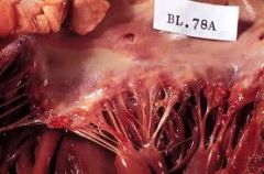
This image demonstrates what disease process?
A. Infective endocarditis B. Libman-Sacks endocarditis C. Calcific heart disease D. Rheumatic heart disease |
D. Rheumatic heart disease shows fibrosis, shortened and thickened chordae tendineae and small verrucous vegetations along the valve closure edge. (B) would show flat, pale tan, spreading vegetations over the valve surface with spread onto the chordae tendineae. (A) would show destructive, heaped lesions.
|
|
|
Which statement is most true about this cardiac tumor?
A. Most common in the right atrium and secretes IL-6 B. Most common in the left atrium and are common in children C. Seen in Carney's Triad and always benign. D. Seen in Carney's Syndrome and secretes IL-6 |
D is the most correct. The true statements are as follows. Seen in Carney's syndrome, secretes IL-6, always benign. Most common in the left atrium (near fossa ovalis). Carney's triad is GISTs, extra adrenal paragangliomas and pulmonary chondromas. Carneys syndrome, AKA carney complex involves myxomas of heart and skin, skin hyperpigmentation and endocrine overactivity.
|
|
|
Rhabdomyoma is the most common cardiac tumor of childhood. Which statement is not true regarding this tumor?
A. It can spontaneously regress B. It is associated with tuberous sclerosis C. It is most often found in the atria. D. PAS is useful to highlight the abundant glycogen content of the cells. |
C. They are actually most often found in the ventricular septum or the left ventricle. Its relationship with tuberous sclerosis is noteworthy. TS additionally manifests as intracranial hamartomas, facial angiofibromas, subungal fibromas, epidermal nevi and renal angiomyolipomas.
|
|
|
|
What is the most common lymphoma seen in the heart?
|
DLBCL
|
|
|
|
Where are myxomas and angiosarcomas most commonly found in the heart?
A. Right atrium, right atrium B. Right atrium, left atrium C. Left atrium, right atrium D. Left atrium, left atrium |
C
|
|
|
|
What is the normal volume of the pericardial sac?
A. 5 cc B. 10 cc C. 30 cc D. 100 cc |
C - It can contain liters of fluid if the fluid accumulates slowly. Fast expansion is often deadly.
|
|
|
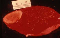
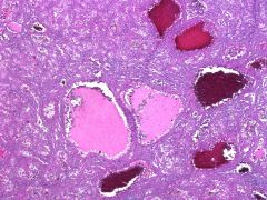
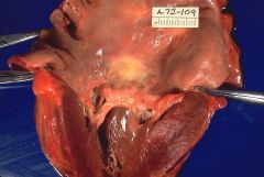
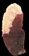
The lesion corresponds with which of the following statements?
A. The lesion is likely a metastatic tumor B. The lesion most likely resulted from a murantic vegetation. C. It is the most common primary tumor of the spleen. D. This spleen most likely came from a teenager with sickle cell disease. |
B. This is a splenic infarct. They usually result from emboli from the heart.
|
|
|
|
Among persons with sickle cell disease, what is the most common cause of splenic abscess?
A. Staph spp. B. Strep spp. C. Enterobacter spp. D. Salmonella spp. |
D. Salmonella
|
|
|
|
Splenic peliosis is associated with all the following except?
A. Hepatic peliosis B. Anabolic steroids C. Malignancy D. Congenital malformations E. Splenic rupture |
D.
Hepatic peliosis is more common. Additional associations include tuberculosis, aplastic anemia and CML |
|
|
|
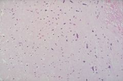
Which entity is characterized by periarteriolar concentric fibrosis in the spleen?
A. TTP B. Sickle cell disease C. Castleman's disease D. Infarction E. Amyloidosis |
A. This was a board question. TTP causes thrombi in arteries and arterioles without inflammation. Also there is PAS+ hyaline subendothelial deposits of platelets. Periarteriolar concentric fibrosis is seen in 58% TTP spleens. Other findings are hemosiderin laden macrophages and hemophagocytosis
|
|
|
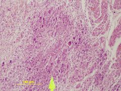
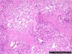
The image depicted here most likely represents what?
A. Metastatesis B. Large cell lymphoma C. Angiosarcoma D. Infarct E. Miliary TB |
B - this was a board question. This picture is consistant with a large cell lymphoma or hodkin lymphoma.
|
|
|
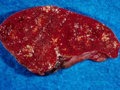
The image depicted here most likely represents what?
A. Metastatesis B. Large cell lymphoma C. Angiosarcoma D. Infarct E. Miliary TB |
D. This was a question on a board exam. This lesion represents a remote splenic infarct.
|
|
|
|
Patient has Hodkin's lymphoma of the mediastinum with bilateral adenopathy and involvement of the spleen. What is the Ann Arbor stage?
A. Stage 1S B. Stage 2S C. Stage 3S D. Stage 4S |
C - this was a board question
Stage I- the cancer is located in a single region, usually one lymph node and the surrounding area. Stage II- The cancer is located in two separate regions, an affected lymph node or organ and a second affected area, and that both affected areas are confined to one side of the diaphragm - that is, both are above the diaphragm, or both are below the diaphragm. Stage III -The cancer has spread to both sides of the diaphragm, including one organ or area near the lymph nodes or the spleen. Stage IV- Diffuse or disseminated involvement of one or more extralymphatic organs, including any involvement of the liver, bone marrow, or nodular involvement of the lungs. S: is used if the disease has spread to the spleen. |
|
|
|
All of the following are true of syphilitic aortitis except?
A. Proximal aortic aneurysms B. Perivascular and medial inflammation C. Destruction of medial elastic tissue D. Medial necrosis E. Adventitial fibrosis |
E. This was on the boards. The wording of this is questionable. Adventitial fibrosis does happen however it is usually more adventitial inflammation. The concept is more important. The disease affects the vasa vasorum with inflammation and eventual destruction of the media and possible aortic aneurysms. The media takes on a "tree bark" appearance.
|
|
|
|
What is the most common palpable vascular tumor of the breast?
A. Cavernous hemangioma B. Capillary hemangioma C. Kaposi's sarcoma D. Angiosarcoma |
A. This was a board question
|
|
|
|
What is the significance of a positive pANCA in ulcerative colitis and primary sclerosing cholangitis?
A. No significance B. Worse outcome C. Better outcome D. Possible accompanying vasculitis. |
A. This was on boards. Patients with a number of other diseases, such as ulcerative colitis and ankylosing spondylitis, will commonly have ANCA as well. However in these cases there is no associated vasculitis, and the ANCA are thought to be incidental or ARTIFACT rather than part of the disease itself.
|
|
|
|
What are the diagnostic antibodies in polyarteritis nodosa and wegener's granulomatosis?
A. Proteinase 3 and nuclear B. MPO and nuclear C. MPO and Proteinase 3 D. Nuclear and MPO E. Proteinase 3 and MPO |
C. A varient of this was a board question.
p-ANCA = MPO c-ANCA = Proteinase 3 |
|