Reading...
![]()
Play button
![]()
Play button
![]()
Use LEFT and RIGHT arrow keys to navigate between flashcards;
Use UP and DOWN arrow keys to flip the card;
H to show hint;
A reads text to speech;
77 Cards in this Set
- Front
- Back
|
When does the heart start beating?
|
7-10 somites
2.2 mm in length 23 days old |
|
|
What is the pericardium?
|
A double walled fibroserous sac which surrounds and covers the heart and roots of the great vessels.
|
|
|
What is the serous pericardium? What are its two layers?
|
A serous lining layer of the pericardium. Its two layers are the parietal layer and th visceral layer (epicardium).
|
|
|
What does the parietal layer line?
|
Lines inside of fibrous pericardium.
|
|
|
What does the visceral layer cover?
|
The outside of the heart.
|
|
|
What is the fibrous pericardium? Where is it? What does it form?
|
A CT layer outside of the parietal serous layer. IT forms the bulk of the gross pericardial sac - fused to adjacent CT planes.
|
|
|
What is the pericardial space? What is in it?
|
Potential space between parietal and visceral layers - contains only a moistening layer of fluid.
|
|
|
Describe the layers of the pericardium from most external to internal. (6)
|
Fibrous pericardium (parietal layer), Serous pericardium (parietal layer), pericardial space, serous pericardium (visceral layer epicardium), myocardium, endocardium
|
|
|
When are the pericardial sinuses formed?
|
Formed during folding of heart tube development.
|
|
|
What is the transverse percirdial sinus?
|
A space behind the aorta that could be used to stop flow to the aorta.
|
|
|
What is the oblique sinus?
|
A sinus on the posterior aspect of the heart were fluid can acumulate. It is continuous withhe pericardial cavity.
|
|
|
What is the superior pericardial extent?
|
Fuses with adventitia of great vessels at the level of the sternal angle.
|
|
|
What is the inferior pericardial extent?
|
Fuses with fascia of diaphragm.
|
|
|
What is the lateral pericardial extent?
|
Fuses with mediastinal pleura
|
|
|
What is the posterior pericardial extent?
|
Fuses with adventitia of esophagus.
|
|
|
Name the arteries that supply the pericardium. (6) Also name any specific sources or nerves associated with those arteries.
|
1)Pericardiacophrenic (runs with the phrenic nerve) from the internal thoracic.
2) muculophrenic 3) internal thoracic 4) esophageal aa. (from aorta) 5) bronchial aa. 6) Pericardial -directly from aorta |
|
|
What is the innervation of the pericardium? (3) Which is the main innervation?
|
Phrenic nerves (main)
Vagi nerves Sympathetic trunk |
|
|
What is the function of the pericardium?
|
Contains approx. 10-15 ccs of serous fluid. The function of the pericardium is to limit or prevent acute pathological distension of the heart once the pericardial reserve volume has been used up and the pericardium is stretched. They also maintain a constant heart position and cancel any external forces on the heart.
|
|
|
What are the 3 layers of the heart wall?
|
Epicardium, myocardium, endocardium.
|
|
|
What is the epicardium?
|
Visceral layer of pericardium - external covering of heart.
|
|
|
What is the subepicardium? What does it contain?
|
The subepicardium is between the epicardium and the myocardium. It contains fat and the major vessels to the heart.
|
|
|
What is the myocardium? What sort of fibers does it have? What is its purpose?
|
Middle muscular layer - oblique - fibers -torsion of chambers
|
|
|
What is the endocardium?
|
Inner lining layer of the heart, composed of endothelium backed by thin CT
|
|
|
What is the subendocardium? What pathological condition initiates here?
|
Just outside the endocardium, contains tissues of the conducting system (All MI starts here)
|
|
|
What are auricles?
|
Primitiv atriums that become vestigial (auricle = ears)
|
|
|
What section of the heart contains the apex?
|
Left ventricle.
|
|
|
What defines the lateral extent of the heart? What pathological condition exists if it extends beyond this?
|
THe midclavicular line. Enlargement of the heart if it extends beyond this.
|
|
|
Where is the oblique sinus? What happens here?
|
It is on the posterior aspect of the heart and is the pericardial insertion
|
|
|
Where is the posterior interventricular sulcus? What runs in it?
|
On the diaphragmatic surface near the right margin. In it run the posterior interventricular artery and middle cardiac vein.
|
|
|
Where is the anterior interventricular sulcus? What runs in it?
|
On the sternocostal surface of the heart, close to its left margin. The Anterior interventricular sulcus contains the anterior interventricular a. and the great cardiac v.; it marks the location of the interventricular septum
|
|
|
Where is the coronary sulcus (atrioventricular groove)? What runs in it?
|
It is the groove on the surface of the heart that seperates the atria from the ventricles. The coronary sulcus contains the coronary sinus, circumflex a., and right coronary a.
|
|

Identify all the numbered structures.
|
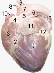
1. left ventricle
2. anterior interventricular sulcus 3. right ventricle 4. conus arteriosus 5. pulmonary artery 6. Ligamentum arteriosum 7. aortic arch 8. brachiocephalic artery 9. left subclavian artery 10. right auricle 11. left auricle 12. fat 13. pulmonary vein |
|
|
Why is the right atrium so important?
|
Everything that happens in development happens here.
|
|
|
What is the fossa ovalis? Where is the fossa ovalis? What is it the remanant of?
|
The fossa ovales is the remnant of the foramen ovales (allows blood through in embryo/fetus). It is in the right atrium.
|
|
|
What does the tricuspid valve seperate?
|
The right atrium and Right ventricle. (AKA right atrioventricular valve)
|
|
|
What does the pulmonic valve seperate?
|
The right ventricle and the pulmonary artery. (consists of 3 semilunar valveules or cusps)
|
|
|
What does the mitral valve seperate?
|
The left atrium and ventricle. (AKA left AV valve)
|
|
|
What does the aortic valve seperate?
|
The left ventricle and the aorta. (consists of 3 semilunar valvules or cusps)
|
|
|
What are chordae tendineae?
|
Points of attachement for papillary muscles to the tricuspid and mitral vavles.
|
|
|
Which chamber has 3 papillary muscles? Which chamber has 2?
|
The right ventricle has 3 the left ventricle has 2 papillary muscles.
|
|
|
Where is the obtuse margin?
|
On the left pulmonary surface.
|
|
|
Where is the acute margin?
|
On the right pulomary surface.
|
|
|
What are the musculi pectinati? Where are they most likely found?
|
In the right atrium, behind the crest the internal surface of the atrium is smooth, while in front of it the muscular fibers of the wall are raised into parallel ridges resembling the teeth of a comb, and hence named the musculi pectinati (pectinate muscles).
|
|
|
Where/what is the crista terminalis? What is its origin?
|
In the development of the human heart, the right horn and transverse portion of the sinus venosus ultimately become incorporated with and form a part of the adult right atrium, the line of union between it and the right auricle being indicated in the interior of the atrium by a vertical crest, the crista terminalis of His. (back of the comb) (is of embryologic origin)
|
|
|
Where is the valve of the coronary sinus? Why is it so important?
|
The valve of the coronary sinus is just superior to the septal leaflet of the tricuspid valve. It is also just to the left of the valve of the inferior vena cava.
|
|
|
What is the valve of the inferior vena cava? Why is important in embryology? Where is it located?
|
The valve of the inferior vena cava seves to direct blood through the foramen ovale into the left atrium.
It is located in the right atrium. |
|
|
Where is the valve of the foramen ovale? What is its origin?
|
In the left atrium. Septum premium.
|
|
|
Where is the infundibulum or conus arteriosus? What important structure arises from here?
|
The conus arteriosus is in the upper and left angle of the right ventricle and forms a conicle pouch from which the pulmonary artery arises.
|
|
|
What is just inferior to the conus arteriosus?
|
The supraventricular crest which is important for the functioning of the right ventricle.
|
|
|
What is the septomarginal trabecula? Where is it?
|
A moderator band in the right ventricle that is part of the electrical conduction of the heart.
|
|
|
Where is the trabeculae carnae large/small?
|
Large in the right ventricle not in the left.
|
|
|
Where is the anterior papillary muscle and why is it so important?
|
This muscle is in the left ventricle (anterior and posterior b/c they only have 2!). They hod the cordae tendineae very tight and prevent regurgitation.
|
|
|
Which ventricle is thicker/stronger?
|
The left ventricle is thicker/stronger.
|
|
|
What separates the right AV (tricuspid) valve and the pulonary valve?
|
The supra ventricular crest.
|
|
|
What Seperates the left AV valve (mitral) and the aortic valve?
|
Not much they are very close.
|
|
|
What is the skeleton of the heart?
|
A fibrous CT to which valves and muscle of the heart are attached. They provide support and rigidity. They also serve as an electrical insulator preventing conduction between atria and ventricles.
|
|
|
Where is the SA node? What does it do? What supplies it?
|
The SA node = pacemaker. It is located along the upper end of the sulcus terminalis near superior vena cava. It initiates teh heart beat. It is supplied by both sympathetic and parasympathetic nerves.
|
|
|
Where is the AV node?
|
located in interatrial septum adjacent to ostium of coronary sinus.
|
|
|
Where does the AV bundle of His extend?
|
From the a-v node alon the interventricular septum to purkinje fibers. IT divids into left right and left bundle branches inteh septum (near the junction of membranous and muscular part of septum).
|
|
|
Where is the right coronary artery? What does it arise from?
|

Arises from the right aortic sinus. It courses in the coronary sulcus (Right AV groove) from the aorta toward the region of the posterior IV sulcus.
|
|
|
Where is the left coronary artery? What does it arise from?
|
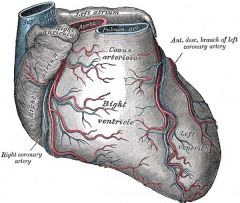
The left coronary artery arises from teh aortic sinus and courses in the anterior IV sulcus where it divides.
|
|
|
Name the branches of the right coronary artery. Give them in the order which they arise from most proximal to the origin of the right coronary artery.
|
SA nodal branch, AV nodal branch, marginal branch, posterior interventricular branch
|
|
|
What percentage of the posterior IV branch artery is supplied by the right coronary artery, the left?
|
Right = 80%
Left = 20% |
|
|
What percentage of the SA branch artery is supplied by the right coronary artery, the left?
|
Right 55%
Left 45% |
|
|
What percentage of the AV (nodal) branch artery is supplied by the right coronary artery, the left?
|
Right 85%
Left 15% |
|
|
Why is the left anterior descending artery so important?
|
It is called the widowmaker artery b/c most infarctions happen here.
|
|
|
When does the blood supply of the hear occur?
|
During diasystolic action.
|
|
|
What are the branches of the left coronary artery? Give them in order that they divide off from the origin of the left coronary artery.
|
SA nodal branch, AV nodal branch, Circumflex branch -> left marginal branch, anterior IV branch (LAD)-> diagonal branch of AV branch, posterior IV branch
|
|
|
Where does the coronary sinus drain?
|
Into the right atrium.
|
|
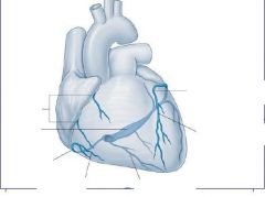
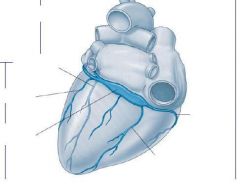
Identify the veins.
|
See above
|
|
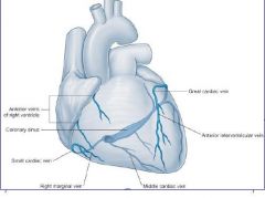
Identify the veins.
|
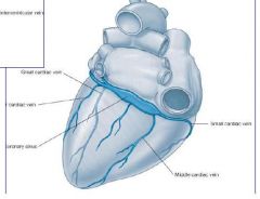
See above
|
|
|
What is the sympathetic innervation to the heart? (2) What is their preganglionic level? What major sensations reach the CNS through the sympathetic routes?
|
1)From cervical sympathetic trunk (cervical cardiac sympathetic nerves)
2) From direct branches of thoracic trunk (thoracic cardiac sympathetic nerves) 3) Preganglionic level (T1-5)-most ascend to synapse Ischimic pain and sensations reach CNS via sympathetic routes. |
|
|
What is the parasympathetic innervation of the heart? (2) What activity reaches the CNS via the vagus?
|
Parasympathetic-
1) from vagus as cervical branches (superior and inferior) 2) from vagus as direct brancehs in thorax (thoracic) Visceral reflex activity reach CNS via vagal route. |
|
|
What/where are aortic bodies? Are they para or sympathetic innervated?
|
Aortic bodies are in the wall of the ascending aorta adn they monitor pCO2/pO2 and change respiration rate.
|
|
|
What nervous systems contribute to the cardiac plexus?
|
Sympathetic and parasympathetic contributions.
|
|
|
Where is the superficial cardiac plexus? What innervates it?
|
The superficial plexus is located near the aortic arch. IT recieves input from the left inferior cervical branch of CN X (vagus) and superior cervical sympathetic branches.
|
|
|
Where is the deep plexus? What innervates it?
|
The deep plexus is located near the tracheal bifurcation (carina). Its sympathetic = superior, middle, and inferior (right) and middle and inferior left.
Parasympathetic = CN X (vagus) |