Reading...
![]()
Play button
![]()
Play button
![]()
Use LEFT and RIGHT arrow keys to navigate between flashcards;
Use UP and DOWN arrow keys to flip the card;
H to show hint;
A reads text to speech;
25 Cards in this Set
- Front
- Back
|
Branches from what cervical spinal nerves come together to form the phrenic nerve in the horse, ox and small ruminant?
What muscle does the phrenic nerve course over? |
Horse - C6, C7 (+/- C5)
Ox - C5, C6, C7 Small ruminant - C5, C6 (+/- C7) Phrenic nerve courses over the scalenus muscle then enters the thoracic cavity |
|
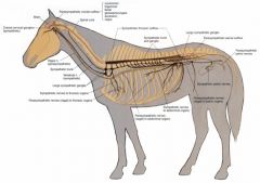
What are the yellow and red nervous systems shown? Where do they originate?
|

Yellow - para - from 3,7,9,10 and S1,2,3 (which become pelvic n.)
Red - symp - from thoracolumbar spinal nerves |
|

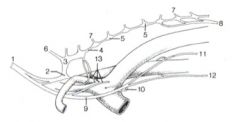
Name these 1-7
|
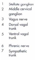
Key
|
|
|
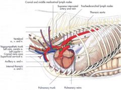
What do the recurrent laryngeals wrap around?
|
Right recurrent laryngeal nerve wraps around the right subclavian artery
Left recurrent laryngeal nerve wraps around the aortic arch |
|

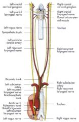
Name these
|
1 Vagosympathetic trunk
2 Middle cervical ganglion 3 Cervicothoracic ganglion 4 Sympathetic trunk 5 Sympathetic ganglia 6 Vertebral nerve 7 Rami communicantes 8 Major splanchnic n. 9 Vagus n. 10 Recurrent laryngeal n. 11 Dorsal vagal trunk 12 Ventral vagal trunk 13 Cardiac nn. |
|
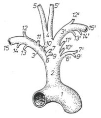
Name these structures on the horse (left is left, right is right).
|

Brachiocephalic trunk (2)
-Left subclavian a. (3) -Bicarotid trunk (4) --Left common carotid a. (5) --Right common carotid a. (5’) -Right subclavian a. (3’) |
|
|
What are the first two branches of the subclavian artery in the horse?
|

Costocervical trunk
-Supreme intercostal a. (Supplies 1st – 3rd dorsal intercostal aa. - the rest come right off of the aorta) -Dorsal scapular a.(Supplies 1st dorsal intercostal a. and) muscles and skin in the area of the withers Deep cervical a. -From costocervical trunk on the right side, subclavian a. on the left side -Courses toward the head to supply cervical structures and musculature in the caudal neck region |
|
|
What is the difference in the source of the deep cervical a. in the horse right side from left side?
|
From costocervical trunk on the right side, subclavian a. on the left side
|
|
|
Where does the vertebral a. course?
|
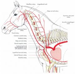
Courses cranially through the transverse foramina of the cervical vertebrae
Gives rise to spinal brs and muscular brs. Courses through the alar foramen and lateral vertebral foramen of the atlas and enters the vertebral canal. Right and left vertebral aa. form basilar a. (Horse) |
|
|
What are the branches off of the internal thoracic a. of the horse?
|
Ventral intercostal aa.
Supply ventral part of the thoracic wall Cranial epigastric a. Supplies the ventral abdominal wall |
|
|
Name the branches off of the horse subclavian artery.
|
Costocervical trunk
Deep cervical a. vertebral a. Superficial cervical a. internal thoracic a. Axillary a. |
|
|
What are the branches off of the descending thoracic aorta of the horse?
|
Descending thoracic aorta
-Bronchoesophageal a. --Bronchial br. --Esophageal br. -Dorsal intercostal aa |
|
|
How can you differentiate between the internal thoracic vein and costocervical vein in the horse?
|
Internal thoracic courses ventrally
costocervical courses dorsally |
|
|
What is the venous counterpart to the bicarotid trunk on the large animal?
|
Bijugular trunk (1)
|
|
|
What veins meet to form the cranial vena cava in the horse and ox?
|
Subclavian veins and bijugular trunk.
|
|
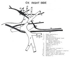
Arteries in white, veins in black
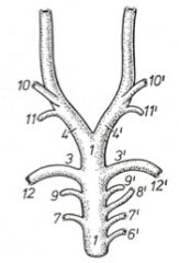
Can you name these arteries and nerves on the right side of the ox? Go ahead and look at the key |
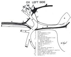
Arteries in white, veins in black
Can you name these arteries and nerves on the left side of the ox? Go ahead and look at the key |
|
|
Where do the right and left azygous drain from and into?
|
Right azygous to cranial vena cava
Left azygous to heart and coronary sinus They drain dorsal part of thoracic wall |
|
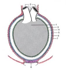
Name these layers of the pericardium
|

1. Heart
2. Great vessels 3. Visceral pericardium (epicardium) 4. Pericardial cavity 5. Parietal pericardium 6. Fibrous pericardium 7. Pericardial mediastinal pleura 8. Sternopericardial ligament |
|

Name these areas of the equine heart
|
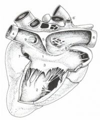
1 Right atrium
2 Right ventricle 3 Right atrioventricular valve 4 Caudal vena cava 5 Intervenous tubercle 6 Cranial vena cava 7 Coronary sinus 8 Terminal crest 9 Fossa ovalis |
|
|
Which of the layers of the heart produce serous fluid?
|

Pericardial mediastinal pleura, parietal pericardium and visceral pericardium.
|
|
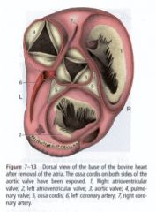
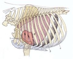
This is a dorsal view of the base of an ox heart - name what you can.
|
This is a dorsal view of the base of a horse heart - name what you can.
|
|
|
What are the coronary arteries and the branches off of them?
|
Left coronary artery
-Paraconal interventricular br. -Circumflex br. --Intermediate br. --Subsinuosal interventricular br. (ruminant) Right coronary artery -Circumflex br. --Subsinuosal interventricular br. (horse) |
|
|
Where is the best place to auscultate the pulmonary valve, aortic valve, left AV valve and right AV valve in the horse?
|
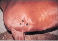
Pulmonary valve
Left side, 3rd or 4th IC space, caudodorsal to the olecranon Aortic valve Left side, 4th IC, just ventral to the shoulder joint Left AV valve Left side, 5th IC space, caudodorsal to the olecranon Right AV valve Right side, 4th IC space, caudodorsal to the olecranon |
|
|
Where are the puncta maxima of bovine hearts?
|
Pulmonary valve
Left side, 3rd IC space, medial to the medial epicondyle of the humerus Aortic valve Left side, 4th IC, just ventral to the shoulder joint Left AV valve Left side, 4th IC space, medial to the medial epicondyle of the humerus Right AV valve Right side, 4th IC space, medial to the medial epicondyle of the humerus |
|
|
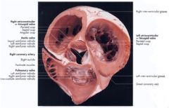
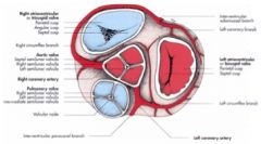
What are the heart vascular grooves and what courses through them in the large animal heart?
|
Coronary groove - great and middle cardiac veins, right and left coronary arteries
Paraconal groove - great cardiac vein and paraconal interventricular branches of left coronary artery Subsinuosal interventricular groove - middle cardiac vein and subsinuosal interventricular branch (from right coronary artery (horse) and left coronary artery (ruminant)) Intermediate groove - caudal cardiac vein (ruminant) |