![]()
![]()
![]()
Use LEFT and RIGHT arrow keys to navigate between flashcards;
Use UP and DOWN arrow keys to flip the card;
H to show hint;
A reads text to speech;
191 Cards in this Set
- Front
- Back
|
What are the functions of the respiratory system? |
*Supplies oxygen and removes carbon dioxide *Filters and conditions inspired air *Produces sound *Contains receptors for smell *Rids the body of excess water and heat *Helps regulate blood pH |
|
|
What produces the superior boundary of the thorax? |
*Jugular notch *Sternoclavicular joint *Superior border of clavicle *Spinous process of C7 |
|
|
What produces the inferior coundary of the thorax? |
*Xiphoid process *Costal arch *Vertebra T12 *11th and 12th ribs |
|

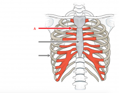
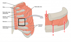
What is A? |
Thoracic Inlet |
|

What is B? |
Inferior thoracic aperature |
|
|
How many ribs do we have in total? |
12 |
|
|
How many true ribs are there? |
7 |
|
|
How many false ribs are there? |
3 |
|
|
How many floating ribs? |
2 |
|
|
What are true ribs |
Individually connected to the sternum |
|
|
What are false ribs? |
Connect indirectly to sternum |
|
What is A? |
Costal cartilage |
|

What is A? |
Manubrium |
|

What is B? |
Sternal Angle |
|
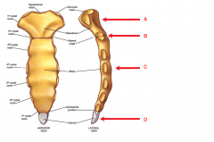
What is C? |
Body |
|

What is D? |
Xiphoid Process |
|
|
What are the three parts of the sternum? |
*Body *Manubrium *Xiphoid proces |
|
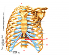
What is A? |
1st Thoracic Vertebra |
|

What is B? |
Infrasternal (Subcostal) Angle |
|

What is C? |
Costal Margins |
|

What is D? |
12th thoracic vertebra |
|

What is A? |
Pericardial Sac |
|

What is B? |
Thymus Gland |
|
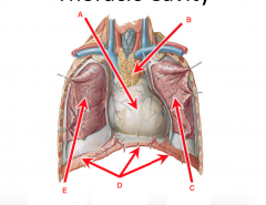
What is C? |
Left Lung |
|

What is D? |
Diaphragm |
|

What is E? |
Right Lung |
|
|
Are thymus gland found in adults? |
No, mostly found in young kind to help develop the immune response (Afterwards it turns into fat) |
|
|
What is the mediastinum? |
Between right and left lung, it makes up the middle of thorax |
|
|
What are the boundaries of the superior mediastinum? |
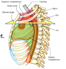
*Anterior: Manubrium *Posterior: T-1 to T-4 *Superior: Plane of thoracic inlet *Inferior: Sternal angle and T-4 *Sides: Mediastinal pleura |
|

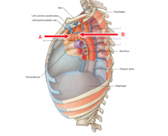
What is A? |
Left common Carotid Artery |
|

What is B? |
Left Subclavian Artery |
|
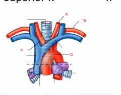
What is C? |
Arch of Aorta |
|

What is D? |
Brachiocephalic artery |
|

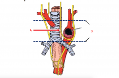
What is A? |
Trachea |
|

What is B? |
Esophagus |
|

What is C? |
Right Brachiocephalic vein |
|
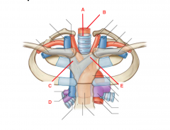
What is D? |
Superior Vena Cava |
|

What is E? |
Left Brachiocephalic Vein |
|
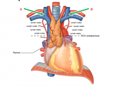
What is A? |
Right lymphatic Duct |
|

What is B? |
Thoracic Duct |
|
|
What is the brachiocephalic vein made up by |
Internal jugular and subclavian vein |
|
|
Where does the internal juglar and subclavian vein come together at? |
Venous angle |
|
|
Where do the right lymphatic duct and thoracic duct come into |
Venous angle |
|
What is A? |
Phrenic Nerve |
|

What is B? |
Vagus Nerve |
|
|
Which is more superifical and anterior, the vagus or the phrenic nerve? |
Phrenic nerve |
|
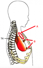
What is A? |
Left Recurrent laryngeal nerve |
|

What is B? |
Boundaries of superior mediastinum |
|

What is A? |
Superior Mediastinum |
|
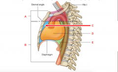
What is B? |
Inferior Mediastinum |
|

What is C? |
Anterior Mediastinum |
|

What is D? |
Middle Mediastinum |
|

What is E? |
Posterior Mediastinum |
|
|
What are the boundaries of the anteiror mediastinum? |
*Anterior: Body of sternum (because below the sternal angle) *Posterior: Pericardium *Sides: Mediastinal pleura *Superior: Sternal angle *Inferior: Diaphragm |
|
What is A? |
Thymus (in children) |
|

What is B? |
Sternopericardial ligament |
|
|
The thymus straddles between which mediastinums? |
Superior and anterior |
|
|
What does the sternopericardial ligaments do |
Helps heart stay in right spot. connects the pericardium to the sternum |
|
|
What are the boundaries of the middle mediastinum? |
*Anterior: Pericardium *Posterior: T5 to T12 *Superior: Level of sternal angle *Inferior: Diaphragm *Sides: Mediastinal pleura |
|

What is A? |
Pulmonary trunk |
|
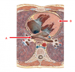
What is B? |
Heart in pericardium |
|
|
Where is the tracheal bifurcation found? |
Right behind manubrium |
|
|
If you want to just hit one branch of the trachea where should you hit? |
Below the sternal angle because the tracheal bifurcation right behind the manubrium |
|

What is A? |
Superior Vena Cava |
|
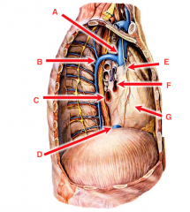
What is B? |
Arch of Azygos |
|

What is C? |
Pulmonary Vein |
|

What is D? |
Inferior Vena Cava |
|

What is E? |
Phrenic Nerve |
|

What is F? |
Pulmonary Artery |
|

What is G? |
Pericardium |
|
|
What can be found in the posterior mediastinum |
*Esophagus *Descending thoracic aorta *Vagus nerve *Sympathetics *Lymph Nodes |
|

What is A? |
Accessory Hemiazygous Vein |
|

What is B? |
Azygos vein |
|
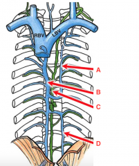
What is C? |
Thoracic Duct |
|

What is D? |
Hemiazygous Vein |
|

What is A? |
Sympathetic Trunk |
|

What is B? |
Vagus Nerve |
|
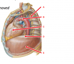
What is C? |
Phrenic Nerve |
|

what is D? |
Central Tendon of Diaphragm |
|

What is E? |
Diaphragm |
|
|
What is the midclavicular line? |
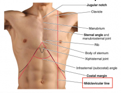
Down from the middle of calvicle |
|
|
What are the respiratory organs? |
*Nose, nasal cavity, and paranasal sinuses *Pharynx and larynx *Trachea and bronchi *Lungs and alveoli |
|
|
What is ventilation |
Bulk movement of air into and out of lungs |
|
|
What is respiration |
Process of gas exchange |
|
|
If ventilation is good but oxygen saturation is low what should you do |
Take in more pure oxygen so oxygen saturation increase |
|
|
If oxygen saturation not good what can you do |
Breathe deeper or breathe faster |
|
|
Breathing rate normally controlled by level of _____ in blood |
CO2 |
|
|
Can breathing be consciously influenced? |
Yes |
|
|
How do you measure ventilation? |
Times/min |
|
|
How can you measure respiration? |
Oxygen saturation |
|
|
What is the normal breathing rate |
12 breathes per minutes |
|
|
What is tachypnea |
Faster rate |
|
|
What is bradypnea |
Slower rate |
|
|
What is total lung capacity |
Volume after maximal inspiration |
|
|
How much is total lung capacity? |
6000 mL |
|
|
What is tidal volume? |
Volume of each normal breath |
|
|
How much is the tidal volume? |
500 mL |
|
|
What is residual volume? |
Volume remaining in lungs |
|
|
How much is the residual volume |
1200 mL |
|
|
What is the upper respiratory tract |
Nose to larynx |
|
|
What does the upper resipratory tract? |
Conducting portion, transports the air |
|
|
What is the lower respiratory tract |
Trachea, bronchi |
|
|
What does the lower respiratory tract do? |
Conducting portion, transports the air down to lungs |
|
|
What is part of the lower respiratory tract |
Alveolar ducts Alveoli (Respiratory portion, gas exchange) |
|
|
What does the larynx do? |
*Produces sounds *Phonation |
|
|
What does the pharynx do? |
*Conducts gas to lower airways *Passage of food to esophagus |
|
|
What does the nose do? |
*Provides an airway for respiration *Resonating chamber for speech *Filters, warm, and moistens inhaled air |
|
|
What do the concha do? |
Increase the surface area of nasal mucosa to warm and filter air because covered in mucous so little things get stuck in it so you don't breathe it in Nose hairs filter out bigger particles |
|
|
What comes together as the pharynx? |
Back of nose and mouth |
|
|
What is the pharynx |
Funnel-shaped passageway |
|
|
What does the pharynx connect |
Nasal cavity and mouth |
|
|
What are the different parts of the pharynx |
*Nasopharynx *Oropharynx *Laryngopharynx |
|
|
What is the nasopharynx |
Behind nose |
|
|
What is the oropharynx |
Behind the mouth |
|
|
What is the laryngopharynx |
Bifuration of larynx and esophagus Right next to the larynx |
|
|
What does the larynx do? |
*Properly routes air to lungs and food to esophagus *Voice production |
|
|
Where are vocal cords found? |
Inside larynx |
|
|
How does the vocal cords work? |
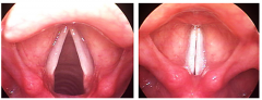
When vocal cord is open, can breathe and goes through When closed this is how it is when we speak. Vocal cord is in this position so they can produce vibration |
|
|
What is the valsalva maneuver |
When vocal cord closed completely can't move any air in or out. When lift something heavy you are holding your breath which is valsalva maneuver and have to strain against the closed cords. It increases the pressure in thorax and might help to lift things. This will also increase blood pressure and could lead to a hemorrhage inside the eye (subconjunctival hemorrhage) |
|
|
What is the epiglottis |
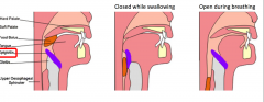
When air pass through. When swallowing something epiglottis (lies on top of glottis) flops down to deflect food away from trachea into esophagus so can swallow it |
|
|
Which is the front tube, trachea or esophagus |
Trachea (breathing) |
|
|
When putting tube for breathing what do you have to do |
Have to move epiglottis out of the way so can visualize the vocal cords to move those out of way and into the trachea |
|
|
Where is the trachea found? |
On midline, inferior to larynx Anterior to esophagus Posterior to great vessels |
|
|
What are the tracheal cartilages |
Ensure open airway (to make sure the trachea is not squished between great vessels/esophagus/anything else that is there) 15-20 |
|
|
The cartilage rings of the trachea are ____ shaped |
C |
|
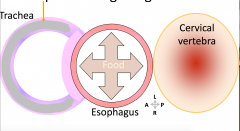
Explain this picture? |

As food goes down esophagus, esophagus has to expand (pushing forward on track when swallow) |
|

What is A? |
Carina |
|

What is B? |
Right main bronchus |
|
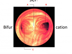
What is C? |
Left main bronchus |
|
|
The right main bronchus is _______ than the left |
Shorter, wider and more vertical |
|
|
Are foreign objects more likely to enter the right or left main bronchus |
Right |
|
|
What happens when the main bronchi branches |
It becomes narrower, diverge throughout the lungs and terminate as terminal bronchioles |
|
|
What do the smaller bronchi have |
*Less cartilage *More smooth muscles (vary diameter) |
|
|
Bronchodilation is ____ activation |
Sympathetic (If during exertion) |
|
|
When does bronchoconstriction happen with |
Histamine release |
|
|
As the bronchi get smaller how does it change |
Starts to lose cartilage and gain more muscle |
|
|
Most bronchioles in the lungs are in the ____ zone |
Conduction |
|
|
Gas exchange takes place in the ____ zone |
respiratory |
|
|
Do the lungs fill most of the thorax? |
Yes |
|
|
What separates the two lungs? |
Mediastinum |
|
|
How many lobes does the right lung have |
3 lobes |
|
|
How many lobes does the left lung have |
2 |
|
|
What are the lobes of the right lung? |
*Superior lobe *Middle lobe *Inferior lobe |
|
|
What are the lobes of the left lung |
*Superior lobe *Inferior lobe |
|
|
What are the fissure(s) of the right lung |
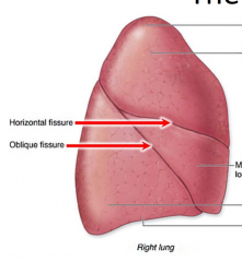
*Horizontal fissure *Oblique fissure |
|
|
What are the fissure(s) of the left lung? |
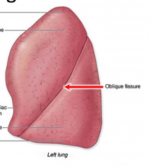
Oblique fissure |
|
|
Which lung is the cardiac notch found on |
Left lung |
|
|
Which is smaller the left or right lung |
Left |
|
|
Which is located more superiorly, the right or left lung? |
Right lung because of the liver |
|
|
What is the hilum of the lung |
Where everything passes in and out of the lungs |
|

What is A? |
Bronchus |
|

What is b? |
Hilum |
|

What is C? |
Pulmonary artery |
|
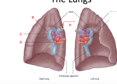
What is D? |
Pulmonary veins |
|
|
What is the pleural cavity |
Potential space between the visceral and parietal pleurae. In between the two pleura have a little bit of fluid that is lubricating and also holds the surface of lung against the chest wall |
|
|
Which is larger the pleural cavity or the lung itself |
Pleural cavity (therefore creates a costodiaphragmatic recess --> space between ribs and diphragm) |
|
|
What keeps lungs stuck to thoracic wall during respiration? |
surface tension (Necessary for proper ventilation) |
|
|
If have excess fluid in pleural cavity and person is standing upright where would the fluid collect |
In costodiaphragmatic recess |
|
|
Are there muscles connected to the ribs? |
Yes Several layers of muscles run just betwene ribs (The "VAN" supplies the muscles) |
|

What is A? |
Innermost intercostal |
|
What is B? |
Internal Intercostal |
|

What is C? |
External intercostal |
|
|
How does the external intercostal move |
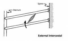
Run down and forward, when contracts it pulls the lower ribs closer to the upper ribs. Moves the sternum up and out and raises the ribs (it helps with inhalation) |
|
|
How does the internal intercostal |
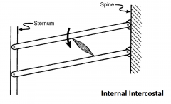
Will pull two ribs closer together (top towards bottom rib) and will help with exhalation |
|
|
Is the external intercostal located more anterior or posterior? What does that cause |
Posterior They pivot at their attachment to the psine, it pull close togethr close to pivot point will slowly make up and out |
|
|
Is the internal intercostal located more anterior or posterior? |
Anterior |
|
|
What is the internal intercostal for, exhalation or inhalation? |
Exhalation |
|
|
What is the external intercostal for, exhalation or inhalation? |
Inhalation |
|
|
What is the major muscle of inspiration? |
Diaphragm |
|
|
What innervates the diaphragm? |
Phrenic nerve |
|
|
Which part of the vertebraes does the phrenic nerve comes from> |
C3, C4, C5 |
|
|
If your neck is broken at the level of C6 can you still breathe? |
Yes, because below the level of where phrenic originates |
|
|
If your neck is broken at the level of C2 can you still breathe? |
No, because broken above level where phrenic originates |
|
|
What is the middle part of the diaphragm |
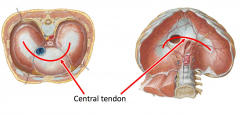
Central tendon |
|
|
Where does the diaphragm insert |
Inserts onto self |
|
|
Is inhalation or exhalation an active process? |
Inhalation |
|
|
Is inhalation or exhalation a passive process? |
Exhalation |
|
|
What happens to the diaphragm during inhalation? |
Diaphragm contracts and flattens during inhalation, air rushes in |
|
|
What happens to the diaphragm during exhalation |
Diaphragm relaxes during exhalation, air is pushed out (don't need any muscles, it just relaxes. The normal elasticity of muscle just forces air out) |
|
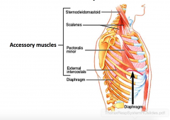
|
What is needed for foreceful inspiration |
|
|
|
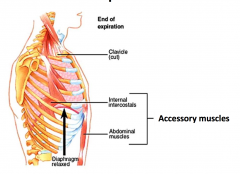
What is needed for forceful expiration? |
|
|
|
How do abdominal muscles help with forceful expiration |
Abdominal muscles push the diaphragm up and will squeeze organs up into chest cavity to help to expell air |
|
|
How does pulmonary edema come about |
Heart can't pump back out so it backs up. Capillary only 1 cell layer then fluid leaks ot of the capillaries and fills chest cavity and alveloli. And decreases the lung capacity of patient which makes it difficult to breathe |
|
|
How many places on the chest do you listen for the right lung? |
4 (for 3 lobes and apex) |
|
|
How many places on the chest do you listen for the left lung? |
3 different places (2 lobes and apex) |
|
|
Chronic Obstructive Pulmonary Disease (COPD) |
Ventilation diffiuclt, more accessory msucles use |
|
|
Asthma |
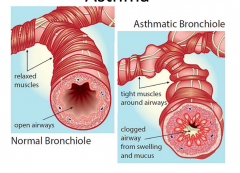
In asthma, muscles get tigheter and smaller lumen of the airway. Also wall of bronchioles are inflammed and will ahve mucous clogging up the airway. Problem getting air in because of obstruction and may also have problems getting air out. Depending on types of asthma can have problems inhaling because of thickened wall of airway and mucous in pathway and muscles around bronchioles are constricting. Treatment is steroid (to decrease swelling) or bronchiodilator (stop muscle constrincting by blocking sympathetics) |
|
|
What is pneumothorax |
Lung is collapsed. But the lung should be airtirght so when expand thoracic cavity pressure decreases and atmospheric pressure forces air in. If hole in chest wall and air leaks into pleural cavity outside of lung membrane you don't have decrease pressure to allow lung to fold |
|
|
What is a chest tube for? |
Tube put into pleural cavity and drain out whatever suppoed to be in there. Could either be excess air or fluid. From the mid-axillary line between the 4th and 5th ribs Go between the two ribs from the superior border |
|
|
Do you stick the tube up or down to suck up more air |
Up (because air typically on top) |
|
|
Do you stick the tube up or down to suck more fluid |
Down (because typically gathers in bottom) |