![]()
![]()
![]()
Use LEFT and RIGHT arrow keys to navigate between flashcards;
Use UP and DOWN arrow keys to flip the card;
H to show hint;
A reads text to speech;
208 Cards in this Set
- Front
- Back
|
Where does the heart sit? |
In the middle of the chest, a little off center (off to the left side) |
|
|
Where can the apex of the heart be found |
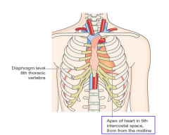
In 5th intercostal space, 9 cm from the midline |
|
|
Does the left or the right lung have a little bit cut out of it to allow room for the heart? |
Left |
|
|
Is the heart surrounded by other structures? |
Yes |
|
|
What is the diaphragm level? |
8th throacic vertebra |
|
|
Where does hte heart rest on |
Diaphragm |
|
|
Does the heart follow where the diaphragm goes? |
Yes, IE if diaphragm moves down so does the heart. Because the heart rests on the diaphragm , |
|
|
Where does the apex of the heart sit right against |
The anterior chest wall |
|
|
The aorta arches from _____ to _____ |
Anterior to poster |
|

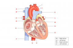
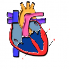
What is A? |
Superior Vena Cava |
|

What is B? |
Right pulmonary artery |
|

What is C? |
Right Coronary artery |
|

What is D? |
Inferior vena cava |
|

What is E? |
Aorta |
|

What is F? |
Left pulmonary artery |
|
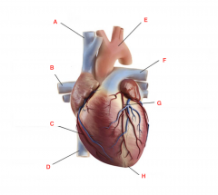
What is G? |
Left coronary artery |
|

What is H? |
Apex |
|
|
How many chambers does the heart have |
4 |
|
|
The heart is a ______ pump |
Dual |
|
|
Why does te heart have pumps? |
Because has to pump 1 way and not the other |
|
|
Do chambers of the heart have valves? |
Yes |
|
|
What are the chambers of the heart for? |
To allow 1 way flow |
|

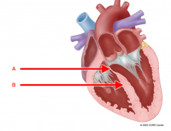
What is A? |
Superior Vena Cava |
|

What is B? |
Right Pulmonary Artery |
|

What is C? |
Right Pulmonary veins |
|

What is D? |
Pulmonary Valve |
|

What is E? |
Right Atreoventricular valve |
|

What is F? |
Inferior Vena Cava |
|
What is G? |
Aorta |
|

What is H? |
Papillary Muscle with chordae tendinae |
|

What is I? |
Arch of aorta |
|

What is J? |
Pulmonary artery |
|

What is K? |
Left Pulmonary Artery |
|

What is L? |
Left Pulmonary Veins |
|

What is M? |
Aortic Valve |
|

What is N? |
Left Atrioventricular valve |
|

What is O? |
Noel |
|
|
Are arteries away or towards the heart |
Away |
|
|
Are veins away or towards the heart? |
Towards |
|
|
Where does gas exchange occur? |
Capillary beds |
|
|
What is the ascending aorta |
Carries oxygenated blood from left ventricle to body organs |
|
|
The ascending aorta heads ____ as exits |
Superiorly |
|
|
The pulmonary trunks carries ______ from ____ to _____ |
*Deoxygenated blood *Right ventricle *Lungs |
|
|
The pulmonary trunk splits into _______ |
Right and left pulmonary arteries |
|
|
What do the coronary arteries do? |
Feed the heart muscle, they stay on the body |
|
|
The right and left pulmonary veins open into ______ |
the left atrium |
|
|
The right and left pulmonary veins return ______ from _____ |
*Oxygenated blood *Lungs |
|
|
The superior and inferior venae cava open into _______ |
Right atrium |
|
|
The superior and inferior vena cava return _____ blood from ____ |
*Deoxygenated *Body cells |
|
|
Where do the veins of the heart empty into? |
Coronary sinu |
|
|
The coronary sinus open into? |
Right atrium |
|
|
The coronary sinus retuns ____ blood from ______ |
*Deoxygenated *Heart muscle |
|
|
What is the fibrous pericardium |
Dense, inelasticl ayer of connective tissue that anchors and protects heart. It's a tough membrane that holds the heart in place so it does not move around a lot |
|
|
Which is thicker, the fibrous pericardium or serous pericardium |
Fibrous |
|
|
What is the serous pericardium |
Thinner, more delicate lines inside of the fibrous pericardium and makes a "sac" |
|
|
How many layers are there of the serous pericardium? |
2 |
|
|
What are the 2 layers of the serous pericardium |
*Parietal *Visceral |
|
|
What is the parietal layer of the serous pericardium |
*Lines inside of the fibrous layer |
|
|
What is the visceral layer of the serous pericardium |
Lines the heart surface (epicardium) |
|
|
What is between the parietal and visceral layers of the serous pericardium |
Fluid |
|
|
What is the pericardial sac |
Double-walled sac around the heart |
|
|
What does the pericardial sac allow |
Allows the heart to work in relatively friction free environment with the help of the pericardial fluid (it acts as a lubricant) |
|
|
What does the pericardial sac help to prevent |
Overfilling of the heart with blood |
|
|
What is cardiac tamponade |
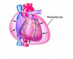
Fluid in the pericardial sac |
|
|
What happens with cardiac tamponade |
If the pericardial sac fills with fluid, it can push against the heart |
|
|
what do you need to do if get cardiac tamponade |
Need to drain |
|
|
What is the epicardium |
Visceral pericardium The visceral layer of the serous pericardium |
|
|
What is the myocardium |
Cardiac muscle layer |
|
|
What is the endocardium |
Endothelial layer of the inner myocardial surface It lines the inside of the heart so blood won't stick to it and clot |
|
|
what are the layers of the heart wall? |
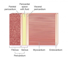
*Epicardium *Myocardium *Endocardium |
|
|
What is the bulk of the heart? |
Myocardium |
|

What is A? |
Trabeculae carnae |
|

What is B |
Endocardium |
|

What is C? |
Myocardium |
|
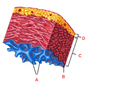
What is D? |
Epicardium |
|

What is A? |
Myocardium |
|
What is B? |
Epicardium |
|

What is C? |
Endocardium |
|
|
What is the inter-atrial septum |
Separates the right and left atria |
|
|
What can be found on the inter-atrial septum as a fetus? |
Foramen ovale |
|
|
What is the foramen ovale |
Fetal opening in inter-atrial septum |
|
|
What can be found on the inter-atrial septum after birth |
Fossa Ovalis |
|
|
What is the Fossa ovalis |
Remnant of foramen ovale (it's a depression) |
|
|
Why do we have foramen ovale instead of fossa ovalis in fetus |
Because in utero, blood does not have to get to lungs to oxygenate, goes instead to placenta |
|
|
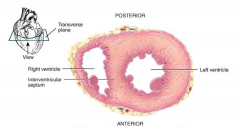
What is the inter-ventricular septum |
Separates right and left ventricles |
|
|
How many parts does the inter-ventricular septum have |
2 |
|
|
What are the two parts of the inter-ventricular septum |
Membranous part Muscular part |
|
What is A? |
Membranous part of the inter-ventricular septum |
|

What is B? |
Muscular part of the inter-ventricular septum |
|
|
What is the anulus fibrosus |
Fibrous skeleton of the heart Fibrous rings of connective tissue |
|
|
What does the anulus fibrosus do for the heart |
*Gives it structure *Electrical insulator *Muscle and valves attached |
|
|
What are the different types of muslce in the body |
*Cardiac muscle cell *Skeletal muscle cell *Smooth muscle cell |
|
|
The muscle fibers of the heart are arranged around what |
Atria and ventricles |
|
|
How do the muscles work when the heart beats |
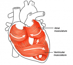
They just squeeze the heart which functions to expell as much of the blood as possible |
|
|
Do cardiac muscle cells have really high or really low demand for oxygen and nutrients |
High |
|
|
Where do coronary arteries originate |
At base of ascending aorta |
|

What is A? |
Opening to right coronary artery |
|
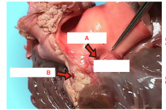
What is B? |
Left coronary artery |
|
|
What are the first arteries off the base of the aorta |
coronary arteries |
|
|
How do coronary arteries fill |
Fill on backflow (when muscle not beating) Valves at the base of the aorta so when blood injected into aorta, it does not backup into the heart. When ventricles eject blood into aorta, valve leaflets open and when start contracting, blood starts to fill back and fills the little pocket which closes the valves |
|
|
What is the anterior interventricular artery a branch off of |
Left coronary artery |
|
|
Where does the anterior interventricular artery lie? |
Between the 2 ventricles, it lies in the anterior interventricular sulcus |
|
|
What is the anterior interventricular artery also known as |
The left anterior descending coronary artery |
|
|
What is the circumflex artery a branch off |
Left coronary artery |
|
|
What is the circumflex artery |
Extends around the left side of the heart to the posterior surface (circles around the back of the heart and feeds the muscle there) |
|
|
What is the posterior interventricular artery a branch off of |
Right coronary artery |
|
|
What is the posterior interventricular artery |
Runs in the posterior interventricular sulcus |
|
|
What is the marginal artery a branch off of |
Right coronary artery |
|
|
What is the marginal artery |
Extends inferiorly along the lateral wall of the right ventricle Branch along the margin of the heart at the base |
|
|
What is the sinoatrial nodal artery a branch off of |
Right cornary artery |
|
|
What is the atrioventricular nodal artery a branch off of |
Right coronary artery |
|
|
Do veins generally run parallel with arteries? |
Yes |
|
|
Most drain blood into ______ |
The coronary sinus but some drain directly into the right atrium |
|
|
What are the different heart chambers? |
*Right atrium *Left atrium *Rigth ventricle *Left ventricle |
|
|
What kind of wall does the atria have |
Small, thin walled chambers |
|
|
What does the atria act as |
Receiving chambers for blood returning the circulation |
|
|
The atria pushes blood into the _____ |
ventricles |
|
|
What are auricles |
Outer poriton of each atrium It looks like a lumpy wrinkled flap, when not filled with blood |
|
|
The right atrium receives _____ blood from ____ |
*Deoxygenated *venous system |
|
|
What is the inflow to the right atrium? |
*Coronary sinus *Superior vena cava *Inferior vena cava |
|
|
What is the outflow of the right atrium |
Tricuspid valve into the right ventricle |
|
|
The left atrium receives ____ blood from _____ |
*Oxygenated *Lungs |
|
|
What is the inflow of the left atrium |
4 pulmonic veins |
|
|
What is the outflow of the left atrium? |
Bicuspid valve |
|
|
What makes up the most of the mass of the heart, the ventricles or atrium |
Ventricles |
|
|
What do ventricles act as |
Discharging chambers |
|
|
Where do ventricles propel blood into |
*Pulmonary trunk (Right) *Aorta (Left) |
|
|
What do the ventricle walls have? |
Papillary muscles Traveculae carnae |
|
|
What are trabeculae carneae |
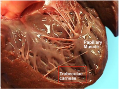
Irregular muscular ridges on inner surface They are covered with an endothelial lining and help it contract (makes it harder to stick to the wall) |
|
|
What are papillary muscles |
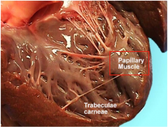
*Contract with the ventricles contract *Prevent valve vanes from bulding back into atria |
|
|
What are the chordae tendinae |
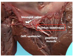
Connect valve leaflets to papillary muscles |
|
|
What does the chordae tendinae and papillary muscles help to do |
Because the valves are big and need a little bit of support, these help to hold the cusp closed. These help to prevent valve cusps from bulding back into the atria |
|
|
The right ventricle delivers ____ blood to the _____ |
*Deoxygenated *The lungs |
|
|
What is the inflow of the right ventricle |
Tricuspid valve |
|
|
What is the outflow of the right ventricle |
Pulmonic valve |
|
|
Is the moderator band found in the right or left ventricle |
Right |
|
|
What is the moderator band |
Little bit of tissue that runs between the septum and papillary muscle. It carries conducting fibers from the conducting system of the heart to the papillary muscle. So when the right ventricle contracts those papillary muscles already have some tension on them and already pulling those three cusps closed before ventricles build up full force |
|
|
The left ventricle delivers _____ blood to the ____ |
*Oxygenated *Body |
|
|
What is the inflow of the left ventricle |
Bicuspid (mitral) valve |
|
|
What is the outflow of the left ventricle |
Aortic valve |
|
|
Is the left ventricle a high or low pressure system |
High pressure |
|
|
Does the left ventricle need more or less forceful contractions than the right ventricle |
More |
|
|
Which ventricle wall is 3x thicker than the other. The left or right? |
Left |
|
|
What do heart valves do? |
Ensure unidirectional blood flow *Prevent backflow of blood (either from ventricles to atria or from ateries to ventricles) |
|
|
What are the valves between the atria and ventricles bigger than the ones between ventricles and arteries |
Because they have to pass lots of blood betwene atria and ventricles |
|
|
How do heart valves work |
When atria contracts blood pushes against to the cusps of AV valves to open so can flow into the ventricles. Then atria relaxes (ventricles contract) and the blood flows against the cusps and forces them together to close it |
|
|
What do the atrioventricular (AV) valves do |
Prevent backflow into atria when ventricles contract |
|
|
What does the chordae tendinae do |
Anchor valve leaflets to papillary muscles |
|
|
How many cusps does the right atrioventricular valve have |
3 |
|
|
What does the right atrioventricular valve do |
Separates right atrium from left ventricle |
|
|
What does the left AV valve do |
Separates left atrium from the ventricle |
|
|
What do semilunar valves do? |
Prevent backflow of blood into ventricles |
|
|
Do semilunar valves have choradae tendinae attachments? |
No |
|
|
What does the pulmonic valve do |
Separates right ventricle from pulmonary arteries |
|
|
what does the aortic valve do |
Separates left ventricle from aorta |
|
|
How many cusps does the pulmonic valve have |
3 |
|
|
How many cusps does the aortic valve |
3 |
|
|
Each cusp of the semilunar valves has ____ |
A free border |
|
|
What is the pattern of how the heart valves work? |
Depending on what part of heart is contracting, certain valves will be closed and certain ones will be open. When ventricles are contracting, you want blodo to go out the arteries not back into the atria. So AV valves are going to be closed because ventricles contract, forcing blood against the back side of the leaflets and force it closed. But semilunar valves are open because have blood flowing through them into the arteries |
|
|
What is systole |
When the heart is contracting |
|
|
What is diastole |
When heart not actively contracting, blood back flows and closes the semilunar valves, fills coronary arteries and now AV valves open because blood flows into atria and ventricles |
|
|
What are heart sounds due to? |
Valves closing |
|
|
What is the "Lub" sound from? |
Closing of AV valves (Systole) |
|
|
What is the "Dub" sound from? |
Closing of semilunar valves (Diastole) |
|
|
What are heart sounds like in the chest? |
Different heart sounds project onto different parts of the chest. So if put stethoscope anywhere on the chest will hear heart sounds. If want to pick up sounds that a particular valve makes, specfici parts on the chest can listen to them |
|
|
Heart sounds are synchronized to what? |
Pulse |
|
|
Why are heart sounds synchornized to pulse? |
Because its valves opening and closing as heart injects into blood into arterial tree |
|
|
What is the conduction system of the heart |
Specialized cardiac muscle cellsthat signal cardiac muscle cells to contract rhythmatically |
|
|
What is the electrocardiogram (EKG) |
Recording of electrical changes resulting from *Action potential traveling through conduction system *Contraction of the cardiac muscle |
|
|
What does an electrocardiogram graph look like? |
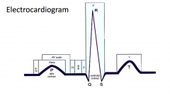
Each blip corresponds to a phase of the cardiac cycle |
|
|
What does the conduction system consist of |
*Sinoatrial node *Atrioventricular node *Atrioventricular bundle *Purkinje fibers |
|
|
Where is the sinoatrial (SA) node found |
Junction of right atrium and superior vena cava |
|
|
What does the sinoatrial (SA) node do? |
Sets the inherent rate of contraction *Acts of pacemaker *Auto-rhythmic fibers |
|
|
What part of the P QRS T graph is the SA node? |
P Wave |
|
|
When does the sinoatrial (SA) node initiate action potentials |
Every 0.6 seconds 100 times/ minute |
|
|
What does the sinoatrial (SA) node cause |
Atria to to contract |
|
|
Where is the arterioventricular node? |
Posterior wall of the right atrium (by the tricuspid valve) |
|
|
What does the atrioventricular node do? |
Delays impulse from atria to ventricles to allow time for atria to empty into the venticles before the ventricles contract |
|
|
What part of the EKG graph is the atrioventricular node correspond to? |
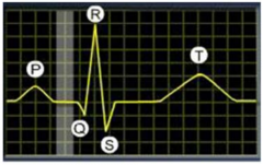
|
|
|
What are the various parts of the atrioventricular bundle? |
*Bundle of His *Right and left branches |
|
|
What does the bundle of his do? |
Passes through fibrous tissue separating atria from ventricles |
|
|
What is the right and left branches |
Muscular portion of the inter-ventricular septum |
|
|
What is the purkinje fibers |
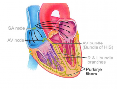
Rapid transmission of action potential Distributes to all ventricular muscle cells to cause ventricles to contract rhythmically |
|
|
What happens in diastole |
Both atria and ventricles are relaxed and chambers passively fill with blood |
|
|
What happens in systole |
Atria contracts first and top off ventricles with an extra 25 mL of blood Then ventricles contract and eject about 70 mL into the aorta on the left side and the pulmonary trunk on the right |
|
|
What is cardiac output |
Volume of blood pumped each minute |
|
|
How do you calculate cardiac output |
Stroke volume x Heart Rate |
|
|
What is stroke volume? |
Volume of blood pumped each contraction |
|
|
What is heart rate |
Number of heartbeats each minute |
|
|
Eahc minute the heart moves ____ of blood around the body |
5L |
|
|
How does the aorta help with blood flow? |
The left ventricle pumps into the aorta.The aorta distends and when the ventricles are done contracting, the aortic valve closes the muscle in the aorta, then brings it back to the normal size and helps repeal blodo through arterial tree. Left ventricles get help from muscular walls of the arteries. |
|
|
When feeling a pulse what are you feelling |
Artery as it distends and contracts as moves blood along arterial tree |
|
|
What is the division of one cardiac cycle |
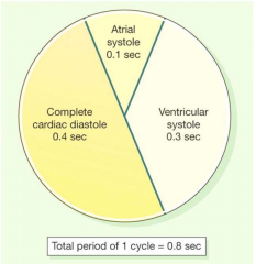
|
|
|
Atrial Septal Defect/Ventricular septal defect |
There is a hole in the septum between the left and right atria (or ventricle). Therefore, blood will flow from the left to the right side because more pressure on left side. Less blood is pumped out also the left atria has oxygenated blood while right contains deoxygenated blood sosquriting oxygenated into deoxygenated which is then going to the lungs to be oxygenated. Therefore, heart is less efficient and will decrease the oxygen capacity to the body |
|
|
Patent Ductus Arteriosus |
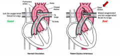
There is a duct between arteries (aorta and pulmonary arteries) in utero because ond't need to go through the lungs This is suppose to close off once born and become a ligmaent (ligamentum arteriosus) however if it doesn't have mxiing of blood from aorta to the pulmonary artery (because aorta has higher pressure) Therefore less deoxygenated blood getting to the lungs and lowers effectivity of the heart |
|
|
Patent Foramen Ovale |
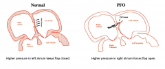
Foramen ovale in utero supposed to allow blood to flow betwene the right and left atrium but when baby first born, now lungs arei nvolved so blood needs to go to the lungs. Foramen ovale is supposed to close off but some people dont and so typically since left atria has high pressure keeps flap closed but if the pressure in the right atrium is higher than the left atrium then could have some blood squirt into the left atrium and therefore deoxygenated blood to the systematic circulation which can also decrease the efficienty of the left atrium |
|
|
What is valve stenosis |
Doesn't open properly |
|
|
What is valve regurgiation |
Does not close properly |
|
|
What is heart valve regurgitation |
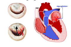
With regurgitation does not close all the way so when ventricles contracts some blood goes back into the atria therefore lowering the efficiency of the heart |
|
|
What is valve prolapse |
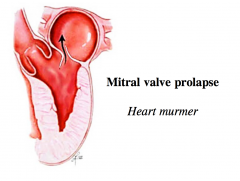
Little extra flap of tissue can prolapse into atrium and cause a flutter (sound of heart murmur) |
|
|
Is a valve prolapse more likely to occur in the mitral valve or tricuspid valve |
Mitral valve |
|
|
What is a valve stenosis |
Valve does not open all the way. Therefore, none of blood getting through, so heart has to work a lot harder to get through |
|
|
What can valve stenosis lead to |
Ventricular hypertrophy |
|
|
What does hypertrophy lead to |
Cardiac output decrease because can't allow as much blood in and gets worse |
|
|
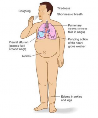
What is heart failure |
Heart cannot supply enough oxygenated blood for the body's metabolic demands *Pressure overlaod *Volume overload *Cardiomyopathy |
|
|
If there is heart failure on the left side where does blood back up into? |
Lungs (can lead to pulmonary edema) |
|
|
If there is a right heart failure where does blood back up into |
Systemic circulation (can lead to peripheral edema) |
|
|
What is congestive heart failure? |
Left and right heart failure |