Reading...
![]()
Play button
![]()
Play button
![]()
Use LEFT and RIGHT arrow keys to navigate between flashcards;
Use UP and DOWN arrow keys to flip the card;
H to show hint;
A reads text to speech;
79 Cards in this Set
- Front
- Back
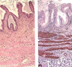
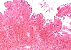
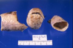
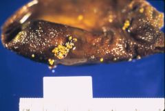
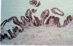
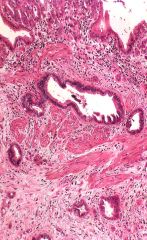
Describe this gall bladder biopsy, is this normal or pathologic?
|
Normal gall bladder
- mucosa: tall columnar epithelium (mucin secreting) - lamina propria: lymphocytes, no lymphocytes - fibromuscular layer: mixed fascicles of smooth muscle and fibrous tissue |
|
|
Composition of bile solutes.
|
- bile salts (67%): cholates, chenodeoxycholates, deoxycholates, lithocholates, ursodeoxycholates
- phospholipids (22%) - cholesterol (4%) - protein (4.5%): secreted to conjugate drugs - bilirubin (0.3%) |
|
|
Compostion of bile secreted by hepatocytes.
|
- 97% water
- 3% solutes |
|
|
What are some functions of gall bladder?
|
- concentrate bile by 5-10 fold: active absorption of eletrolytes, waters follows sodium
- release bile when stimulated by CCK (I cell in duodenum and jejunum) in response to lipid and protein in small intestine. |
|
|
Where are most of secreted bile (95%) salts reabsorbed?
|
ileum
|
|
|
What is a very sensitive screen for gall bladder calculi?
|
ultrasound
- can detect stones as small as 2-3mm diameter |
|
|
What is ERCP useful for?
|
- directly visualize bile ducts
- inject dye into pancreatic and extrahepatic bile ducts - take biopsies - place stents to relieve obstructions |
|
|
What imaging step is required after cholecystectomy?
|
Intraoperative cholangiogram
- make sure there is no left over gall stone in common bile duct |
|
|
Name three general categories of congenital gall bladder anomalies.
|
- cystic diseases
- extrahepatic biliary atresia - anatomic variants |
|
|
What are some classes of cystic diseases of bile ducts (5)?
|
- choledochal cyst: dilation of common bile duct
- diverticulum of bile duct - choledochocele: cysts protrudes into duodenum - multiple cysts (caroli disease) - fusiform cysts: intra- and extra- hepatic |
|
|
What is this cystic disease of gall bladder?
- dilations of common bile ducts |
choledochal cyst
|
|
|
What is this cystic disease of gall bladder?
- cysts attached to common bile duct |
diverticulum
|
|
|
What is this cystic disease of gall bladder?
- cysts protrudes into duodenum |
choledochaocele of intraduodenal bile duct
|
|
|
What is this cystic disease of gall bladder?
- multiple intrahepatic cysts |
caroli disease
|
|
|
What is this cystic disease of gall bladder?
- intra- and extra-hepatic cysts |
fusiform cysts
|
|
|
What are some complications of cystic disease?
|
- obstruction
- perforation -> bile peritonitis - ascending cholangitis (caroli disease) - hepatic abscess - secondary biliary cirrhosis - carcinoma (rare) |
|
|
What is a common complication associated with caroli disease?
|
ascending cholangitis
|
|
|
What is this gall bladder disease?
- complete obstruction od bile flow due to destruction/absence of all or part of extrahepatic bile ducts |
extrahepatic biliary atresia
- neonatal jaundice (conjugated) - 10% surgically curable. others need liver transplant |
|
|
Treatment for extrahepatic atresia.
|
- 10% surgically curable
- others need liver transplant |
|
|
Describe the morphology of early sequalae of extrahepatic biliary atresia.
|
- inflammation and/or necrosis of bile duct cells
|
|
|
Describe the morphology of late sequalae of extrahepatic biliary atresia.
|
- fibrosis/obliteration of bile duct -> biliary cirrhosis
|
|
|
Name some anatomic variations of gall bladder (3).
|
- hourglass gallbladder
- double and bilobed gallbladder - aberrant locations of gall bladder: intrahepatic gallbladder, and left sided gallbladder. |
|
|
Name some anatomic variations of bile ducts (3).
|
- abnorsmally long
- accessory hepatic ducts - low fusion of hepatic ducts -> double common bile duct |
|
What is this anatomic variation of gallbladder?
|
Hourglass gallbladder
- septation |
|
|
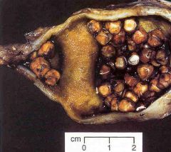
Definition: stones in gallbladder.
|
cholelithiasis
|
|
|
Definition: stones in common bile duct.
|
choledocholithiasis
|
|
|
Three varieties of gallstones.
|
- pure cholesterol: 10%
- pure pigment (calcium bilirubinate): 10% - mixed: 80% |
|
|
What percentage of all gallstones contain cholesterol?
|
90%
|
|
|
What percentage of all gallstones contain pure cholesterol?
|
10%
|
|
|
What makes up the pure pigment gallstone?
|
Calcium bilirubinate
|
|
|
Pathogenesis of gallstone.
|
- deficient bile salts or excess cholesterol -> cholesterol supersaturation -> crystals
- gallbladder hypomotility and hypersecretion of mucus -> accretion of crystals - calcium salts + cholesterol crystals => stone |
|
|
Why is flat plate abdominal xray not used anymore to diagnose gallstones?
|
not very sensitive
- only 10-20% stone are opaque. |
|
|
Which gender has more risk for cholesterol containing gallstones?
|
premenopausal women
- estrogen promotes uptake and synthesis of cholesterol by hepatocytes (OCP and pregnancy) |
|
|
What are some risk factors for cholesterol containing gallstones?
|
- native american, inductrialized societies
- elderly - premenopausal women - estrogen - obesity - hypercholesterolemia - family history - GI disorder that interferes with bile salt reabsorption in ileum (Crohn's) |
|
|
What are some risk factors for pure pigment gallstones?
|
disorders with elevated unconjugated bilirubin in bile
- hemolytic syndromes - disease or removal of ileum - bacterial infection of biliary tree (E coli): beta-glucuronidase convert conjugated yo unconjugated bilirubin - parasitic infection: clonorchis, ascaris lumbricoides |
|
|
How does bacterial infection such as E. coli cause pure pigment stones of gallbladder?
|
beta-glucuronidase convert conjugated yo unconjugated bilirubin
|
|
|
People who had ileum removal is at risk for what gallbladder conditions?
|
gall stones
- less bile salts in bile |
|
|
Which two parasites may cause pure pigment stones?
|
- Clonorchis sinensis
- Ascaris lumbricoides |
|
|
What are some complications of gallstones?
|
- common bile duct obstruction
- biliary colic - acute cholecystitis with sepsis - acute pancreatitis (obstriction of pancreatic duct) - gallstone ileis: fistula from gallbladder to small bowel - Mirizzi syndrome: stone in bile duct causing stricture |
|
|
What is this complication of gallstones?
- fistula from gallbladder to small bowel |
gallstone ileus
|
|
|
What is this complication of gallstones?
- stone in bile ducts causing stricture |
Mirizzi syndrome
|
|
|
Is medical treatment useful in gallstones?
|
only for pure cholesterol stones
|
|
|
How to treat gallstones?
|
- medicine for pure cholesterol stones
- surgery: laproscopic, open cholecystectomy, ERCP |
|
|
What is this gallbladder disease?
- intermittent pain, recently worse RUQ pain - fever, leukocytosis - palpable dilated gallbladder - jaundice |
acute cholecystitis
|
|
|
Pathogenesis of acute cholecystitis.
|
- 90% calculous
|
|
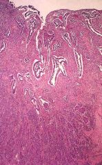
What is this gallbladder disease?
- edema, hemorrhage, mucosal necrosis |
acute cholecystitis
- transmural infiltrate of neutrophils, hemorrhage, edema, necrosis |
|
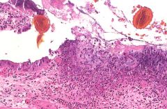
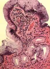
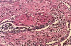
What is this gallbladder disease?
- ulcerated epithelium with dense infiltrate of neutrophils extending through fibromuscular layer |
acute cholecystitis
|
|
|
What are some treatments for acute cholecystitis?
|
- 25% require risky surgery because of pain, stones or sepsis
- others: antibiotics followed by elective surgery |
|
|
What is this gallbladder disease?
- intermittent nausea, belching, discomfort - epigastric or RUQ pain - symptoms worse after meals (fatty or large meals) "flatulent dyspepsia" |
chronic cholecystitis
|
|
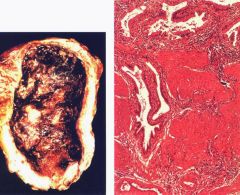
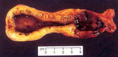
What is this gallbladder disease?
gross - stones and thickened wall micro - hyperplasia of fibromuscular layer |
chronic cholecystitis
|
|
|
What are some variants of typical chronic cholecystitis?
|
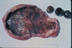
- Porcelain gallbladder: lithiasis gradually erode mucosa, leaving a leathery wall
- hydrops: stone obstructing cystic duct -> watery fluid in lumen - mucocele: stone obstructing cystic duct -> thick mucoid fluid in lumen |
|
What is this gallbladder disease?
- leathery thicj all and eroded mucosa - stone found at orifice of cystic duct |
porcelain gallbladder (variant of chronic cholecyctitis)
|
|
What is this gallbladder disease?
- watery fluid in lumen - stone obstructing cystic duct |
hydrops variant of chronic cholecystitis
|
|
What is this gallbladder disease?
- thick mucoid fluid in lumen - stone obstructing cystic duct |
mucoid variant of chronic cholecystitis
|
|
|
Name some non-neoplastic disorders of the gallbladder.
|
- cholesterolosis
- cholesterol polyps - diverticular disease - torsion - metaplasia (gastric type) and dysplasia |
|
What is this gallbladder disease?
- accumulation of lipid laiden macrophages in lamina propria |

cholesterolosis
- diffuse linear streaks in mucosa |
|
What is this gallbladder disease?
- accumulations of cholesterolosis |
cholesterol polyps
|
|
What is this gallbladder disease?
- diffuse mural thickening with cysts in the wall - cysts lined by normal columnar epithelium embedded within fibromuscular layer |
diverticula: localized adenomyomatous hyperplasia type
|
|
|
Name the 2 types of diverticular diseases of the gallbladder.
|
- diffuse type: adenomyomatosis
- localized type: adenomyomatous hyperplasia |
|
|
Pathogenesis of diverticular disease of gallbladder.
|
pulsion diverticulum
|
|
|
Torsion of the gallbladder leads to _____.
|
ischemia -> necrosis (urgent surgery to avoid rupture with bile peritonitis
|
|
Neoplastic or non-neoplastic gallbladder disease?
|
non-neoplastic
- metaplasia (gastric type) due to chronic inflammation |
|
Neoplastic or non-neoplastic gallbladder disease?
|
non-neoplastic
- dysplasia due to chronic inflammation |
|
|
Are gallbladder neoplasms common?
|
NO!
but if one has one, it is more likely to be malignant. |
|
|
Benign neoplasm of the gallbladder. (4)
|
- papillary adenoma
- heterotopias: gastric, pancreatic - non-epithelial neoplasms: lipoma, fibroma - adenomyomatous hyperplasia (diverticulum) |
|
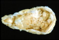
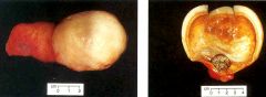
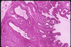
What is this gallbladder disease?
gross - soft, polypoid, exophytic lesion protruding into lumen micro - papillary frons of epithelium supported by fibrovascular stroma - no invasion of wall |
papillaty adenoma
|
|
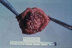
Which type of gross appearance is this gallbladder carcinoma?
|
diffusely infiltrating (70%)
|
|
What type of gross appearance is this gallbladder carcinoma?
|
localized fungating (30%)
|
|
|
Which is more common?
carcinoma of the gallbladder or carcinoma of the bile duct |
carcinoma of the gallbladder
|
|
|
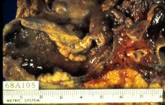
What is the observation of the correlation between gallstone and gallbladder carcinoma?
|
- 80-90% gallbladder carcinoma have gallstones
- 0.5% patients with gallstones develop carcinoma |
|
|
3 microscopic features of gallbladder carcinomas. Which one is most common?
|
- adenocarcinoma in situ (5%)
- invasive adenocarcinomas (90%) - squamous cell carcinoma or mixed (5%) |
|
|
What is the clinical presentation of gallbladder carcinoma?
|
- often late and invading liver with regional/distant metastases
- poor prognosis: 5-10% 5 yr survival |
|
What is this gallbladder disease?
|
Adenocarcinoma
- well differentiated cell invading wall |
|
What is this gallbladder disease?
|
carcinoma with glandular and squamous features
|
|
What is this gallbladder disease?
|
carcinoma (mixed)
- adenocarcinoma (top) - squamous carcinoma (lower) |
|
|
What is the most common site of carcinoma of bile ducts?
|
1. ampula
2. common bile duct 3. hepatic duct 4. junction hepatic and common duct (Klatskin tumor) |
|
|
What is this called?
- carcinoma at the junction of hepatic and common bile duct |
Klatskin tumor
|
|
|
What is this biliary disease?
- obstructive jaundice - predisposition: gallstones, parasitic infections, sclerosing cholangitis, chronic ulcerative colitis |
- carcinoma of bile ducts
- stones in common bile duct * need to differentiate |
|
|
How to surgically treat carcinoma of bile duct?
|
Whipple procedure for lesion <2cm big
|