![]()
![]()
![]()
Use LEFT and RIGHT arrow keys to navigate between flashcards;
Use UP and DOWN arrow keys to flip the card;
H to show hint;
A reads text to speech;
34 Cards in this Set
- Front
- Back
- 3rd side (hint)
|
Radiograph of the abdomen is termed as |
KUN or flat plate |
|
|
|
Xray examination of the phRynx, esophagus, stomach and duodenum |
GI series |
|
|
|
Upper GI series uses this fluorscopy contrast material |
barium |
|
|
|
In addition to giving barium, patients are also given baking soda crystals to improve image |
air contrast, dpible contrast GI series |
|
|
|
fundus of the stomach is protruded to the thoracic region |
hiatal hernia |
|
|
|
normal mucosal pattern of the esophagus and stomach |
parallel |
|
|
|
appearance of gastric ulcer with barium swallow |
spoke wheel appearance |
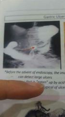
hollow portion where barium is collected |
|
|
Parts of the duodenum |
D1- bulb D2- descending D3- transverse D4- ascending |
|
|
|
landmark for duodenojejunal junction |
ligament of trietz |
|
|
|
normal mucosal pattern of the duodenum |
parallel but becomes feathery |
|
|
|
the c shaped configuration of the duodenum is useful in the detection of what |
pancreatic tumor |
|
|
|
radiologic examination of the distal duodenum to the ileocecal valve |
small bowel series |
|
|
|
mucosal pattern of jejnum and ileum |
jejunum- feathery ileum-smooth |
|
|
|
a typical endoscope cannot view what |
majority of the jejunum and ileum |
|
|
|
Imaging used to view jejunum and ileum |
capsule endoscope small bowel follow through enterocolysis |
|
|
|
capsule endoscope visualize what structure |
lumen of the jejunum and ileum |
|
|
|
Abnormality in the motility of small intestines can be assessed by small bowel follow through by assessing what? |
if it takes more than an hour for the barium sulfate to reach the ileum |
|
|
|
specialized study that allows radiographic visualisation of the small intestine that uses double contrast |
small bowel enterocolysis |
|
|
|
a narrowed and irregular ileum may indicate what |
crohns disease |
|
|
|
an apple core appearance of the ileum suggest what |
local thickening of the wall |
|
|
|
what are the indications for use of small bowel enterocolysis |
suspected neoplasm hematochezia positive fecal occult blood |
|
|
|
radiologic exam of the large intestine |
barium enema |
|
|
|
contrast study used to detect abnormalities of the lumen |
air contrast barium enema |
|
|
|
In barium enema what should be the position of the patient to assess the left part of the colon and transverse colon |
right decubitus |
|
|
|
In barium enema what should be the position of the patient to assess the ascending colon and hepatic fexure |
left decubitus |
|
|
|
position of the patient for the cross sectional view to evaluate the rectum |
lateral prone |
|
|
|
This is the view of the cecum and terminal ileal region using double contrast |
spot view |
|
|
|
what is the main risk of barium enema |
may cause impaction and constipation |
|
|
|
inflamed areas in the colon because of barium |
barium granuloma |
|
|
|
hemangioma cannot be diagnosed through ultrasound. true or false |
false |
|
|
|
Ultrasound has 99% sensitivity in the diagnosis of stones in the gallbladder. true or false |
true |
|
|
|
normal bile duct diameter |
6mm in ap diameter |
|
|
|
a bile duct that is more than 6mm in ap diameter indicates what |
cholidocolithiasis |
|
|
|
uneven out line of the gland and heterogenous structure of pancreatic tissue in ultrasound indicates |
chronic pancreatitis |
|