![]()
![]()
![]()
Use LEFT and RIGHT arrow keys to navigate between flashcards;
Use UP and DOWN arrow keys to flip the card;
H to show hint;
A reads text to speech;
16 Cards in this Set
- Front
- Back
|
What does the term allelic and allelic heterozygosity and apply it DMD (Duchenne )and and BMD (Beckers)...
|
Xp21.2
Allelic heterozygosity- Many mutations in one gene that causes disease -BMD- no frameshifts, quantitavely or qualitatively altered dystrophin DMD- frameshifts- no detecteble dystrophin - Females could have cells with normal X chromosome inactivated --> mild clinical forms shown of MD |
|
|
What disorder is characterized by mutations to the following genes?
a. Xp21.2 |
a. X-linked recessive becker and duchenne Muscular dystrophy
b. |
|
|
Explain the significance of a gene having multiple promoters
|
creates one protein but has multiple facets in its creation?
|
|
|
Describe the genetic basis of DMD...
|
a. usually a frameshift mutation that makes a premature stop codon--> dystrophin less than it should be
|
|
|
Describe the structure and function of dystrophin
Describe structure of mitochondrial gene and what parts are more likely to mutate... |
a. intracellular protein
b. all muscles c. links extracellular matrix with actin cytoskeleton 2. DNA polymerage gamma (POLG)- responsible for mtDNA replication has 2 domains a. catalytic domain b. exonuclease domain- recognizes and removes DNA--- area of higher mutation |
|
|
Clinical presentations of Duchenne (DMD) and Becker (BMD) muscular dystrophies
|
DMD-
a. no dystrophin b. early childhood d. walking is waddling, difficult to climb steps e. Gower Maneuver (getting up using arms) f. pseudohypertrophy g. wheelchair by 12 h. smooth muscle complications i. increased infections j. respiratory cardiac failure BMD- a. some dystrophin b. left ventricle |
|
|
What are the diagnostic tools for muscular dystrophy?
|
1. EMG- CRDs
2. Blood- Creatine kinase (50-100x higher) 3. biopsy- to find dystrophin levels, mosaicism 4. MRI 5. DNA- looking for gene deletions, PCR for mutations in hot spots (western blot) |
|
|
Describe the genetic basis and clinical presentations of
1. LHON, 2. MERRF 3. MELAS |
1. LHON- vision loss, retinal ganglion cell and optic nerve, smoking correlation
2. mutation to np8344 tRNA lys, myoclonus, random muscle twitches, ragged red fiber, 3. lactic acidosis, stroke-like symptoms, ragged red fiber, tRNAleu (missense mutations) 2-10 years old |
|
|
Describe differences between mitochondrial disease caused by mutations in mitochondrial genes and nuclear genes
|
Energy difficiency disease may be either mitochondrial or nuclear down path
1. NADH dehydrogenase Complex I (45 subunits) 2. Nuclear DNA (nDNA) or Mitochondria DNA (mtDNA) 3. cellular energy |
|
|
The function of mitochondria for the electron transport chain and oxidative phosphorylation
|
Has complexes of electron transport chain
Things are oxidized by TCA and B-oxidation and NADH + H are oxidized in Complex I |
|

What type of inheritance and why?
|
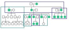
mitochondrial due to males never passing it on to further generations
|
|
|
Name the type of muscular dystrophy by the following descriptions...
a. gowers sign b. no frameshift mutation c. no dystrophin d. wheel chair bound by 12 |
a. DMD
b. BMD c. DMD d. DMD |
|
|
What are the three most important mitochondral myopathies?
|
1. LHON,
2. MERRF 3. MELAS |
|
|
What causes a mitochondria disease?
What is replicative segregation? |
1. no recombination- mtDNA does not edit like nDNA so mutations can accrue
2. Heteroplasmy- many different types of mtDNA in muscles means certain mutations chould affect while others may not 3. Threshold Expression- # of mutations required for serious deficiency to show (replicative seregation) |
|
|
Describe the etiology and clinical significance of “ragged red fibers”.
|
- marker for dysfunction of mtDNA
- denote absence of cytochrome oxidase staining in fibers (shows blue in H&E stain |
|
|
What is the difference between Kearns-Sayre syndrome and Chronic Progressive External Ophthalmoplegia (CPEO)?
|
Both have- mild to moderate mitochondrial myopathy (drooping eyes)
_Kearns_ is more severe with at least one of the following 1. Cardiac 2. Cerebellar 3. Cerebral spinal protein high |

