![]()
![]()
![]()
Use LEFT and RIGHT arrow keys to navigate between flashcards;
Use UP and DOWN arrow keys to flip the card;
H to show hint;
A reads text to speech;
245 Cards in this Set
- Front
- Back
- 3rd side (hint)
|
Paracetamol antidote |
N-acetyl cysteine |
|
|
|
Pathology of steatosis in alcohol consumption (needs editing) |
1)Increased alcohol metabolism over fat metabolism 2) acetyldehyde by product causes hc damage and inflammatory rxn results in fa/tg build up 3) alcohol stimulates collagen synthesis which cause fibrosis
|
3 points |
|
|
Clinical signs of hepatic steatosis |
1) increased bilirubin 2)Increased apl 3)hepatomegally
|
3 points |
|
|
Alcoholic steatohepatitis sx and lft |
Hepatomegally Fever Leukocytosis Liver failure: -Anorexia -Ruq abdo pain -Malaise
Ast>alt x2 unl (alt <300) Raised serum bilirubin Raised apl and ggt
|
|
|
|
Nafld association and diagnosis |
Ass. Diabetes, obesity, hyperlipideamia, tpn, drugs, Wilson's ECT... Dx: 1) Liver biopsy showing steatosis, hepatocyte ballooning degeneration, acute/chronic inflammation, perisinusoidal collagen deposit.
2) limited alcohol consumption
3) absence of other aetiology
(Disease of exclusion) |
|
|
|
Isolated unconjugated hyperbilirubineamia without haemolysis |
Crigler najar syndrome |
|
|
|
Isolated conjugated hyperbilirubineamia |
Dubin Johnson or rotor syndrome |
|
|
|
Biliary Atresia clinical signs |
Cholestasis within the first 3 months of life with normal weight and normal weight gain |
|
|
|
Fibropolycystic disease clinical presentation |
Hepatosplenomegaly and portal hypertension in the absence of liver dysfunction |
|
|
|
Primary biliary cirrhosis signs and associations |
>50 yrs Female Sjogren syndrome ass. Ama+ |
|
|
|
Primary sclerosing cholangitis signs |
+/- 30 years Male Ass. Inflammatory bowel disease such as ulcerative colitis |
|
|
|
Budd chiari syndrome cause: |
Hepatic vein thrombosis |
|
|
|
Wilson's disease clinical signs and dx |
Clinical signs: Young px Hepatomegaly/splenomegaly Hepatitis (fulminant or chronic) Portal Ht Neuro sx Fanconi syndrome Dx = 2 or more of the following: 1) kayser Fleischer rings 2) serum caeruloplasmin <20mg/dl 3) hepatic copper >/= 250ug/g |
|
|
|
Wilson's disease treatment |
D-penicillamine Zinc supplementation Trientine Ammonium tetrathiomolybdate |
|
|
|
Enzyme responsible for fe regulation |
Hepcidin |
|
|
|
Pathogenesis of Fe accumulation in hereditary heamochromatosis |
1) Gene mutations 2) reduced hepcidin expression 3) excess iron entry to the plasma compartment 4) subsequent binding with transferrin 5) tissue iron loading |
|
|
|
Treatment of hereditary haemochromatosis |
Therapeutic phlebotomy |
|
|
|
Alpha1 antitrypsin deficiency clinical features |
Emphysema Liver disease: presents as cholestasis in infancy Cirrhosis and HCC in adults |
|
|
|
Dx of alpha-1-antitrypsin |
Decreased serum alpha 1 antitrypsin level Phenotyping +ve acid Schiff on biopsy |
|
|
|
Insulin resistance in the pathogenesis of fatty liver |
1)Insulin resistance results in increase peripheral fat break down (lipolysis) 2) this results in increased hepatic uptake of fatty acids and subsequent accumulation of hepatic triglycerides 3)this results in a preferential increase of FFA beta oxidation over carbohydrates. 4)FFA induce cytochrome p450 lipoxygenases 5) these lipoxygenase produce hepatotoxic free oxygen radical species 6) these radicals cause hepatocellular injury and fibrosis |
|
|
|
Cause of insulin resistance |
Adipokine secretion by adipocytes (secretion is proportional to mass thus higher ass. In obese px) |
|
|
|
Veno-occlusive disease is caused by? (3) |
Pyrrolizidine alkaloids Antineoplastic drugs Irradiation |
|
|
|
Veno-occlusive disease definition |
Occlusive fibrosis of small intrahepatic veins causing severe congestion of pericentral sinusoids
|
|
|
|
Veno-occlusive disease clinical picture and Dx |
Clinical picture: Sudden onset Hepatomegally Severe ascites- without clinical jaundice Dx: biopsy (dx of exclusion) |
|
|
|
Reyes syndrome clinical presentation |
-Children > 5 and < 16 -Hx of salicylate (aspirin) use in varicella or flu-like disease -Non-Inflammatory encephalopathy -Fatty degeneration of the liver |
|
|
|
Laboratory findings in Reyes syndrome |
Elevated s-hepatic transaminases 2-3× uln Elevated S-ammonia 2-3× uln |
|
|
|
Cystic fibrosis results in: |
Focal nodular fibrosis (dx for cf) Biliary cirrhosis Hepatic steatosis Neonatal cholestasis |
|
|
|
Ascites definition |
Pathological accumulation of free fluid in the peritoneal cavity |
|
|
|
Ascites main causes: |
1) Cirrhosis 2) Malignancy 3) Heart failure 4) Tuberculosis 5) Pancreatitis |
|
|
|
Pathophysiology of ascites |
1) Initiated by a total body increase of sodium and water
2) ascites associated with an increase in catecholamines and SNS outflow which stimulates the renin-angiotensin system to conserve sodium and water
3) the ras reduces sensitivity to atrial nutriuretic peptide (which causes sodium excretion)
4) portal ht increased hydrostatic pressure in splanchnic bed
5) concomitant hypoalbuminaemia and decreased plasma oncotic pressure also occur
6) all resulting in extravasation of fluid from plasma to the peritoneal cavity |
|
|
|
Ascites fluid ADA above 60 indicates? |
TB |
|
|
|
Ascites neutrophils count above 250 cells/mm3 indicates? |
Spontaneous bacterial peritonitis |
|
|
|
High ascitic amylase indicates? |
Pancreatitis |
|
|
|
High serum ascites albumin gradient (SAAG) indicates? |
>/= 11g/dl Cirrhosis Cardiac failure |
|
|
|
Low SAAG indicates? |
<11g/dl Malignancy Pancreatitis TB Nephrotic syndrome |
|
|
|
Tx of ascites in the cirrhotic px |
1)Decreased salt intake 2) spirinolactone (1st line) 3) furosemide (if spirinolactone fails) 4) therapeutic paracentesis (high grade) |
|
|
|
Refractory ascites definition and tx |
Drug resistant ascites or drug contraindicated ascites Tx: therapeutic paracentesis Then TIPS |
|
|
|
TIPS, use: |
Transjugular intrahepatic portosystemic shunt Used to alleviate pressure between portal and systemic circulation in portal hypertension |
|
|
|
Hepatorenal syndrome is due to? |
Cirrhosis Liver failure Ascites Alcoholic hepatitis |
|
|
|
Hepatorenal syndrome caused by? |
Intrarenal vasoconstriction due to liver disease and circulatory dysfunction |
|
|
|
Spontaneous hepatorenal syndrome is due to? |
Worsening liver function |
|
|
|
Secondary hepatorenal syndrome precipitated by? |
Bacterial infection
Large volume paracentesis without albumin supplementation |
|
|
|
Dx of hepatorenal syndrome (6) |
1) cirrhosis with ascites 2)no evidence of shock 3) no parenchymal kidney disease (No blood/ protein in urine) 4) no nephrotoxic substances in hx 5)serum creatinine above 133mmol/l 6)no improvement of serum creatinine after 2 days of: - diuretic withdrawal - albumin volume expansion |
|
|
|
Hepatorenal syndrome TX |
Terlipressin Albumin |
|
|
|
Spontaneous bacterial peritonitis (SBP) signs and sx |
Px with cirrhotic ascites presenting with: -Fever -Abdo pain and tenderness -Encephalopathy (confusion) -Diarrhoea -Renal failure
|
|
|
|
Secondary vs spontaneous bacterial peritonitis 3 points: |
Spontaneous: Neutrophils in hundreds Monomicrobial Protein less than 10g/dl
Secondary: Neutrophils in thousands Polymicrobial Protein higher than sbp |
|
|
|
Causes of secondary bacterial peritonitis (2): |
Perforation peritonitis: rupture bowel/ stomach Non-perforation: perinephric abscess |
|
|
|
Most commone Microbiological causes of SBP |
E. Coli Streptococcus Enterococcus |
|
|
|
Tx of SBP(4) |
3rd generation cephalosporin: -cefotaxime -ceftriaxone -ceftazidime -co-amoxiclav |
|
|
|
High risk alcoholic units per week in males and females |
1 unit= 1 glass wine, half a pint of beer >60 in males >40 in females |
|
|
|
Maddrey index for prognosis of alcoholic steatohepatitis : |
[Px PT - control time+ serum bili] >32= poor <32= good |
|
|
|
Cirrhosis definition: |
Irreversible injury characterised by Extensive fibrosis and nodular regeneration |
|
|
|
Alcoholic cirrhosis clinical picture (8): |
-bilateral parotid enlargement -dupuytrens contracture -palmer erythema -gynecomastia -testicular atrophy -splenomegaly -ascites -caput medusa (sign port ht)
|
|
|
|
What is Dupuytrens contracture: |
Fibromatosis in the hands causing permanently bent fingers |
|
|
|
What is palmer erythema: |
Red rash on the palms due to nitric oxide and associated with cirrhosis |
|
|
|
Metabolites causing gout in steatohepatitis: (2) |
Raised triglycerides Raised urea |
|
|
|
Mx of alcoholic steatohepatitis: (3) |
-abstinence -diet and vit supplements -corticosteroids |
|
|
|
Drug treatment for alcoholic hepatitis: |
Indicated with a maddrey index over 32 -Corticosteroids -Pentoxifylline ( muscle analgesic) |
|
|
|
Cirrhosis tx: |
-treatment is directed at cx - only 'cure' is liver transplant |
|
|
|
Pyogenic liver abscess, biliary causes: |
Acute cholangitis Acute cholecystitis |
|
|
|
Pyogenic abscess, portovenous (pyelophlebitis) causes: |
Diverticulitis GIT perforation Appendicitis |
|
|
|
Pyogenic abscess, hepatic arterial causes: |
Iv drug use Endocarditis Systemic bacteraemia |
|
|
|
Pyogenic abscess CT scan will reveal? |
Enhancing capsule |
|
|
|
Hydatid (echinococcal) cyst CT scan will reveal? |
Non-enhancing, multiple layered, thick walled cyst |
|
|
|
Hydatid cyst hx must include? |
Contact with a sheep or dog |
|
|
|
Hydatid cyst structural signs: (5) |
-honeycomb or rosette-like (ce2) -water Lily or floating membrane (ce3) -ball of wool (ce4) |
|
|
|
Drug tx for hydatid cysts : |
Albendazole or Mebendazole (C/I in pregnancy) |
|
|
|
Pair as Tx for hydatid cysts: |
Pair = percutaneous aspiration injection and re-aspiration Performed after 1 week albendazole Injection= scolicidal agent |
|
|
|
Amoebic abscess causative organism: |
Entamoeba histolytica |
|
|
|
Signs of amoebic dysentery with entamoeba histolytica: |
Tear drop or flask shaped ulcerations in the colon |
|
|
|
Signs of amoebic liver abscess: |
Anchovy sauce aspirate |
|
|
|
Radiological findings of amoebic abscess: |
Vague pseudocapsulated abscess in liver |
|
|
|
Tx of amoebic liver abscess: |
Metronidazole Dilanoxanide |
|
|
|
Caroli's disease definition: |
Type 5 choledochal cyst disease characterised by segmental dilatation of intra-hepatic biliary ducts |
|
|
|
Caroli's disease pathognomic sign: |
'Dot sign' : portal v branching into cyst |
|
|
|
Complications of caroli's disease: |
-Cholangiosarcoma -hepatolithiasis - liver abscess -cholangitis - hepatic amyloidosis |
|
|
|
In paediatrics secondary Hepatic malignancies are most common with? (3) |
-Wilms- nephroblastoma -Neuro blastoma -Leukaemia |
|
|
|
Hemangioma's sx developed in 1/3 of px, what are the sx?(HAPI) |
- high output cardiac failure - anaemia, oj -platelet trapping ( kassabach meritt) -intra-peritoneal haemorrhage |
|
|
|
Hemangioma's present with what on ct scan? |
Multiple well encapsulated homogenous masses |
|
|
|
Hemangioma's are usually benign what is the mx if sx occur? |
Sx are H.A.P.I Mx: High dose prednisone Beta blockers |
|
|
|
Paediatric liver tumour dx by age of onset: |
- <3 yrs = hepatoblastoma -0-19 yrs= hcc |
|
|
|
Hepatoblastoma common associations in children: |
Japanese Vlbw infants <3 yrs |
|
|
|
Hepatoblastoma disease associations: |
1- beckwith-weidemann syndrome 2- hemihypertrophy 3- familial adenomatous polyposis 4- feotal alcohol syndrome |
|
|
|
Hepatocellular carcinoma disease associations in children? ( normally occurs following underlying condition) |
1- hep b/c (from parent) 2- alpha 1 antitrypsin deficiency 3- biliary atresia 4- glycogen storage disease (1 and 3) 5- methotrexate therapy 6- wilms tumour |
|
|
|
What are the 3 most important factors associated with HCC? |
1- Hep b and c 2- aflatoxin (aspergillus fungus) 3- alcohol abuse |
|
|
|
Elevated tumour marker In HCC? |
Alpha-fetoprotein |
|
|
|
Rx treatment for HCC? |
Sorafenib |
|
|
|
Most common primary sites for secondary liver malignancy? (8) |
1) rectum 2) colon 3) stomach 4) pancreas 5) lung 6) breast 7) oesophagus 8) melanoma |
|
|
|
Red man syndrome is due to? |
Iv vancomycin use |
|
|
|
Red man syndrome is due to? |
Iv vancomycin use |
|
|
|
In hyposplenism a peripheral blood smear will contain: (3) |
Howell jolly bodies Heinz bodies Pappenheimer bodies |
|
|
|
Clinical sign of wandering spleen: |
Extreme left hypochondriac pain |
|
|
|
Hereditary spherocytosis is associated with? |
Gallstones |
|
|
|
Tx for Hereditary spherocytosis? |
Folate supplementation Splenectomy if sever and px >6yrs |
|
|
|
What Encapsulated organisms cause overwhelming sepsis post-splenectomy? (3) |
Pneumoccoccus Menigococcus Haemophilus pneumonia |
|
|
|
Features of asplenia: |
Howell jolly bodies + vacterl |
|
|
|
Tx autoimmune haemolytic anaemia? |
Prednisone, immunosuppresents Folate If severe splenectomy |
|
|
|
Causes of acute pancreatitis ( GET SMASHED) |
Gallstones Ethanol Trauma Steroids Mumps Auto-immue Scorpions Hypertriglyceraemia Ercp Drugs |
|
|
|
Pathogenesis of acute pancreatitis, 3 main pathways: |
1: acinar cell injury 2: metabolic injury 3: duct obstruction |
|
|
|
Mechanisms of pathogenesis of acute pancreatitis basic concept: |
1)Duct obstruction: causes blood flow problems, ischemia, then acinar cell injury 2) metabolic injury: proenzymes and hydrolases mix inducing enzyme activation and pancreatic autolysis 3: acinar cell injury: enzyme activation and pancreatic autolysis |
|
|
|
Processes which result in acute pancreatitis: |
Interstitial inflammation Oedema Proteolysis Fat necrosis BV damage Haemorrhage |
|
|
|
Autoimmune pancreatitis is associated with: |
IgG4 secreting plasma cells |
|
|
|
Autoimmune pancreatitis tx: |
Responds to steroids |
|
|
|
Main rf's for pancreatic carcinoma: |
Smoking Fat rich diet Chronic pancreatitis/ DM |
|
|
|
Most common anatomical position of pancreatic carcinoma is: |
Head of the pancreas |
|
|
|
Pancreatic exocrine insufficiency clinical features: |
1) fat maldigestion: steatorrhea 2)protein maldigestion 3)carbohydrate maldigestion: flatulence/ bloating/cramp 4) decreased fat soluble vitamins (ADEK) 5) vitamin b-12 deficiency 6) decreased bicarb/ protease/ amylase/ lipase |
|
|
|
Trypsinogen changes in a direct PFT indicate: |
decreased trypsinogen indicates: - cystic fibrosis in neonates (screening test) - pancreatic exocrine insufficiency
Increased trypsinogen indicates: -acute pancreatitis
|
|
|
|
Decreased faecal chymotrypsin indicates: |
Advanced pancreatic exocrine insufficiency |
|
|
|
Decreased faecal elastase indicates: |
Sever PEI |
|
|
|
Test to confirm mucosal dysfunction: |
D-xylose absorption test (It indicates disease that presents with malabsorption due to mucosal dysfunction) |
|
|
|
Factors causing pathogenesis of gallstones: |
1) abn bile secretion 2) increased cholesterol in bile 3)low bile salt pool 4)interruption of enterohepatic circulation 5) GB dysfunction 6)cholesterol crystals from saturated vesicles |
|
|
|
Black pigment gallstone: |
Associated with haemolytic conditions ie sickle cell/ thalassemia |
|
|
|
Brown pigment gallstone |
Associated with infection and stasis found in CBD |
|
|
|
Complications of Gallstones (rare): |
Biliary colic Acute cholecystitis Obs jaundice Gallstone pancreatitis Gallstone ileus |
|
|
|
To alleviate sx of peptic ulcer disease drink: |
Milk |
|
|
|
A px drinks milk after suspected peptic ulcer pain and the pain worsens, what's the dx? |
Biliary colic ( milk is a fat) |
|
|
|
Clinical presentation of biliary colic: |
Severe RUQ pain lasting 2-3 hrs with normal inflammatory markers and radiological confirmation of cholelithiasis |
|
|
|
Biliary colic is defined as gallbladder outflow obstruction due to a stone in: |
-hartman's pouch or -cystic duct |
|
|
|
Mx of biliary colic: |
1) analgesics 2) anti-spasmodics 3) exclusion of other dx 4) elective laparoscopic cholecystectomy |
|
|
|
Acute cholecystitis definition: |
Blockage of the cystic duct which fails to resolve leading to: -bile infection - inflammation -ischemia -necrosis -perforation -rupture |
|
|
|
Clinical picture acute cholecystitis: |
RUQ pain and tenderness Nausea and vomiting Murphy's sign (Arrest of inspiration following gentle palpation RUQ) |
|
|
|
Tx of acute cholecystitis |
Iv broad spec antibiotics Fluid resus Analgesics Cholecystectomy |
|
|
|
Percutaneous transhepatic cholecystostomy indicated in the treatment of acute cholecystitis if px: |
Elderly DM Immune compromised |
|
|
|
Management of choledocholothiasis: |
ERCP ( endoscopic retrograde cholangiopancreatography) with stone extraction Elective Cholecystectomy (Is a stone blocking common bile duct) |
|
|
|
Gallstone ileus is causes by: |
A gall stone reaching the terminal ileum through a fistula between the 2nd part of the duodenum and the gallbladder causing a small bowel obstruction |
|
|
|
Mx of gallstones ileus: |
Resus Ngt Urine Cath Explorative lap |
|
|
|
Gall stone ileus abdominal x-ray triad: |
1) signs small bowel obstruction 2) stone in ileum 3) air in RUQ |
|
|
|
Which two types of fistula can cause Bouveret's syndrome: |
Cholecystogastric Cholecystoduodenal |
|
|
|
What is the anatomical position for a Gall stone in Bouveret's syndrome: |
Duodenal bulb |
|
|
|
What is the anatomical position of the Gall stone in mirrizi syndrome: |
Neck of the gallbladder |
|
|
|
Rigler's triad for dx of Gall stone ileus: |
1) partial/complete bowel obs 2) pneumobilia 3) aberrant Gall stone in intestine Ct to confirm |
|
|
|
Clinical presentation of acute pancreatitis: |
Fever RUQ/epigastric pain radiating to back N/V Tachycardia Respiratory sx Distension Dehydration |
|
|
|
Atlanta classification for dx of acute pancreatitis: |
1) pain- acute onset, persistant, epigastric or RUQ radiating to back 2) serum amylase/ lipase: >3× uln 3)radiographic findings |
|
|
|
Definition of acute pancreatitis: |
Local inflammatory response characterised by: 1) parenchymal autodigestion 2) peri-pancreatic oedema 3) necrosis 4) thrombosis of parenchymal vessels |
|
|
|
Mx of mild acute pancreatitis: |
1) nil per os or only as much as the px can tolerate 2) fluid resus iv (R/L) 3) opioid analgesics 4) ward observation |
|
|
|
Mx of moderate to severe pancreatitis: |
1) ICU admission : - heart monitor -catheter -ventilator 2) adequate fluid resus 3) invasive pressure monitoring 4) nutrition: ng tube distal to pancreas 5) mx of complications |
|
|
|
Clinical presentation of chronic pancreatitis: |
1) pain - epigastric (relieved by leaning forward) - radiates to back - worsened by meals 2) steatorrhea/ malabsorption - stool= bulky, pale, foul smelling - decreased lipase and proteolytic enzyme 3) DM in late stage |
|
|
|
Imaging results in chronic pancreatitis: |
Axr: calcification around pancreas Sonar/CT: fibrosis around head of pancreas |
|
|
|
Mx of chronic pancreatitis: |
1) pain - medical: nsaids, enzyme replacement, celiac block -endoscopic: sphincterotomy, remove stones, stent -surgical drainage: puestow
2)malabsorption: - decrease fats - decrease protein - enzyme- creon
3) mx cx: - ascites- ocreoltide for 6 wks then surgery -pseudocysts- drain
|
|
|
|
Rx tx for pancreatic ascites: |
Ocreoltide ( only 6 wks) |
|
|
|
Pancreatic tumours mucinous vs serous: |
Serous: usually benign Mucinous: commonly malignant |
|
|
|
IPMN- intraductal papillary mucinous neoplasm cause and risk increase: |
Epithelium prone to dysplastic changes Risk increases if: - >3cm - growth >1cm/ year -mural nodules |
|
|
|
Mucinous cystadenoma (MCN) cause and association: |
-Estrogen/progesterone receptor +ve ovarian like subepithelial stroma - exclusive to women |
|
|
|
Solid pseudopapillary tumours features and associations: |
- both cystic and solid parts - mc in young women |
|
|
|
PDAC- pancreatic ductal adenocarcinoma sx: |
Non-specific sx: - early satiety - obs jaundice - unexplained weight loss - endoscopy -ve epigastric/ back pain - DM (late presentation) - malabsorption signs - courvoisier sign |
|
|
|
PDAC- pancreatic ductal adenocarcinoma investigations: |
Serology - normocytic/chromic anaemia - increased ALP and bili Tumour markers - 50% px's = increased Ca 19-9 U/s: 95% sensitivity if >3cm CECT: > 90% sensitivity if >2cm |
|
|
|
PDAC- Pancreatic ductal adenocarcinoma 1st line treatment by stage: |
Stage 1/2 -Surgical: resection
-Adjuvant chemo: single agent gemcitabine Stage 3: - non-resectable -neo-adjuvant chemo role negligible but: gemcitabine alone or combined with 5-flourouracil Stage 4: palliative |
|
|
|
PNET: pancreatic neuroendocrine tumour associated hereditary syndromes: |
- von hippel-lindau - multiple endocrine neoplasis-1 |
|
|
|
Premalignant conditions for GB cancer: |
1)GB polyps 2)GB wall calcifications 3)cholelithiasis >1cm |
|
|
|
GB cancer T staging and tx: |
T1: cholecystectomy T2: cholecystectomy, adjacent hepatic resection, lymphadenectomy and bile duct resection T3- cholecystectomy, hepatic resection, porta hepatis lymphadenectomy T4: palliation |
|
|
|
Surgical procedure and position of pancreatic adenocarcinoma: |
Head of pancreas: Pancreaticoduodenectomy Tail/ body of pancreas: Distal pancreatectomy and splenectomy |
|
|
|
Second line treatment for pancreatic ductal adenocarcinoma: |
- gemcitabine plus capecitabine -5-flourouracil plus oxaliplatin/irinotecan |
|
|
|
Rx treatment for pancreatic ductal adenocarcinoma with no co-morbidities and decreased bilirubin: |
Fulfirinox |
|
|
|
Rx treatment for pancreatic ductal adenocarcinoma with no co-morbidities and increased bilirubin: |
Folfox |
|
|
|
Rx treatment for pancreatic ductal adenocarcinoma with co-morbidities and decreased bilirubin: |
Gemcitabine |
|
|
|
HIV has two effects on the GIT: |
Infection Neoplasia |
|
|
|
Aids defining Infection in the HIV associated GIT: |
Tb |
|
|
|
Non-aids defining infection in the hiv associated GIT: |
Viral -herpes- esophagus -CMV- colon
Fungal - candidiasis- oral/ oeso
Protozoa -giardiasis- colitis
|
|
|
|
Aids defining neoplasm in the hiv associated GIT: |
Lymphoma- EBV, small bowel Kaposi sarcoma- HHSV8- oral/gastric |
|
|
|
Non-aids defining neoplasm in the hiv associated GIT: |
Squamous cell carcinoma- HPV, oropharanyx/ anus HCC- HBV/ HCV |
|
|
|
GALT: gut associated lymphoid tissue: |
1) tonsils 2) peyer's patches 3) appendix 4) kuppfer cells 5) spleen: white pulp |
|
|
|
Faecal lactoferrin assays: |
Differentiate inflammatory diarrhoea(colitis) from non-inflammatory diarrhoea(IBS) |
|
|
|
Faecal elastase: |
Detect fat in stools |
|
|
|
Faecal osmotic gap results to distinguishes secretary from osmotic diarrhoea: |
FOG: <50 = SECRETORY (infections, laxative abuse, endocrine tumours) FOG: >50= OSMOTIC ( carb malabsorption, mg induced) |
|
|
|
GIT role in HIV pathogenesis: |
1- hiv breaks down mucosal barrier in GIT 2- microbial products translocate 3- microbial products cause systemic immune response 4- results in influx of activated CD4 t cells to mucosa 5- HIV targets activated CD4
|
|
|
|
Candida esophagitis (hiv ass.) Sx and dx: |
Sx: -Odynophagia -Retrosternal pain -fever Dx: endoscopy - whitish plaque with mucosal ulcerations |
|
|
|
CMV esophagitis (hiv ass.) Sx and dx: |
Sx: - CD4 less than 50 - dysphagia - odynophagia - retrosternal pain Dx: endoscopy - large, shallow hemorrhagic ulcers |
|
|
|
Generalised Colonic cancer sx based on position: |
Right: tired/ anemic Left: bowel habit changes |
|
|
|
Basic features of diverticular disease: |
-due to increased pressure - form where arteries enter colonic wall - left side predominance (narrower) - white population most affected Cx: bleed/ sepsis/ infection |
|
|
|
Hirschsprungs aetiology: |
Aganglionosis of bowel segment due to failure of caudal Neural crest cell migration |
|
|
|
Acute appendicitis mc cause in children and adults: |
Children: lymphoid hyperplasia Adults: faecolith |
|
|
|
Clinical picture acute appendicitis in teens and young adults: |
-Visceral Pain around the umbilicus which migrates to the R. Iliac fossa - vomiting - anorexia |
|
|
|
Cx's of appendicitis: |
-inflammatory mass - perforation -peritonitis -abscess |
|
|
|
Structure of colon: |
1) mucosa- crypts of lieberkuhn -lamina propria- reticular lymph fol -muscularis mucosae- glands, circ/long mm 2) submucosa- connective tissue 3) muscularis propria -muscularis externa- circ/ long mm 4) subserosa 5) serosa
|
|
|
|
R. Paracolic gutter spreads infection to: |
Diaphragm |
|
|
|
L. Parabolic gutter spreads infection to: |
Pelvis |
|
|
|
McBurney's point- surface anatomy of appendix: |
1/3 of the way between the umbilicus and ASIS on an oblique line |
|
|
|
Blood supply foregut, midgut, hindgut: |
Foregut: coeliac - mouth to middle of the 2nd part duodenum - liver, pancreas, lungs Midgut: Sup. Mesenteric - middle 2nd part duodenum to proximal 2/3rds transverse colon Hindgut: inf. Mesenteric - distal 1/3rd transverse colon to proximal part anal canal |
|
|
|
Gastroileal reflex: |
Dilates ileocaecal valve Increases ileal peristalsis |
|
|
|
Vago-vagal reflex: |
Dilates ileocaecal valve Increases ileal peristalsis |
|
|
|
Gastrin: |
Dilates ileocaecal valve |
|
|
|
Caecum reflex: |
Caused by distention/ irritation Contracts ileocaecal valve Inhibits ileal peristalsis |
|
|
|
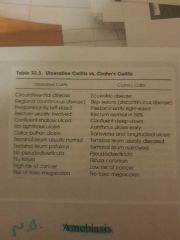
Ulcerative colitis vs crohns |
|
|
|
|
Melanosis coli is due to: |
Anthraquinone laxatives |
|
|
|
What is melanosis coli? |
Deposition of dark melanin like pigment in colonic macrophages after laxative use |
|
|
|
Most common etiology of pseudomembranous colitis: |
Disruption of gut flora by broad spectrum antibiotics and subsequent colonization by clostridium difficile |
|
|
|
Sx of pseudomembranous colitis and radiological findings: |
Chronic diarrhoea post anti-biotic use
Abdominal films: dilated colon, nodular haustra, ascites
Barium enema: thumbprinting (superficial ulcers, plaque like filling defects)
CT: wall thickening, halo/target appearance, accordion sign
|
|
|
|
Definition of inflammatory bowel disease: |
Chronic idiopathic, persistent activation of mucosal immune system |
|
|
|
What presenting feature is associated with ulcerative colitis and not crohns? |
Bloody diarrhoea |
|
|
|
Hirschsprungs disease is associated with what gene? |
RET gene |
|
|
|
Which plexuses are absent in hirschsprungs: |
Meissner and Auerbach myenteric plexuses |
|
|
|
Histological feature of hirschsprungs: |
Hypertrophy of non-myelinated nerve fibres |
|
|
|
What ligament differentiates between a upper and lower GI bleed: |
Ligament of Treitz: distal to = lower, proximal to = upper |
|
|
|
Blood features during haematemesis : |
Red/ clotting = ongoing bleed Dark "coffee ground" = stopped or slow |
|
|
|
Resus of GI bleed in children: |
-NG tube - 20ml/kg r/l iv -Blood products if: 1) no rsp to crystalloids 2) on-going bleed 3)pre-existing heart or lung condition |
|
|
|
Hemorrhagic disease of the newborn is due to: |
Vit K and vit K dependent clotting factor deficiency Dx: coagulation studies |
|
|
|
Sx's of hemorrhagic disease of the newborn: |
Coffee ground gastric aspirate Melena |
|
|
|
Dx test for swallowed maternal blood: |
APT: if mother's blood will turn rusty brown |
|
|
|
Stress gastritis cause, Sx, features and treatment |
Due: Stressful delivery Sx: coffee ground Feat: -ve APT and normal coag study Tx: NG tube, gastric irrigation, IV H2 blockers |
|
|
|
Necrotizing enterocolitis rf's and cause: |
Rf's: -prematurity - formula feeds (Ig's in breast milk prevent NEC) Is due to a bowel wall bacterial infection caused by an immature mucosal barrier |
|
|
|
Sx of necrotizing enterocolitis: |
Blood per rectum Sepsis Billious vomiting Abdo distension Abdo wall erythema or mass Feeding intolerance |
|
|
|
Axr features in necrotizing enterocolitis: |
Pneumatosis intestinalis Bowel wall thickening Portal venous gas Pneumoperitoneum |
|
|
|
Tx of Necrotizing enterocolitis: |
NPO Ngt Antibiotics Total parenteral feeds |
|
|
|
Clinical presentation of malrotation with midgut volvulus: |
Sudden onset blood pR Billious vomiting Abdo distension Previous healthy baby |
|
|
|
Clinical picture of intussusception: |
-abdo pain, vomiting -abdo distension -palpable mass -mucoid bloody stools (red currant jelly) |
|
|
|
Intussusception is associated with: |
- haematochezia btw 6-18 months -lymphoid hyperplasia of peyers patches |
|
|
|
U/s features in intussusception: |
Reveals bullseye sign |
|
|
|
Meckels diverticulum definition and common features: |
It is a true intestinal diverticulum involving all layers of the intestine and is a vitelline duct abnormality Normally on antimesenteric border of small bowel within 90cm of the ileocaecal valve |
|
|
|
Clinical presentation of meckels diverticulum: |
Painless episodic bleeding which spontaneously resolves |
|
|
|
Rome III criteria for dx of constipation : |
Any 2 or more of the following: 1) incomplete evacuation >25% 2)hard stool >25% 3) Straining >25% 4) manual manoeuvres >25% 5) sense of anorectal obs >25% 6) <3 bowel movements/ week |
|
|
|
If a patient presents with the co-existance of rectal bleed and weight loss think? |
Colorectal cancer |
|
|
|
IBS mx : |
Gluten free diet Soluble over insoluble fibres Peppermint oil Anti-depressants Psychotherapy Anti-spasmodics Probiotics Serotonegic agents (tegaserod) Pro-secretory agents (linaclotide/lubiprostone) |
|
|
|
CiC laxatives mx: |
Osmotic laxatives: PEG/ Lactulose Stimulant laxatives: senna/ bisacodil |
|
|
|
Amebiasis is due to: |
Trophozoites which have migrated to the colon and formed small abscesses submucosally |
|
|
|
Radiology of amebiasis: |
Mimics crohns disease: - aphthous/ deep ulcers - assymetrical with skip lesions |
|
|
|
Cx of amebiasis: |
Stricture Ameboma formation Tox megacolon Fistula Liver abscess Pleural effusion |
|
|
|
Aids related colitis features and causes? |
CD4 Less than 200 Right colon disease with ulceration and colitis Caused by: CMV, cryptosporidium, or hiv |
|
|
|
Radiation colitis radiography and features |
Radiological features: -Thickened walls -Ulceration -Stricture -Spiculation Mimics early ulcerative colitis and is caused by radiation ass. Endarteritis Later presents as fibrotic rigid bowel |
|
|
|
Cathartic colon radiographic features and eitiology: |
Caused by laxatives (senna) Ahaustral dilated colon Right side dominance |
|
|
|
Definition of diverticulosis: |
Acquired state where mucosa and muscularis mucosa herniate through muscularis propria |
|
|
|
Contraindications and complications of colonoscopy |
C/I: -severe ulcerative colitis -shock - perforation - ischemic heart disease -severe acute infection Cx: -Cardiopulmonary depression -bleeding and perforation -infective endocarditis |
|
|
|
Tx of pruritis: |
Cholestyramine (1st line) Rifampicin (2nd line- may cause hepatotoxicity) Bezafibrate |
|
|
|
Dx of sever ulcerative colitis True love and Witts criteria |
1) bowel movements- >/= 6/day 2) blood in stool- visible 3) pyrexia- >/= 37.8 4) anaemia- yes 5) pulse rate- >/= 90 bpm 6) ESR- >30 |
|
|
|
Defecation reflex: |
1) faeces enter rectum 2) rectum distends sending afferent signals: 3) intrinsic reflex- afferent signals through myenteric plexus causing: colonic peristalsis, internal sphincter relaxation. 4) parasympathetic reflex: afferent signals to spinal cord then efferent reflex neurons which augment intrinsic reflex action 5) afferent signals also facilitate defecation by increasing intrabdominal pressure and relaxing the puborectal mm 6) defecation then achieved through voluntary external anal sphincter relaxation by pudendal nerve |
|
|
|
Features of high cord injury constipation: |
Reflexes intact and can be triggered by digital anal stimulation |
|
|
|
Features of low cord injury: |
Lumbosacral/ sacral nerve damage resulting in dysfunction of the defecation reflex |
|
|
|
Disease states causing constipation |
1) MS 2) Parkinson's 3) hirschsprungs |
|
|
|
Colonic pacemaker cells: |
Interstial cells of cajal (myenteric plexus) |
|
|
|
Large bowel obstruction tx by cause: cancer: |
Resect and anastomose or stoma |
|
|
|
Large bowel obstruction tx by cause: intussusception: |
Pneumatic reduction |
|
|
|
Large bowel obstruction tx by cause: volvulus right side: |
Resect with anastomosis |
|
|
|
Large bowel obstruction tx by cause: stricture: |
Resect and anastomose |
|
|
|
Large bowel obstruction tx by cause: herniation |
Repair hernia |
|
|
|
Large bowel obstruction tx by cause: sigmoid volvulus: |
Uncomplicated: deflate, elective resection Complicated: resection with anastomoses/ stoma |
|
|
|
In a large bowel obstruction elevated wcc and CRP indicate: |
Ischemic bowel |
|
|
|
Perianal abscess is also called: |
Crypto glandular abscess |
|
|
|
Pathogenesis of perianal abscess: |
1) anal canal has 6-14 glands which lie in the intersphincteric plane and drain into crypts at the dentate line 2) when the crypts are occluded stool and bacteria get trapped in the glands. 3) abscesses form
|
|
|
|
Causative organisms for perianal abscesses: |
1) E. Coli 2) enterococci 3) anaerobes |
|
|
|
Dx of perianal abscess: |
Clinical dx: -tender bulging mass pointing externally -tender digital exam -hx of pain/ swelling/ pus discharge |
|
|
|
Perianal abscesses classification by location: |
1) supralevator 2) intersphincteric 3) peri-anal 4) ischiorectal |
|
|
|
Tx of perianal abscess |
Incision and drainage: - incise abscess - drain pus - debride cavity
Antibiotics: augmentum |
|
|
|
Cx of perianal abscesses: |
Necrotizing fasciitis (dm= high risk) Fistula-in-ano |
|
|
|
Features of anal fissure: |
1) severe pain during defecation 2) mc at pos. Midline distal to dentate line 3) skin tag - indicates chronic 4) hypertrophied anal papilla |
|
|
|
Conservative Mx of anal fissure: |
1) stool softners/ bulk agents (isphagula husk) 2) botulinum toxin 3) nitroglyceryl ointment |
|
|
|
Surgical management of anal fissure: |
Lateral internal sphincterotomy |
|