Reading...
![]()
Play button
![]()
Play button
![]()
Use LEFT and RIGHT arrow keys to navigate between flashcards;
Use UP and DOWN arrow keys to flip the card;
H to show hint;
A reads text to speech;
17 Cards in this Set
- Front
- Back
|
Embryonic Period
|
Fertilization to 8 weeks
|
|
|
Fetal period
|
8 weeks after fertilization to birth
|
|
|
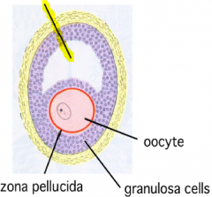
Granulosa cells
|

Cells that surround the oocyte release glycoproteins that form the zona pellucida.
|
|
|
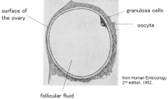
Antrum
|
The space that develops in the granulosa cells as the follicle continues to develop.
|
|
|
Zona Pellucida
|
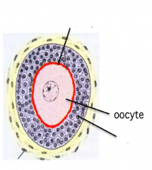
A jelly like glycoprotein coat around the oocyte, secreted by the granulosa cells.
|
|
|
Mature Follicle
|
|
|
|
Oocyte development
|
Arrested in first meiosis prior to birth and only
complete 1st meiosis as they progress towards ovulation (after puberty). |
|
|
Oocyte second meiotic division
|
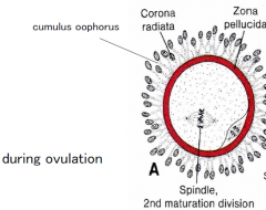
Commences only during ovulation to generate haploid cell.
Second meiosis only completes upon fertilization. |
|
|
Polar body development
|
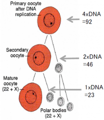
Complete meiotic development of an oocyte results in 3 nonviable polar bodies;
All 4 cells have all chromosomes 22+X chromosome In general, only one oocyte released per ovulation cycle. |
|
|
Sperm cell development
|
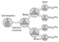
Spermatogonium cells undergo the two standard meiotic divisions to produce 4 viable spermatids, which later mature into spermatozoa
2 with 22+X chromosomes 2 with 22+Y chromosomes |
|
|
Seminiferous tubule
|
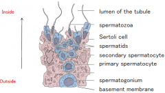
Site of sperm development.
Organized with precursor cells on the outside, with more differentiated cells closer to the lumen. |
|
|
Chromosome abnormalities
|
Due to abnormal meiotic division; either too few or many chromosomes
Most commonly observed are trisomies; 3 copies of a chromosome Loss of a chromosome (other than sex) is invariably lethal |
|
|
Trisomy 21
|
Down's syndrome
|
|
|
Turner Syndrome
|
Female inherits only one X chromosome
Short stature, webbed neck, reduction in secondary female characteristics, ovaries do not develop normally leading to sterility. |
|
|
Klinefelter syndrome
|
Males with extra X chromosome (XXY)
No major medical problems, though they may posses some feminine characteristics (higher voice, breast enlargement, reduced body hair). Thought to be significantly under-diagnosed. |
|
|
Male infertility
|
Absence or reduction of sperm count
Defective sperm Blockage of seminiferous tubules |
|
|
Female infertility
|
Failure of oocyte maturation/release
Blockage of egg transport in fallopian tubes. Polycystic ovarian syndrome (PCOS) often results in increased male hormones, inhibiting oocyte maturation/release. |