Reading...
![]()
Play button
![]()
Play button
![]()
Use LEFT and RIGHT arrow keys to navigate between flashcards;
Use UP and DOWN arrow keys to flip the card;
H to show hint;
A reads text to speech;
42 Cards in this Set
- Front
- Back
|
What are the bones of the foot?
|
7 tarsal bones: talus, calcaneous, navicular, cuboid, and 3 cuneiforms
5 metatarsal bones (1-5) 14 phalanges, only 2 in the first toe |
|
|
What are the bones of the ankle joint and its movements?
|
-Talus, tibia, and fibula. The weight of the body rests on the talus.
-Movements include dorsiflexion (raising toes off the ground) and plantar flexion (raising the heal off the ground) |
|
|
Describe the joints of the foot.
|
-Gliding joints in the tarsal and metatarsal areas
-Condyloid joints in the metatarsal-phalangeal and interphalangeal areas -Movements such as inversion and eversion of the foot occur at the subtalar and mid-tarsal joints. |
|
|
What is the deep fascia of the foot called?
|
The plantar aponeurosis. It is the deep fascia that covers the musculature in the sole of the foot.
|
|
|
How are the muscles of the foot arranged?
|
4 layers and consist of both intrinsic and extrinsic muscles, which help to maintain the arches of the foot.
|
|
|
Extensor digitorum brevis
|
O: dorsal surface of calcareous
I: middle phalanges 2, 3, 4 Inv: deep fibular n. A: extends digits 2, 3, 4 |
|
|
Extensor hallucis brevis
|
O: calcaneous
I: proximal phalanx of digit 1 Inv: deep fibular n. A: extends hallux |
|
|
How many layers of muscles are on the sole of the foot?
|
Four
|
|
|
What muscles are in the first layer of the sole of the foot?
|
-Abductor hallucis
-Flexor digitorum brevis -Abductor digiti minimi |
|
|
What muscles are in the second layer of the sole of the foot?
|
-Quadratus plantae
-Lumbricals (4) -Tendon of flexor digitorum longus -Tendon of flexor hallucis longus |
|
|
What muscles are in the third layer of the sole of the foot?
|
-Flexor hallucis brevis
-Adductor hallucis -Flexor digiti minini brevis |
|
|
What muscles are in the fourth layer of the sole of the foot?
|
-Plantar interossei (3)
-Dorsal interossei (4) |
|
|
Abductor hallucis
|
O: calcaneal tuberosity
I: medial side- base of proximal phalanx of hallux Inv: medial plantar n. A: abducts big toe |
|
|
Flexor digitorum brevis
|
O: calcaneal tuberosity
I: middle phalanx Inv: medial plantar n. A: flexes toes 2-5 at PIP joints |
|
|
Abductor digiti minimi
|
O: calcaneal tuberosity
I: lateral side- base of proximal phalanx of 5th toe Inv: lateral plantar n. A: abducts the 5th toe |
|
|
Quadratus plantae
|
O: calcaneus (medial surface)
I: tendon of flexor digitorum longus Inv: lateral plantar n. A: flexes toes |
|
|
Lumbricales (4)
|
O: tendons of flexor ditiorum longus
I: extensor expansions of lateral 4 toes Inv: 1st toe - medial plantar n., toes 2, 3, & 4 - lateral plantar n. A: flexes MP joints, extends IP joints |
|
|
Tendon of flexor digitorum longus
|
O: posterior tibia
I: distal phalanx toes 2-5 Inv: tibial n. A: flexes toes 2-5 |
|
|
Tendon of flexor hallucis longus
|
O: posterior fibula
I: distal phalanx of hallux Inv: tibial n. A: flexes big toe |
|
|
Flexor hallucis brevis
|
O: plantar surface of cuboid
I: medial and lateral plantar sides of proximal phalanx of hallux Inv: medial plantar n. A: flexes big toe |
|
|
Adductor hallucis
|
O: transverse part - heads of metatarsals, oblique part - plantar surface of metatarsals 2, 3, & 4.
I: lateral side of proximal phalanx of hallux Inv: lateral plantar n. A: adducts and assists flexion of hallux |
|
|
Flexor digiti minimi brevis
|
O: plantar surface of 5th metatarsal
I: base of proximal phalanx toe 5 Inv: lateral plantar n. A: flexes proximal phalanx of toe 5 |
|
|
Plantar interossei (3)
|
O: plantar surfaces of metatarsals 3, 4, & 5
I: medial side of proximal phalanges 3, 4, & 5 Inv: lateral plantar n. A: adductors of toes 3-5, flexes MP joints |
|
|
Dorsal interossei (4)
|
O: adjoining parts of metatarsals 1-5
I: both sides of proximal phalanges of 2nd toe, lateral side of proximal phalanges toes 3 and 4 Inv: lateral plantar n. A: abductors of toes 2-4, flexes MP joints |
|
|
Where is the center line of the foot?
|
Goes through the 2nd toe.
|
|
|
Sesamoid bones in foot
|
Near flexor hallucis brevis. Helps increase mechanical advantage when it contracts.
|
|
|
What are the nerves that provide innervation of the foot?
|
Medial and lateral plantar nerves, both branches of the tibial nerve.
|
|
|
What muscles are supplied by the medial plantar nerve?
|
Abductor hallucis, flexor digitorum brevis, flexor hallucis brevis, and the 1st lumbrical.
Also has cutaneous branches that supply both sides of hallux, toes 2 & 3, and the tibial side of toe 4. |
|
|
What muscles are supplied by the lateral plantar nerve?
|
Abductor digiti minimi, quadratus plantae, flexor digiti minimi brevis, adductor hallucis, lateral 3 lumbricals and the interossei muscles.
Also has cutaneous branches that supply the lateral side of toe 4 and both sides of toe 5. |
|
|
What is the principal artery of the sole of the foot?
|
Lateral plantar artery, a continuation of the posterior tibial artery (which enters the foot via the tarsal tunnel).
|
|
|
What does the lateral plantar artery form?
|
The plantar arch, which branches off into the plantar metatarsal arteries.
|
|
|
What artery supplies the dorsum of the foot?
|
The dorsalis pedis artery, a continuation of the anterior tibial artery.
|
|
|
What makes up the longitudinal arch of the foot?
|
All the tarsal and metatarsal bones, and is divided into a medial and lateral arch.
|
|
|
Describe the medial arch (part of the longitudinal arch).
|
Highest side of longitudinal arch. It is more pronounced and is made up of a short posterior pillar formed by the calcaneus and body of the talus, a keystone formed by the head of the talus and a long anterior pillar formed by the navicular, cuneiforms, and 3 medial metatarsals.
|
|
|
What is the keystone of the foot arches?
|
Talus.
|
|
|
What supports the keystone of the foot?
|
-tendons of the deep muscles on the back of the leg
-small muscles in the sole of the foot -plantar aponeurosis -plantar ligaments of all the joints (especially the plantar calceno-navicular ligament, or "spring" ligament, and the long and short ligaments) |
|
|
What are the principal support of the arches?
|
Muscles.
Ligaments are only used when temporary and excessive strain is put on them. |
|
|
Describe the transverse arch.
|
Found in the region of the cuboid and cuneiform bones and is maintained by the peroneus longus and tibialis posterior tendons.
|
|
|
Describe the lateral arch (part of the longitudinal arch).
|
Lower arch than the medial arch.
|
|
|
Describe what happens to your arches in the resting upright position.
|
The weight of the body acts on the talus and tends to flatten the longitudinal and transverse arches.
|
|
|
Describe what happens to your arches during activity.
|
Simultaneous contraction of the tibialis posterior, flexor digitorum longs, peroneus longus, peroneus brevis and small intrinsic muscles of the foot restore and accentuate the arches. This converts the foor into an almost rigid but resilient lever.
|
|
|
What happens when you contract the flexor digitorum and quadratus plantar at the same time?
|
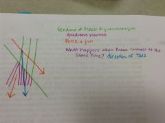
Force and pull will combine to move in the direction of the toes.
|