Reading...
![]()
Play button
![]()
Play button
![]()
Use LEFT and RIGHT arrow keys to navigate between flashcards;
Use UP and DOWN arrow keys to flip the card;
H to show hint;
A reads text to speech;
25 Cards in this Set
- Front
- Back
|
Diaphragm
|
A muscle that appear sonographically as a hypoechoic curved structure separating the more echogenic lung from the liver and stomach.
|
|
|
Liver
|
The Liver is large and occupies most of the upper abdomen in the fetus.
The left lobe is larger than the right lobe |
|
|
Gall Bladder
|
GB is an anechoic fluid fill structure seen in the anterior right abdomen inferior to the liver margin.
|
|
|
Spleen
|
Spleen is seen the upper left abdomen posterior to the stomach
The spleen is echogenic and homogenous |
|

Gastroschisis
|
Relatively small defect involving all 3 layers of abdominal wall thatcallown the intestines to protrude out in the amniotic cavity.
Occurs lateral to umbilical cord insert , usually to the right. Is not covered by an membranous sac. has no associated anomalies |
|
Omphalocele
|
Results from faulire of the intestines to return by to the abdomen during the second stage of intestinal rotation.
Occurs at the level of the umbilical cord insertion. May contain single loops of bowel, or most of abdominal content depending on severity. herniation is covered by a membranous sac which is layers or amnion and peritoneum. Has high association with congenital anomalies. |
|
Songraphic findings of Gastroschisis
|
-variable amounts of bowel floating in amniotic fluid
-no membranous sac covering herniation -cord is seen adjacent to defect |
|

Songraphic findings of Omphalocele
|
-extra-abdominal mass consisting of a combination of bowel, liver or other abdominal content
-membranous sac covering herniation -mass at the level of umbilical cord |
|
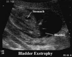
Bladder Extrophy
|
Midline defect that involves the lower abdominal wall and the anterior wall of the urinary bladder.
Isolated defect characterized by the exposure and protrusion of the urinary bladder. May be associated with genital anomalies. |
|

Sonographic findings for Bladder Extrophy
|
-unable to identify the bladder after 30mins of scanning
-mirophallus in male fetuses separation of pelvic bones -possible soft tissue protrusion from lower abdominal wall |
|
|
Esophagus
|
Unable to identify unless the fetus is swollen or stenosis is present.
|
|
|
Stomach
|
In transverse view the stomach is seen as a oviod fluid collection in the upper left abdomen.
The muscular wall is much thinner in fetuses. Occasional echoes my be visualized in the stomach. |
|
|
Intestines
|
Diffect to isolate unless fluid is present to provide specific contrast.
Has a cyctic to mixed echogenicity Peristalsis is seen in the late 2nd trimesters. Meconium fills the colon and it appears highly echogenic. The colon is peripheral and small intestines is centrally located in the fetal abdomen. Colon is most obvious in the 3rd trimester. |
|
|
Esphageal Atresia
|
The discontinuity of the esphagus.
90% are accompanied by a distal traheo-esophageal fistula. Associated with other congenital malformation most commonly cardiovascular, gastrointestinal, genitourinary, musculoskeletal anomalies. |
|
|
Sonographic findings for Esophageal Atresia
|
-small to absent fetal stomach
-unable to identify the stomach on a serial scan -may not be sonographically identified if a TE fistal is not present -polyhydramnios |
|
Duodenal Atresia
|
-most common type of perinatal intestinal obstruction
- 65% of fetuses has a karyotypic abnormality at birth, most commonly vertebral and cardiac. -30% of infant has trisomy 21 |
|

Songraphic findings for Duodenal Atresia
|
- "double-bubble" sign- dilated stomach with proximal duodenum
-may not be apparent until after 24 weeks -polyhydramnios |
|
Intestinal Atresia
|
Obstruction of the intestines.
Dilated bowel loops may occur at any location along intestinal tract or at the level of the anus. |
|

Sonographic findings for Intestinal Atresia
|
-multiple fluid fill bowel loops
-small bowel internal diameter > 7mm -possible perferation due to calification and ascites -polyhydramnios -increased bowel paristalsis |
|
Meconium Peritonitis
|
Sterile chemical peritonitis is cause by small bowel perforation in utero.
Perforation is caused by intestinal atresia, volvulus or meconium ileus. Cystic fibrosis is considered to be the etiology in 35-40% due to thick and sticky meconiun. |
|

Songraphic finding for Meconium Peritonitis
|
-presence of a meconium psudocyst
-calcification that may cast a shaddow -fetal ascites and polyhydramnios |
|

Hyperechoic Bowel
|
Bowel that is similar to or greater than that of adjacent bone.
Lower Fz should be used Located in lower abdomen or pelvic Diffuse or Focal over a well defined area that does not shaddow. Can be identified on a 2nd trimesters scan. |
|
Common etiology for hyperechoic bowel
|
-normal variant
-trisomy 21 -cystic fibrosis -swollowed intra - amniotic blood -CMV infection |
|
Abdominal Ascites
|
Ascites is commonly associated with a condition call hydrops fetalis
|
|
Persistent Right Umbilical Vien
|
When the umbilical vien course toward the left of the fetal abdomen.
umblical vien enters the right portal vien instead of the left portal vien. Not pathological but can be associated with some anomalous conditions |