Reading...
![]()
Play button
![]()
Play button
![]()
Use LEFT and RIGHT arrow keys to navigate between flashcards;
Use UP and DOWN arrow keys to flip the card;
H to show hint;
A reads text to speech;
38 Cards in this Set
- Front
- Back
|
What is the sensation of spinning while actually stationary? |
Vertigo |
|
|
What are the types of vertigo? Which is more common?
|
- Peripheral vertigo (more common)
- Central vertigo |
|
|
What causes peripheral vertigo?
|
Inner ear etiology (eg, semicircular canal debris, vestibular nerve infection, Ménière disease
|
|
|
What causes central vertigo?
|
Brain stem or cerebellar lesion (eg, stroke affecting vestibular nuclei or posterior fossa tumor)
|
|
|
What are the findings in peripheral vertigo?
|
Positional testing → delayed horizontal nystagmus
|
|
|
What are the findings in central vertigo?
|
- Directional change of nystagmus
- Skew deviation - Diplopia - Dysmetria - Postional testing → immediate nystagmus in any direction; may change directions - Focal neurological findings |
|
|
What disease is characterized by a port-wine stain of the face, seizures/epilepsy, intellectual disability, and early-onset glaucoma?
|
Sturge-Weber Syndrome
STURGE: - Sporadic port-wine Stain - Tram track Ca2+ (opposing gyri) - Unilateral - Retardation - Glaucoma and GNAQ gene - Epilepsy |
|
|
What is the cause of Sturge-Weber Syndrome?
|
- Congenital, non-inherited (somatic), developmental anomaly of neural crest derivatives (mesoderm/ectoderm)
- Due to activating mutation of GNAQ gene |
|
What causes the port-wine stain seen in Sturge-Weber Syndrome?
|

Small (capillary-sized) blood vessels have developmental anomalies → port-wine stain of face (non-neoplastic "birthmark" in CN V1/V2 distribution)
|
|
|
What causes the seizures/epilepsy seen in Sturge-Weber Syndrome?
|
Ipsilateral leptomeningeal angioma → seizures / epilepsy
|
|
|
What causes the glaucoma in Sturge-Weber Syndrome?
|
Episcleral hemangioma → ↑ intraocular pressure → early onset glaucoma
|
|
|
What genetic change causes Sturge-Weber Syndrome?
|
Activating mutation of GNAQ gene
|
|
|
What disease causes hamartomas in the CNS and skin, angiofribromas, mitral regurgitation, ash-leaf spots, cardiac rhabdomyoma, mental retardation, renal angiomyolipoma, seizures, and Shagreen patches?
|
Tuberous Sclerosis
|
|
|
What mnemonic helps you remember the characteristics of Turberous Sclerosis?
|
HAMARTOMAS:
- Hamartomas in CNS and skin - Angiofibromas (C) - Mitral regurgitation - Ash-leaf spots - Rhabdomyoma (cardiac) - Tuberous sclerosis - autosomal dOminant - Mental retardation - Angiomyolipoma (renal) (D) - Seizures and Shagreen patches |
|
|
What is there increased incidence of in Turberous Sclerosis?
|
- Subependymal astrocytomas
- Ungual fibromas |
|
|
Which disease is associated with café-au-lait spots, Lisch nodules, neurofibromas in the skin, optic gliomas, and pheochromocytomas?
|
Neurofibromatosis Type 1 (von Recklinghausen disease)
|
|
|
What is the cause of Neurofibromatosis Type 1 (von Recklinghausen disease)?
|
- Mutated NF1 tumor suppressor gene (neurofibromin - a negative regulator of Ras)
- On chromosome 17 |
|
|
What does a mutated NF1 tumor suppressor gene (chr 17) cause? Clinical symptoms?
|
Neurofibromatosis Type 1 (von Recklinghausen disease)
- Café-au-lait spots (E) - Lisch nodules (pigmented iris hamartomas) (F) - Neurofibromas in skin - Optic gliomas - Pheochromocytoma |
|
|
What are skin tumors of NF-1 derived from?
|
Neural crest cells
|
|
|
What disease is associated with cavernous hemangiomas in skin, mucosa, and organs; bilateral renal cell carcinomas; hemangioblastoma in retina, brain stem, cerebellum; and pheochromocytoma?
|
von Hippel-Lindau Disease
|
|
|
What is the cause of von Hippel-Lindau Disease?
|
- Autosomal dominant mutation in VHL tumor suppressor gene on chr 3
- Results in constitutive expression of HIF (transcription factor) and activation of angiogenic growth factors |
|
|
What disease is caused by the autosomal dominant mutation in the VHL tumor suppressor gene on chr 3? What does this mutation cause?
|

von Hippel-Lindau Disease
- Mutation results in constitutive expression of HIF (transcription factor) and activation of angiogenic growth factors - Cavernous hemangiomas in skin, mucosa, and organs - Bilateral renal cell carcinoma - Hemangioblastoma (high vascularity with hyperchromatic nuclei (G)) in retina, brainstem, and cerebellum (H) - Pheochromocytoma |
|
|
Where are there cavernous hemangiomas in von Hippel-Lindau Disease?
|
- Skin
- Mucosa - Organs |
|
|
Where are there hemangioblastomas in von Hippel-Lindau Disease?
|
- Retina
- Brainstem - Cerebellum |
|

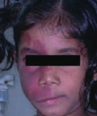
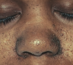
What is wrong with this little girl? Associated with what disease?
|

Port-wine stain on face
- Caused by Sturge Weber Syndrome |
|

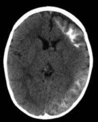
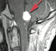
What is wrong with this brain? Associated with what disease?
|

Ipsilateral leptomeningeal angioma → seizures / epilepsy
- Caused by Sturge Weber Syndrome |
|

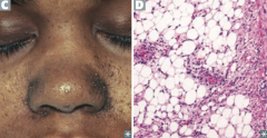
What is wrong with this little boy? Associated with what disease?
|
Angiofibromas of the face
- Caused by Tuberous Sclerosis |
|

What aspect of Tuberous Sclerosis has this histology?
|
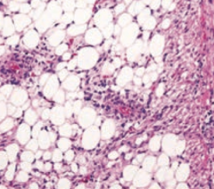
Renal Angiomyolipoma
|
|

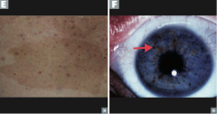
What is this skin finding? Associated with what disease?
|
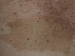
Café-au-lait spot
- Caused by Neurofibromatosis type I (von Recklinghausen disease) |
|

What is this eye finding? Associated with what disease?
|
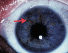
Lisch nodule (pigmented iris hamartoma)
- Caused by Neurofibromatosis type I (von Recklinghausen disease) |
|
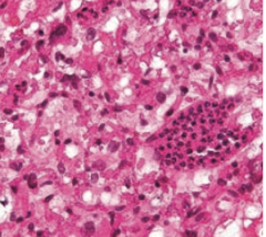
What aspect of von Hippel Lindau disease has this histology?
|

Hemangioblastomas (high vascularity with hyperchromatic nuclei)
|
|
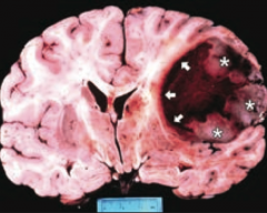
What is this brain finding? Associated with what disease?
|

Hemangioblastoma in cerebellum
- Caused by von Hippel Lindau disease |
|
|
What are the adult primary brain tumors?
|
- Glioblastoma multiforme (grade IV astrocytoma)
- Meningioma - Hemangioblastoma - Schwannoma - Oligodendroglioma - Pituitary adenoma |
|

What tumor is found in the cerebral hemispheres and is known for crossing the corpus callosum?
|
Glioblastoma Multiforme (grade IV astrocytoma)
|
|
|
What is the prognosis of Glioblastoma Multiforme (grade IV astrocytoma)? How common?
|
- Common, in adults
- Highly malignant with ~1 year median survival |
|
|
What is the appearance of a Glioblastoma Multiforme (grade IV astrocytoma)?
|
- Found in cerebral hemispheres
- Can cross corpus callosum ("butterfly glioma") |
|
|
What adult primary brain tumor will stain positively for GFAP? Why?
|
Glioblastoma Multiforme (grade IV astrocytoma) - astrocytes are stained with GFAP
|
|
|
What is the histologic appearance of Glioblastoma Multiforme (grade IV astrocytoma)?
|
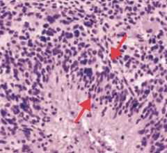
"Pseudopalisading" pleomorphic tumor cells - border central areas of necrosis and hemorrhage
|