![]()
![]()
![]()
Use LEFT and RIGHT arrow keys to navigate between flashcards;
Use UP and DOWN arrow keys to flip the card;
H to show hint;
A reads text to speech;
74 Cards in this Set
- Front
- Back
|
different types of synapse |
-adrenergic synapse employs the neurotransmitter norepinephrine (NE) -GABA-ergic synapse employs γ-aminobutyric acid (GABA) as its neurotransmitter |
|
|
Signals arrive at the synapse by way of the _________ __________, which releases a neurotransmitter. The next neuron, which responds to it, is called the _______ ___________ |
presynaptic neuron and posysynaptic neuron |
|
|
Presynaptic |
neuron may synapse with a dendrite, the soma, or the axon of a postsynaptic neuron, forming an axodendritic, axosomatic, or axoaxonic synapse, respectively |
|
|
electrical synapses |
where adjacent cells are joined by gap junctions and ions diffuse directly from one cell into the next. These junctions have the advantage of quick transmission because there is no delay for the release and binding of neurotransmitter. |
|
|
chemical synapses |
the disadvantage to electrical synapses is that they cannot integrate information and make decisions. The ability to do that is a property of chemical synapses, in which neurons communicate by neurotransmitters. |
|
|
what depolarizes the membrane? |
Excitatory; cholinergic synapse employs acetylcholine (ACh) as its neurotransmitter. ACh excites some postsynaptic cells (such as skeletal muscle) and inhibits others, but this discussion will describe an excitatory action. |
|

Part I |
*The arrival of a nerve signal at the synaptic knob opens voltage-gated calcium channels. Ca2+ enters the knob and triggers exocytosis of the synaptic vesicles, releasing ACh. *Empty vesicles drop back into the cytoplasm to be refilled with ACh, while synaptic vesicles in the reserve pool move to the active sites and release their ACh—a bit like a line of Revolutionary War soldiers firing their muskets and falling back to reload as another line moves to the fore. |
|
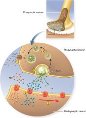
Part II |
*Meanwhile, ACh diffuses across the synaptic cleft and binds to ligand-gated channels on the postsynaptic neuron. These channels open, allowing Na+ to enter the cell and K+ to leave. *As Na+ enters, it spreads out along the inside of the plasma membrane and depolarizes it, producing a local voltage shift called the postsynaptic potential. Like other local potentials, if this is strong and persistent enough (that is, if enough current makes it to the axon hillock), it opens voltage-gated ion channels in the trigger zone and causes the postsynaptic neuron to fire. |
|
|
GABA-ergic |
synapse employs γ-aminobutyric acid (GABA) as its neurotransmitter. Amino acid neurotransmitters work by the same mechanism as ACh—binding to ion channels and causing immediate changes in membrane potential. The release of GABA and binding to its receptor are similar to the preceding case. The GABA receptor, however, is a chloride channel. When it opens, Cl− enters the cell and makes the inside even more negative than the resting membrane potential. The neuron is therefore inhibited, or less likely to fire. |
|
|
second-messenger |
neurotransmitter norepinephrine (NE), also called noradrenaline and other monoamines, and neuropeptides act through second-messenger systems such as cyclic AMP (cAMP). The receptor is not an ion channel but a transmembrane protein associated with a G protein. |
|
|
Transmission at an Adrenergic Synapse. |

The norepinephrine receptor is not an ion channel. It activates a second-messenger system with a variety of possible effects in the postsynaptic cell. Numbered steps correspond to the description in the text. |
|

number steps (part I) |
-The unstimulated NE receptor is bound to a G protein. -Binding of NE to the receptor causes the G protein to dissociate from it. -The G protein binds to adenylate cyclase and activates this enzyme, which converts ATP to cAMP. -Cyclic AMP can induce several alternative effects in the cell. |
|
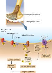
number steps (part 2) |
-One effect is to produce an internal chemical that binds to a ligand-gated ion channel from the inside, opening the channel and depolarizing the cell. -Another is to activate preexisting cytoplasmic enzymes, which can lead to diverse metabolic changes (for example, inducing a liver cell to break down glycogen and release glucose into the blood). -Yet another is for cAMP to induce genetic transcription, so that the cell produces new enzymes leading to diverse metabolic effects. |
|
|
properties of neurons |
1Excitability. All cells are excitable—that is, they respond to environmental changes (stimuli). Neurons exhibit this property to the highest degree.2.Conductivity. Neurons respond to stimuli by producing electrical signals that are quickly conducted to other cells at distant locations. 3.Secretion. When the signal reaches the end of a nerve fiber, the neuron secretes a neurotransmitter that crosses the gap and stimulates the next cell. |
|
|
functional classes (part I) |
-Sensory (afferent) neurons are specialized to detect stimuli such as light, heat, pressure, and chemicals, and transmit information about them to the CNS. -Interneurons (association neurons) lie entirely within the CNS. They receive signals from many other neurons and carry out the integrative function of the nervous system—that is, they process, store, and retrieve information and “make decisions” that determine how the body responds to stimuli. About 90% of our neurons are interneurons. |
|
|
functional classes (part II) |
-Motor (efferent) neurons send signals predominantly to muscle and gland cells, the effectors. They are called motor neurons because most of them lead to muscle cells, and efferent neurons to signify signal conduction away from the CNS. |
|
|
Neurons are classified structurally according to the number of processes extending from the soma |
Multipolar neurons that have one axon and multiple dendrites. This is the most common type and includes most neurons of the brain and spinal cord. Bipolar neurons have one axon and one dendrite. Unipolar neurons have only a single process leading away from the soma. Anaxonic neurons have multiple dendrites but no axon. |
|
|
Multipolar neurons |
that have one axon and multiple dendrites. This is the most common type and includes most neurons of the brain and spinal cord. |
|
|
Bipolar neurons |
have one axon and one dendrite. Examples include olfactory cells of the nose, certain neurons of the retina, and sensory neurons of the ear. |
|
|
Unipolar neurons |
have only a single process leading away from the soma. They are represented by the neurons that carry sensory signals to the spinal cord. |
|
|
Anaxonic neurons |
have multiple dendrites but no axon. They communicate locally through their dendrites and do not produce action potentials. Some anaxonic neurons are found in the brain, retina, and adrenal medulla. In the retina, they help in visual processes such as the perception of contrast. |
|
|
the two-way passage of proteins, organelles, and other materials along an axon is called axonal transport. (2 ways of transport) |
Movement away from the soma down the axon is called anterograde transport and movement up the axon toward the soma is called retrograde transport. |
|
|
Fast anterograde transport |
moves mitochondria; synaptic vesicles; other organelles; components of the axolemma; calcium ions; enzymes such as acetylcholinesterase; and small molecules such as glucose, amino acids, and nucleotides toward the distal end of the axon. |
|
|
Fast retrograde transport |
returns used synaptic vesicles and other materials to the soma and informs the soma of conditions at the axon terminals. Some pathogens exploit this process to invade the nervous system. |
|
|
Slow axonal transport |
is an anterograde process that works in a stop-and-go fashion. |
|
|
Glial cells |
protect the neurons and help them function; The word glia, which means “glue,” implies one of their roles |
|
|
Oligodendrocytes |
Form myelin in brain and spinal cord |
|
|
Ependymal cells |
Line cavities of brain and spinal cord; secrete and circulate cerebrospinal fluid |
|
|
Microglia |
Phagocytize and destroy microorganisms, foreign matter, and dead nervous tissue |
|
|
Astrocytes |
Cover brain surface and nonsynaptic regions of neurons; form supportive framework in CNS; induce formation of blood−brain barrier; nourish neurons; produce growth factors that stimulate neurons; communicate electrically with neurons and may influence synaptic signaling; remove K+ and some neurotransmitters from ECF of brain and spinal cord; help to regulate composition of ECF; form scar tissue to replace damaged nervous tissue |
|
|
Schwann cells |
Form neurilemma around all PNS nerve fibers and myelin around most of them; aid in regeneration of damaged nerve fibers |
|
|
Satellite cells |
Surround somas of neurons in the ganglia; provide electrical insulation and regulate chemical environment of neurons |
|
|
Astrocytes (indepth description) (part I) |
are the most abundant glial cells in the CNS and constitute over 90% of the tissue in some areas of the brain. They cover the entire brain surface and most nonsynaptic regions of the neurons in the gray matter of the CNS.They form a supportive framework for the nervous tissue. They have extensions called perivascular feet, which contact the blood capillaries and stimulate them to form a tight, protective seal called the blood–brain barrier. |
|
|
Astrocytes (indepth description) (part II) |
They monitor neuron activity and regulate blood flow in the brain tissue to meet changing needs for oxygen and nutrients.They convert blood glucose to lactate and supply this to the neurons for nourishment.They secrete nerve growth factors that regulate nerve development.They communicate electrically with neurons and influence synaptic signaling between them.They regulate the composition of the tissue fluid. When neurons transmit signals, they release neurotransmitters and potassium ions. |
|
|
Astrocytes (indepth description) (part III) |
Astrocytes absorb these and prevent them from reaching excessive levels in the tissue fluid.When neurons are damaged, astrocytes form hardened scar tissue and fill space formerly occupied by the neurons. This process is called astrocytosis or sclerosis. |
|
|
saltatory conduction |
Since action potentials occur only at the nodes, this mode of conduction creates a false impression that the nerve signal jumps from node to node. Conduction in myelinated fibers.Think of crowded subway car. The doors open (like the Na+ gates at a node), 20 more people get on (like Na+ flowing into the axon), and everyone has to push to the rear of the car to make room for them. No one passenger moves from the door to the rear, but the crowding and transfer of energy from person to person forces even those at the rear to move a little, like the sodium ions at the next node. |
|
|
possible to repair nerve fibers?
|
yes depending on damage done to nerve, as long as soma (cell body) has not been destroyed. |
|
|
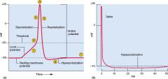
action potential diagram |
|
|
|
action potential diagram (explained)1 |
When the local current arrives at the axon hillock, it depolarizes the membrane at that point. This appears as a steadily rising local potential. For anything more to happen, this local potential must rise to a critical voltage called the threshold (typically about –55 mV)—the minimum needed to open voltage-gated channels. The neuron now “fires,” or produces an action potential. At threshold, voltage-gated Na+ channels open quickly, while K+ channels open more slowly. The initial effect on membrane potential is therefore due to Na+. Initially, only a few Na+ channels open, but as Na+ enters the cell, it further depolarizes the membrane. This stimulates still more voltage-gated Na+ channels to open and admit even more Na+, creating a positive feedback loop that makes the membrane voltage rise rapidly. |
|
|
action potential diagram (explained)2 |
The neuron now “fires,” or produces an action potential. At threshold, voltage-gated Na+ channels open quickly, while K+ channels open more slowly. The initial effect on membrane potential is therefore due to Na+. Initially, only a few Na+ channels open, but as Na+ enters the cell, it further depolarizes the membrane. This stimulates still more voltage-gated Na+ channels to open and admit even more Na+, creating a positive feedback loop that makes the membrane voltage rise rapidly. |
|
|
action potential diagram (explained)3 |
As the rising potential passes 0 mV, Na+ channels are inactivated and begin closing. By the time they all close and Na+ inflow ceases, the voltage peaks at approximately +35 mV. (The peak is as low as 0 mV in some neurons and as high as 50 mV in others.) The membrane is now positive on the inside and negative on the outside—its polarity is reversed compared to the RMP. |
|
|
action potential diagram (explained)4 |
By the time the voltage peaks, the slow K+ channels are fully open. Potassium ions, repelled by the positive ICF, now exit the cell. Their outflow repolarizes the membrane—that is, it shifts the voltage back into the negative numbers. The action potential consists of the up-and-down voltage shifts that occur from the time the threshold is reached to the time the voltage returns to the RMP. |
|
|
action potential diagram (explained)5 |
Potassium channels stay open longer than Na+ channels, so slightly more K+ leaves the cell than the amount of Na+ that entered. Therefore, the membrane voltage drops to 1 or 2 mV more negative than the original RMP, producing a negative overshoot called hyperpolarization (or afterpotential).-- As you can see, Na+ and K+ switch places across the membrane during an action potential. During hyperpolarization, Na+ diffusion into the cell and (in the CNS) the removal of extracellular K+ by astrocytes gradually restore the original RMP. |
|
|
refractory period - |
1. A period of time after a nerve or muscle cell has responded to a stimulus in which it cannot be reexcited by a threshold stimulus. 2. A period of time after male orgasm when it is not possible to reattain erection or ejaculation |
|
|
absolute refractory period |
lasts from the start of the action potential until the membrane returns to the resting potential |
|
|
absolute refractory period vs relative refractory period |
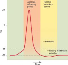
an absolute refractory period in which no stimulus of any strength will trigger a new action potential, followed by a relative refractory period in which it is possible to trigger a new action potential, but only with an unusually strong stimulus |
|
|
why does a nerve signal only go in one direction? Synapses? |
cannot re-excit a nerve; synapses only works in one direction as well. |
|
|
four types of neural circuits (1 & 2) |
1. diverging circuit, one nerve fiber branches and synapses with several postsynaptic cells. Each of those may synapse with several more, so input from just one neuron may produce output through hundreds of neurons. 2. converging circuit is the opposite of a diverging circuit—input from many nerve fibers is funneled to one neuron or neural pool. Such an arrangement allows input from your eyes, inner ears, and stretch receptors in your neck to be directed to an area of the brain concerned with the sense of balance. |
|
|
four types of neural circuits (3 & 4) |
3. everberating circuit, neurons stimulate each other in a linear sequence from input to output neurons, but some of the neurons late in the path send axon collaterals back to neurons earlier in the path and restimulate them. 4. parallel after-discharge circuit, an input neuron diverges to stimulate several chains of neurons. Each chain has a different number of synapses, but eventually they all reconverge on one or few output neurons. |
|
|
neural coding |
the way in which the nervous system converts information to a meaningful pattern of action potentials is called neural coding (or sensory coding when it occurs in the sense organs). The most important mechanism for transmitting qualitative information is the labeled line code. |
|
|
labeled line code. |
This code is based on the fact that each nerve fiber to the brain leads from a receptor that specifically recognizes a particular stimulus type. Nerve fibers in the optic nerve, for example, carry signals only from light receptors in the eye; these fibers never carry information about taste or sound. |
|
|
function of posterior horn? |
Transmission of neural signal; It receives several types of sensory information from the body, including fine touch, proprioception, and vibration. |
|
|
function of anterior horn? |
transmission of neural signal; contains motor neurons that affect the skeletal muscles. |
|
|
in a sensory circuit, how many neurons? |
(3) a first-order neuron that detects a stimulus and transmits a signal to the spinal cord or brainstem; a second-order neuron that continues as far as a “gateway” called the thalamus at the upper end of the brainstem; and a third-order neuron that carries the signal the rest of the way to the cerebral cortex. First order synapse w/ second order, second order w/ third 2nd & 3rd synapse in thalamus, 1st & 2nd synapse in medulla. |
|
|
motor neurons
|
The upper motor neuron begins with a soma in the cerebral cortex or brainstem and has an axon that terminates on a lower motor neuron in the brainstem or spinal cord. The axon of the lower motor neuron then leads the rest of the way to the muscle or other target organ. |
|
|
where to find cell body in upper neuron |
cerebral cortex -- front lobe, precentral gyrus |
|
|
3rd order |
precentral gyrus |
|
|
distal branches |
Immediately after emerging from the intervertebral foramen, the nerve divides into an anterior ramus a posterior ramus, and a small meningeal branch. Thus, each spinal nerve branches on both ends—into anterior and posterior roots approaching the spinal cord, and anterior and posterior rami leading away from the vertebral column. |
|
|
The cervical plexus |
receives fibers from the anterior rami of nerves C1 to C5 and gives rise to the nerves listed below, in order from superior to inferior. |
|
|
The brachial plexus |
is formed predominantly by the anterior rami of nerves C5 to T1 (C4 and T2 make small contributions). It passes over the first rib into the axilla and innervates the upper limb and some muscles of the neck and shoulder. |
|
|
The lumbar plexus |
is formed from the anterior rami of nerves L1 to L4 and some fibers from T12. |
|
|
The sacral plexus |
is formed from the anterior rami of nerves L4, L5, and S1 through S4. It has six roots and anterior and posterior divisions. |
|
|
why enlargements in spine? |
all the neurons that go out to the body parts |
|
|
function of lateral horn |
contains visceral and motor nuclei.transmission of neural signals sympathetic nerves system integration |
|
|
dermatome.
|
dermatome map is a diagram of the cutaneous regions innervated by each spinal nerve. Such a map is oversimplified, however, because the dermatomes overlap at their edges by as much as 50%. Therefore, severance of one sensory nerve root does not entirely deaden sensation from a dermatome. |
|
|
connective tissue for nerves |
Nerve fibers of the peripheral nervous system are ensheathed in Schwann cells, which form a neurilemma and often a myelin sheath around the axon; Nerves have a high metabolic rate and need a plentiful blood supply, which is furnished by blood vessels that penetrate these connective tissue coverings. |
|
|
endoneurium; perineurium; epineurium |
External to the neurilemma, each fiber is surrounded by a basal lamina and then a thin sleeve of loose connective tissue called the endoneurium. In most nerves, the fibers are gathered in bundles called fascicles, each wrapped in a sheath called the perineurium. The perineurium is composed of up to 20 layers of overlapping, squamous, epithelium-like cells. Several fascicles are then bundled together and wrapped in an outer epineurium to compose the nerve as a whole. The epineurium consists of dense irregular connective tissue and protects the nerve from stretching and injury. |
|
|
Reflexes |
are quick, involuntary, stereotyped reactions of glands or muscles to stimulation |
|
|
4 important properties for reflexes |
1.require stimulation—they are not spontaneous actions but responses to sensory input.2. quick—they generally involve few interneurons, or none, and minimum synaptic delay.3. are involuntary—they occur without intent, often without our awareness, and they are difficult to suppress.4. are stereotyped—they occur in essentially the same way every time; the response is very predictable, unlike the variability of voluntary movement. |
|
|
what happens in the medulla oblongata how |
All nerve fibers connecting the brain to the spinal cord pass through the medulla. The ascending fibers include first-order sensory fibers of the gracile and cuneate fasciculi, which end in the gracile and cuneate nuclei, a cross section of the medulla. Here, they synapse with second-order fibers that decussate and form the ribbonlike medial lemniscuson each side. The second-order fibers rise to the thalamus, synapsing there with third-order fibers that complete the path to the cerebral cortex |
|
|
cardiac center; vasomotor center; respiratory center |
In the medulla, it includes a cardiac center, which regulates the rate and force of the heartbeat; a vasomotor center, which regulates blood pressure and flow by dilating and constricting blood vessels; two respiratory centers, which regulate the rhythm and depth of breathing; and other nuclei involved in the aforementioned motor functions. |
|
|
what is the thalamus and what does it do? |
an ovoid mass perched at the superior end of the brainstem beneath the cerebral hemisphere; the “gateway to the cerebral cortex.”Nearly all input to the cerebrum passes by way of synapses in the thalamic nuclei, including signals for taste, smell, hearing, equilibrium, vision, and such general senses as touch, pain, pressure, heat, and cold. Synapse 2nd & 3rd order neurons |
|
|
what is the hypothalamus and what does it do?
|
forms the floor and part of the walls of the third ventricle. also is the major control center of the endocrine and autonomic nervous systems. It plays an essential role in the homeostatic regulation of nearly all organs of the body. emotion control, hormone control, role in memory, part of limbic system, thirst, thermoregulation |
|
|
white & gray matter
|
projection, association |