![]()
![]()
![]()
Use LEFT and RIGHT arrow keys to navigate between flashcards;
Use UP and DOWN arrow keys to flip the card;
H to show hint;
A reads text to speech;
174 Cards in this Set
- Front
- Back
- 3rd side (hint)
|
Lecture 1 |
Tissues and Body Systems |
|
|
|
Tissues |
They are similar cells to perform common functions. 4 types |
|
|
|
4 types of tissues |
1.Connective 2.Muscle 3.Nervous 4.Epithelia |
|
|
|
Functions of Connective Tissue |
A. Physical Support B. Defense against Infection |
|
|
|
Functions of Nervous Tissue |
A. Detect senses B. Control muscles and glands C. Thoughts |
|
|
|
Functions of Muscle Tissue |
A. Movement and Force |
|
|
|
Functions of Epithelia Tissue |
A. Absorbtion B. Secretion |
|
|
|
Connective Tissue |
Cells plus Matrix |
|
|
|
Organization of Connective Tissue |
Cells plus Matrix 1. Ground Substance 2. Fibers |
|
|
|
1. Ground Substance |
1. Matrix surrounding cells a. Consistency: Fluid to rock hard ex. Plasma, Bone |
|
|
|
2. Fibers |
1. Protein fibers in matrix a. Collagen is most common |
|
|
|
4 major types of Connective Tissue |
1. Fibrous connective Tissue 2. Cartilage 3. Bone 4. Blood |
|
|
|
1. Fibrous Connective Tissue |
A. Made by Fibroblasts B. Loose and Dense |
|
|
|
2. Cartilage |
A. Made by Chondrocytes B. Avascular: no blood= slow to heal |
|
|
|
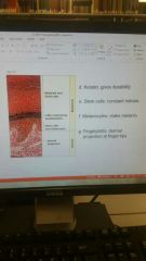
3. Bone |
A. Made by Osteocytes B. Sold matrix: minerals (Ca2+) |
|
|
|
4. Blood |
A. Consists of Formed Elements plus Plasma |
|
|
|
Formed Elements |
Red blood cells, white blood cells, platelets |
|
|
|
Muscular Tissue |
3 types: 1. Skeletal Muscle 2. Smooth Muscle 3. Cardiac Muscle |
|
|
|
1. Skeletal Muscle: Location,Function,Control,Organization |
Location: attached to skeleton
Functions: Movement, posture, and Thermogenesis
Control: Voluntary, motor neurons
Organization - Muscular Fibers (Myotube): Basic Unit -Multiple nuclei/fiber -Striated |
|
|
|
2. Smooth Muscle |
Location: mostly around hollow chambers
Functions: Regulate internal environment (ex. Blood pressure) Control: Involuntary
Organization: - Single cells, one nucleus - Close together or widely spread
|
|
|
|
3. Cardiac Muscle |
Location: Heart
Function: Move blood
Control: Involuntary
Organization: -Striated single cells - One nucleus/cell
|
|
|
|
Nervous Tissue |
Neurons plus Glia |
|
|
|
Neurons |
A specialized cell transmitting nerve impulses, a nerve cell
Function: generate action potentials (AP) and release neurotransmitters (NT)
Organization: 1. Dendrite 2. Axon 3. Nerve Terminal 4. Myelin
|
|
|
|
Dendrite |
Connects with other neurons |
|
|
|
Axon |
Sends (AP) away from cell body |
|
|
|
Nerve Terminal |
Site of (NT) release |
|
|
|
Myelin |
A membrane insulating the Axon |
|
|
|
Glia- 3 kinds |
1. Astroglia (Astrocytes) 2. Oligodendroglia (Oligodendrocytes) 3. Microglia |
|
|
|
Astroglia(Astrocytes) |
Functions: Support neurons |
|
|
|
Oligodendroglia(Oligodendrocytes) |
Function: forms Myelin |
|
|
|
Microglia |
Function: to phagocytize dead cells |
|
|
|
Epithelial Tissue (skin) |
Location: body surfaces A. Apical B. Basal |
|
|
|
Apical |
Surface faces out to aqueous/gaseous environment (Ex. Lining of stomach, lungs) |
|
|
|
Basal |
Surface faces in,contracts basement membrane. |
|
|
|
Epithelial cell types and function. 3 kinds |
1. Squamous 2. Cuboidal 3. Columnar Functions: absorption, secretion, and excretion |
|
|
|
Glands |
Epithelial organs A. Exocrine B. Endocrine |
|
|
|
Exocrine Glands |
- secrete into ducts - secretion directly to apical surface (Ex. Sweat glands) |
|
|
|
Endocrine glands |
- Secrete from cell,picked up by blood - secretion goes through entire body - (ex. Insulin) |
|
|
|
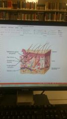
Integumentary System |
Functions: A. Protection: barrier against: infection, loss of compounds (water), and UV light.
B. Senses: pressure, touch, pain, and temperature.
C. Vitamin D Metabolism: precursor activation by UV light
D. Temperature Regulation: Sweat
Organization: a. Epidermis B. Dermis
|
|
|
|
Epidermis |
Outer layer of stratified squamous, epithelium |
|
|
|
Dermis |
Underlying connective tissue, plus epithelial,nerve and muscle cells |
|
|
|
|
|
|
|
Organ systems |
Composed of various organs designed to perform specialized functions. 11 systems function to maintain homeostasis |
|
|
|
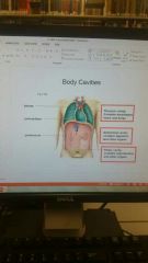
Body cavities |
1. Thoracic 2. Abdominal 3. Pelvic |
|
|
|
Thoracic cavity |
Contains esophagus, heart, and lungs |
|
|
|
Abdominal cavity |
Contain digestive and other organs |
|
|
|
Pelvic cavity |
Contains reproductive and other organs |
|
|
|
Body membranes |
1. Mucous 2. Serous 3. Synovial 4. Meninges |
|
|
|
Mucous membranes |
Cover internal surfaces exposed to air |
|
|
|
Serous membranes |
Cover internal surfaces |
|
|
|
Synovial membranes |
Protect joints |
|
|
|
Meninges |
Protect the central nervous system |
|
|
|
G |
|
|
|
Lecture 2 |
Cardiovascular system: Heart and vessels |
|
|
|
Cardiovascular system: Organization 3 types |
1.Heart: cardiac muscle; pumps blood. 2.Blood vessels: carry blood in circuit to and from heart. 3.Blood transports: a. Plasma: dissolved chemicals b. Formed Elements: cells and platelets -note- both heart and vessels are lined with epithelial cells= endothelium
|
|
|
|
Cardiovascular system: 3 Functions |
*circulates blood throughout the body* 1. Transport chemicals: A. Nutrients, wastes, hormones, ions, etc. 2. Regulate internal environment: A. pH B. Temperature C. Osmolarity 3. Protection: A. Against Infection B. Against blood loss
|
|
|
|
Example |
1. Gas exchange -O2 from air into blood> tissues - CO2 from tissues to blood> out = overcomes limits of diffusion of the gases |
|
|
|
Blood Vessels- 5 kinds |
5 types: 1. Artery 2. Arteriole
3. Capillary 4. Venule 5. Vein
|
|
|
|
Artery |
Carries blood from heart A. Under great pressure B. Thick, muscular walls; smooth muscle |
|
|
|
Arterioles |
A. Carries blood to capillary B. Smooth Muscle sphincters direct flow |
|
|
|
Capillary |
A. Thin walls B. Capillary Bed C. Site of chemical exchange |
|
|
|
Venule |
Connects capillaries and vein A. Low pressure B. Thin walls |
|
|
|
Vein |
Carries blood to the heart
A. Low pressure B. Thin walls C. Valves: prevent back flow D. Bad valves leads to vericose veins |
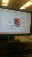
Right Tri, Left Bi |
|
|
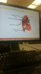
The Heart: Chambers and Valves |
|
|
|
|
The Heart: continued |
|
|
|
|
Blood Flow To and From the Heart |
Superior and Inferior Vena Cava~> Right Atrium~> Right Ventricle~> Pulmonary Arteries~> Lung Capillaries~> Pulmonary Veins (Oxygenated Blood)~> Left Atrium~> Left Ventricle~> Aorta |
|
|
|
Cardiac Cycle- 2 types |
1. Systole 2. Diastole |
|
|
|
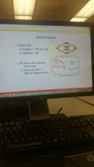
Systole |
Muscle contraction
A. Creates blood pressure B. Measured force due to contraction of left ventricle C. Avg~ 120mm Hg |
|
|
|
Diastole |
Relaxation phase A. Pressure drops B. Measured as pressure in arteries during cardiac relaxation C. Avg~ 80mm Hg |
|
|
|
Cardiac Cycle: Electrical Conductance |
1. Electric signals initiate cardiac muscle contractions = force blood out
2.Conduction Pathway: Sinoatrial (SA) node~> Right and Left Atria~> Atrioventricular (AV) Node~> AV Bundles~> Purkinje Fibers~> Left and Right Ventricle Muscle |
|
|
|
Electrocardiogram (ECG) |
1. Recording of the heart electrical activities at body surface. PWave: atrial depolarization QRS Wave: ventricular depolarization TWave: ventricular repolarization Atrial Fibrillations, Ventricular Fibrillations, Defibrillation |
|
|
|
Heart Rate -4 effects |
1. SA Node 2. Cardiac Center 3. Blood Pressure 4. Other |
|
|
|
SA Node |
SA Node is a pacemaker A. Self-Depolarizes= start cardiac Cycle B. Avg.~ 70 beats per minute |
|
|
|
Cardiac Center |
A. Located in medulla oblongata of the brain B. Sympathetic: activates SA Node= increases rate (fight or flight) C. Parasympathetic: slows SA Node= decreases rate. - rest and repose; relaxed state (ex. After workout) |
Billy Madison |
|
|
Blood Pressure effect on Heart Rate |
A. Increased BP in aorta or carotid arteries slows SA Node= slows rate> decreased BP
B. Increased BP in Right Atrium stimulates SA Node= increases rate= prevents increased venous pressure
|
|
|
|
Other influences on Heart Rate |
A. Circulating chemicals: (ex. Caffeine) B. Temperature: rate affected by body temperature |
|
|
|
Blood pressure- Arteries, Capillaries, Veins |
|
|
|
|
Venous Return |
- Blood in veins under low pressure -Veins have Valves = blood moves in one direction. - Venous Return to heart aided by: 1. Respiratory Pump: inhale causes decreased pressure in thoracic cavity= aids blood to heart 2. Skeletal Muscle Pump: contractions squeeze veins= pushes blood= milking |
|
|
|
Capillaries |
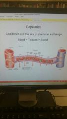
They are the site of chemical exchange |
|
|
|
Atherosclerosis |
Plaque in arteries 1. Effects: blood clots form A. Thrombus in Coronary Artery> heart attack B. Embolus moves to brain> Stroke 2. Causes: high fat diet, obesity, smoking, hypertension. 3. Treatments: arterial bypass, Stent, heart transplant. 4. Prevention: diet, exercise, don't smoke, asprin, floss |
|
|
|
Hypertension |
1. Causes: obesity, diet, smoking, kidney problems. 2. Effects: A. Thickening of arterial walls= reduced flow. B. Kidney failure C. Aneurysm: burst vessel |
|
|
|
Lecture 3 |
Blood and Lymph |
|
|
|
Composition of Blood |
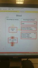
Percentage by volume= 55% plasma, 45% cells and platelets Percentage by weight= 91% water, 7% proteins, 2% other solutions |
|
|
|
Red Blood Cells (Erythrocytes)- Structure and Functions |
1. Structure: a. Biconcave disks b. No nucleus or organelles c. Packed with Hemoglobin (Hb)
2. Functions: a. Transport Oxygen b. Transport Carbon dioxide c. Carbonic anhydrase |
|
|
|
Carbonic Anhydrase |
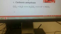
CO2+H2O <<>>H2CO3 <<>>Hplus + HCO3 |
|
|
|
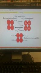
Oxygen and Hemoglobin: Binding |
|
|
|
|
RBC |
1. Formed in bone marrow 2. Lifespan of 120 days; removed by liver and spleen 3. RBC Disorders: A. Anemia: insufficient RBC or Hb B. Sickle cell Anemia: Hb precipitates at low O2. 4. Blood doping: artificial increase in RBC. |
|
|
|
White Blood Cells |
1. Structures: contain nucleus and organelles 2. Functions: Immunity a. *Antigens* 3. Cell types: 2 kinds a. *Phagocytize* b. *Lymphocytes*
|
|
|
|
Antigens |
Foreign material (ex. Bacteria) |
|
|
|
Phagocytes |
Neutrophils and macrophages |
|
|
|
Lymphocytes |
Produce Antibodies |
|
|
|
Platelets |
1. Structures: formed as pieces off Megakaryocytes. a.Make 2 billion a day 2. Function: blood clotting |
|
|
|
Platelets and Blood Clotting |
1. Vessel rupture~> 2. Platelets form plug to slow bleeding~> 3. Platelets and damaged tissue release prothrombin activator~> 4. Prothrombin activator initiates enzyme cascade to throw net over wound to stop bleeding. |
|
|
|
Blood clotting disorders |
1. Thrombocytopenia: lack of platelets. 2. Hemophilia: lack of clotting factor |
|
|
|
Aspirin and Blood Clotting |
1. Reduces platelet aggregation= slows clotting a. Reduce risk of Heart Attack b. May allow for gastric bleeding 2. Acts as an analgesic: reduces inflammation / pain 3. Reduces risk of some cancers and neuro-disorders. |
|
|
|
Blood types |
1. Type A 2. Type B 3. Type AB 4. Type O |
|
|
|
Type A blood |
Has A antigen (Ag) on RBC and Anti- B antibody (Ab) in plasma |
|
|
|
Type B blood |
Has B antigen on RBC and Anti-A antibody in plasma |
|
|
|
Type AB blood |
Has AB antigen on RBC and has no antibody in plasma |
|
|
|
Type O |
Has no antigen RBC and both A and B Ab in plasma |
|
|
|
Blood Transfusions |
Compatibility: 1. Cannot give A blood to B or O 2. Cannot give B blood to A or O 3. Cannot give AB blood to A, B, or O 4. Can give O blood to A, B, AB, and O. *O blood is a universal donor* |
|
|
|
The Rh Factor |
1. Rh Factor: Rh antigen (Ag) on RBC= Rh+ |
|
|
|
Rh Factor and Pregnancy |
1. Problem if Mother is Rh-, but fetus is Rh+ 2. Initial Pregnancy: Rh+ RBC may enter Mother's blood at birth >> vaccinate >> increased Ab against Rh Factor. 3. Later pregnancy with Rh+ fetus: Rh antibody (Ab) from previous pregnancy attacks fetus' blood. |
|
|
|
Lymphatic System - Structure and Functions |
1. Structures: lymph vessels and organs.
2. Functions: a. Returns fluids from tissues to blood
b. Absorb fats in small intestines
c. Produce and maintain lymphocytes; traps and destroys infectious materials = involved in immunity |
|
|
|
Lymphatic Vessels |
1. Closed ened: No Circuit 2. Filled with Lymph Fluid a. Fluid drained from tissues |
|
|
|
Lymphatic Vessels and Organs |
1. Tonsil 2. Lymph Node 3. Spleen 4. Thymus 5. Red Marrow |
|
|
|
Tonsils, Lymph Nodes, and Spleen |
Trap debris in lymph (ex. Bacteria) |
|
|
|
Thymus |
Aid lymphocyte development |
|
|
|
Red Marrow |
Produce blood cells |
|
|
|
Lecture 4 |
Immunity |
|
|
|
Immune system |
The bodies defense against infections organisms and other invaders. |
|
|
|
Pathogens |
Bacteria, Viruses, etc.. that cause disease |
|
|
|
3 Defenses against Invasion |
1. Barriers: Skin and Mucous Membranes 2. Innate immunity: built in defenses; phogocytes and killer cells, protective proteins, inflammatory response 3. Acquired Immunity: adaptive responses; lymphocytes, antibodies |
|
|
|
Barriers to infection |
1. Skin: a. Epidermis is physical barrier b. Sebum, sweat and ear wax have lysozoyme and chemicals to limit the growth of pathogens c. Normal flora on skin exclude harmful microbes 2. Mucous Membranes: a. Mucous protects: traps pathogens for swallowing or excretion, uses lysozoyme b. Normal flora exclude pathogens *note*- Antibiotics and soap can destroy normal flora |
|
|
|
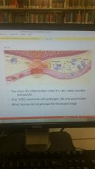
Inflammatory Response |
1. Pathogen enters tissue 2. Mast cells and damaged tissues release histamine= increased blood flow 3. WBC arrive at site a. Neutrophils come first to phagocytize pathogens b. Monocytes enter tissue: - develop into Macrophages, which phagocytize pathogens and release cytokines - Cytokines are proteins that stimulate mitosis = increase response |
|
|
|
Inflammation |
|
|
|
|
Protective Proteins |
1. Complement System 2. Interferons |
|
|
|
Complement System |
a. Serum proteins enhance inflammatory response b. Some bind directly to pathogens to destroy plasma membrane |
|
|
|
Interferons |
a. Released by virus infected cells b. Protects neighbor cells c. Used to treat viral infections (ex. He C) |
|
|
|
Adaptive Immune Defenses - 2 types |
Responses of Lymphocytes to Antigens on surface of pathogens
1. Antibody- meditated immunity 2. T Cell- Meditated immunity |
|
|
|
Antibody- meditated immunity |
a. Antigen on pathogens binds to B Lymphocyte receptors (one type od B cell antigen) b. B cells become: • Plasma cells: secrete Antibodies which bind destroy pathogens - Clonal expansion: fast replication of cells • Memory B Cells: remember Ag to respond at next exposure |
|
|
|
T Cell- Meditated immunity |
a. T Cells activated by pathogen presented by macrophage b. T Cells become: 1. Cytotoxic T Cells 2. Memory T Cells 3. Helper T Cells |
|
|
|
Cytotoxic T Cells |
Binds to virus- infected cells and cancer cells then puts holes in plasma membrane |
|
|
|
Memory T Cells |
Remember for next exposure |
|
|
|
Helper T Cells |
Release cytokines to activate B Cells |
|
|
|
Acquired Immunity 3 parts |
Active immunity: you make Ab against foreign Ag
1. Immunization: Vaccine containing non-active pathogen is injected. 2. Immune response initiated with plasma cells making Ab to destroy Ag = Primary Response 3. Memory B Cells wait for any exposure to active pathogen to respond with even more Ab= Secondary Response 4. Examples: measles, polio, smallpox, tetanus, Hep B |
|
|
|
Hypersensitivity Reactions: Allergies |
1. Allergen 2. Anaphylactic shock 3. Delayed Allergic Response |
|
|
|
Allergen |
A non-pathogen Ag which initiates immune response.
a. 10 times more Ab produced Ex. Cat hair, pollen |
|
|
|
Anaphylactic Shock |
Happens when allergen enters blood a. Excess Histamine>drop in BP>death b. Epinephrine may block effects (Epi-Pen) c. Ex. Bee sting, penicillin shot |
|
|
|
Delayed Allergic Response |
Takes longer due to T Cell activation a. TB test, poison ivy, cosmetics |
|
|
|
Autoimmune diseases- 2 kinds |
1. Rheumatoid Arthritis 2. Multiple Sclerosis |
|
|
|
Rheumatoid Arthritis |
Cartilage components seen as antigens, which starts immune response |
|
|
|
Multiple Sclerosis |
Proteins in CNS myelin seen as antigens |
|
|
|
Infectious Diseases |
1. Epidemic: human spread disease with more cases than expected (ex. swine flu) 2. Pandemic: a global epidemic (ex. malaria) |
|
|
|
Aids/Hiv |
•Acquired Immunodeficiency Syndrome (AIDS) • Human immunodeficiency Virus (HIV)
1. Virus to humans from green monkeys. 2. Infects macrophages and helper T Cells which leads to immune response = infections and cancers 2. Infects macrophages and helper T Cells which leads to immune response = infections and cancers |
|
|
|
Tuberculosis |
•Cause: Mycobacterium tuberculosis • spread in airborne droplets; cough or sneeze • Infects lungs= "consumption" • multiple drugs for treatment • infects 9 million people each year |
|
|
|
Malaria |
• Cause: plasmodium,a prosit passed by mosquitoes • Parasite causes flu like symptoms • more than 350 million cases each year • Treatments: drugs and netting |
|
|
|
Influenza |
•Cause: Virus •Many types: swine flu, bird flu • Treatments: ? • Prevention: vaccines • symptoms: you know them |
|
|
|
Lecture 5 |
Digestive system |
|
|
|
Digestive system- Structure and Functions |
1. Functions: Digestion and Absorbtion of nutrients 2. Structures: A. Alimentary Canal: muscular tube from mouth to anus B. Accessory organs: teeth, tounge, salivary glands, liver, gall bladder, pancreas
|
|
|
|
Basic System Mechanisms |
1. Ingestion- taking in food 2. Secretion- digestive secretions and mucous 3. Motility- movement for mixing and propulsion 4. Digestion- catabolism of complex molecules 5. Absorbtion- nutrients from canal into blood 6. Defication- excretion of waste (****) |
|
|
|
4 layers of the Alimentary Canal |
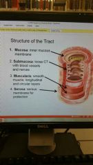
1. Mucosa 2. Submucosa 3. Muscularis 4. Serosa |
|
|
|
Mouth oral cavity |
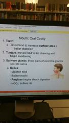
1. Teeth 2. Tounge 3. Salivary Glands |
|
|
|
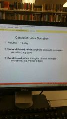
Secretion of Saliva |
|
|
|
|
Pharynx and Esophagus- Swallowing process |
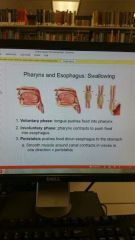
1. Voluntary Phase 2. Involuntary Phase 3. Peristalsis |
|
|
|
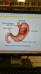
Structure of stomach |
|
|
|
|
Functions and Secretion of stomach |
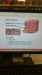
Stomach breaks down food |
|
|
|
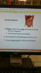
Small Intestine |
|
|
|
|
Suface area and Efficiency |
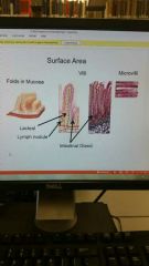
Work together for efficient absorption |
|
|
|
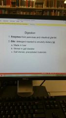
Digestion |
|
|
|
|
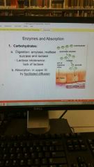
Absorbtion of Carbohydrates |
|
|
|
|
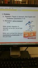
Absorbtion of Proteins |
|
|
|
|
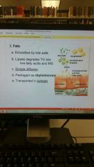
Absorbtion of Fats (Lipids) |
|
|
|
|
Accessory Organs- 3 kinds |
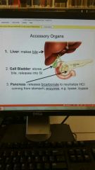
1. Liver 2. Gall Bladder 3. Pancreas |
|
|
|
The Large Intestine |
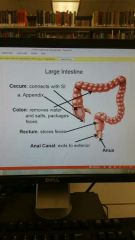
1. Cecum 2. Colon 3. Rectum 4. Anal Canal 5. Anus |
|
|
|
Defication Process |

|
|
|
|
Lecture 6 |
Nutrition |
|
|
|
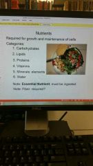
6 Nutrients Categories |
1. Carbs 2. Lipids 3. Proteins 4. Vitamins 5. Minerals 6. Water |
|
|
|
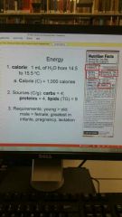
Calorie measurement |
1 mL of H2O from 14.5 to 15.5°C A. Calorie (C)= 1000 calories |
|
|
|
Dietary Sources and Calorie Requirements |
|
|
|
|
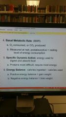
BMR, Specific Dynamic Action and Energy Balance |
|
|
|
|
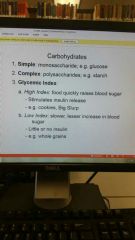
Carbohydrates |
|
|
|
|
High Glycemic Index Related to Blood Sugar and Insulin |
High Index food quickly raises Blood Sugar. High Index food Stimulates Insulin Release. |
|
|
|
Blood Sugar - effects if it drops low |
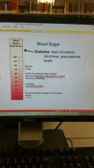
If drops low: 1. Glucagon released, epinephrine, cortisol, sweating, trembling. 2. Lethargy Convulsions, Coma 3 Permanent Brian damage if prolonged, Death |
|
|
|
Dietary Carbs: High Glycemic Index |
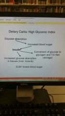
High Glycemic Index= low blood sugar |
|
|
|
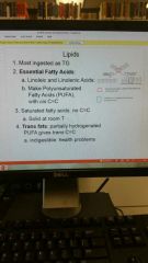
Dietary Lipids |
|
|
|
|
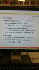
Cholesterol- Source and Transportation |
|
|
|
|
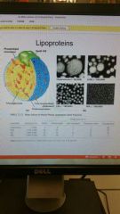
Lipoproteins |
|
|
|
|
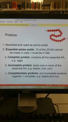
Dietary Proteins |
|
|
|
|
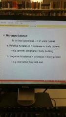
Nitrogen Balance in a Diet |
|
|
|
|
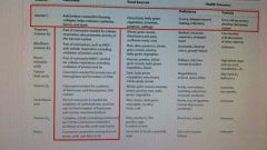
Required Elements |
1. Calcium 2. Sodium 3. Iron |
|
|
|
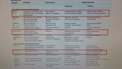
Required Vitamins- Vitamin A |
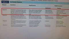
|
|
|
|
Required Vitamins- Vitamin C |
|
|
|
|
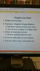
Energy Balance and Body Weight |
|
|