![]()
![]()
![]()
Use LEFT and RIGHT arrow keys to navigate between flashcards;
Use UP and DOWN arrow keys to flip the card;
H to show hint;
A reads text to speech;
141 Cards in this Set
- Front
- Back
|
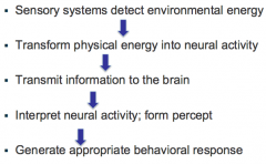
Sensory systems |
Adaptive systems designed to process information from the environment |
|
|
Functional brain systems for processing input |
|
|
|
Sensation leads to... which is ... |
perception: interpretation of sensory signals |
|
|
Johannes Müller |
(1830’s) "Doctrine of Specific Nerve Energies" Each sense has a specific “nerve energy” that generates a specific sensation, different from other senses (proven incorrect). Correctly surmised that sensation was related to sense organ. In fact, each sense has its own specialized receptor organs and pathways, but the “energy” is the same: action potentials. Discovered Labeled Lines |
|
|
Labeled Lines |
different sense modalities use separate nerve tracts. |
|
|
____ pairs of __________ connect directly to the brain
|
Twelve pairs of cranial nerves connect directly to the brain |
|
|
Labeled lines within a sensory domain : |
Different receptor types carry signals related to different stimulus qualities |
|
|
Sensitivity range results from... |
receptor type and threshold |
|
|
Sensory threshold is limited by... |
receptor cell threshold |
|
|
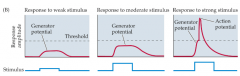
Coding sensory events: intensity |
-Different receptors have different thresholds
-Intensity coded in number, frequency and pattern of action potentials |
|
|
Different receptors code... |
[Within a sensory domain, ] different receptors code different stimulus qualities |
|
|
Common organizational plan across sensory systems |

|
|
|
Sensory neurons... |
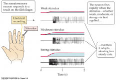
signal change; adapt quickly. |
|
|
McGurk Effect |
Shows that sensory systems interact.
The McGurk effect is a perceptual phenomenon that demonstrates an interaction between hearing and vision in speech perception. The illusion occurs when the auditory component of one sound is paired with the visual component of another sound, leading to the perception of a third sound. |
|
|
Synesthesia |
Shows sensory cross talk. Anomalous blending of the senses Stimulation in one modality produces sensation in another: feel sound, hear color, taste shapes Within modality: letters/numbers colored (consistently so) |
|
|
External organ or structure designed to... |
gather/signal specific type of stimulus energy |
|
|
Receptors specialized for the specific type of energy, they... |
filter energy of a specific range or quality |
|
|
threshold for reception: |
minimum energy required for excitation of sense organ |
|
|
Sensory systems encode the following three: |
quality (e.g. color), quantity (e.g. intensity), source of energy |
|
|
threshold for perception: |
minimum energy required for detection 50% of the time |
|
|
Sensory systems signal "change" : |
adaptation to constant stimulation, unchanging stimulation unimportant to organism |
|
|
Organizational plan is __________________ |
similar across systems |
|
|
Peripheral nerves carry information to ... |
brainstem or thalamus; info is relayed to associated sensory cortex |
|
|
Wilder Penfield |
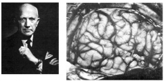
(1891-1976) Mapping of brain function in awake humans |
|
|
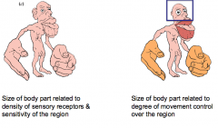
Somatotopic map in primary somatosensory cortex |
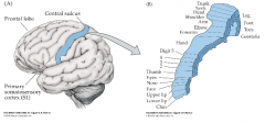
Amount of territory occupied by a given body part is related to its sensitivity |
|
|
Somatosensory Homunculus (“little man”) Size of the body parts of homunculus are related to the ______ & ______ of the region. |
density of sensory receptors & sensitivity of the region |
|
|
___________ differ by species. |
Enhanced areas. e.g. Mouse whisker barrels in S1 |
|
|
Multiple types of somatosensory receptors |
-Vary in form, primary stimulus and action -Mechanoreceptors, thermoreceptors, nociceptors -Firing patterns vary: fast-adapting receptors signal changing conditions, slow-adapting receptors signal ongoing stimulation (stretch) or form information -Fiber types vary in size and myelination; limit speed of information transmission |
|
|
Diversity of somatosensory receptors in the skin. Different receptor types carry... |
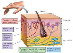
Different receptor types carry signals related to different stimulus qualities. Each type has its own labeled line, transmission speed, sensitivity |
|
|
Primary somatosensory afferents: some are... |
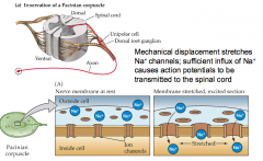
mechanoreceptor. Mechanical displacement stretches Na+ channels; sufficient influx of Na+ causes action potentials to be transmitted to the spinal cord |
|
|
Dorsal Column System (3 steps) |
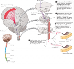
1. Afferents enter at appropriate level of spinal cord; preserving order and location of origin 2. Ascend to brainstem on the same side, synapsing in dorsal root at medulla 3. Postsynaptic cell axons cross midline at medulla |
|
|
Organization of afferent information yields... |
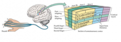
orderly somatotopic maps in cortex. Columnar organization by body part and receptor type |
|
|
Cells in somatosensory cortex have... |
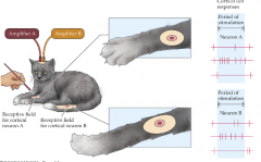
receptive fields on body surface. |
|
|
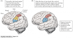
Reorganization of S1 following injury |
|
|
|
Disorders of somatic sensation |
-Hemilateral neglect – reduced or absent awareness of stimuli from one side of the body or space -Agnosia – (common across senses) abnormal perception of sensory stimuli; e.g. may be able to use an object but not identify it by touch -Phantoms – abnormal or excessive sensation from a damaged or missing body part; often painfu |
|
|
Neuropathic pain: |
chronic hyperexcitability or reorganization |
|
|
What is “pain”? |
Unpleasant sensory experience often associated with damaging stimulation Reflex response precedes awareness of painful sensation Reflex is local to the spinal cord; perceptual experience requires the brain |
|
|
Why is pain good? |
Injury avoidance Adaptive avoidance of dangerous food, situations Attract assistance from conspecifics Promote recovery (induce rest/sleep, withdrawal) |
|
|
Congenital Insensitivity to Pain |
Genetic disorder that results from a dysfunctional gene that codes for the particular sodium channel used by “nociceptors” (receptors triggered by extreme/harmful stimuli in the skin) Tend to die young due to repeated, often serious injuries |
|
|
Injury to skin causes... |
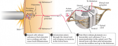
release of chemicals that stimulate free nerve ending |
|
|
Nocireptors |
free-nerve endings with receptors that signal noxious, potentially damaging stimulation. Different varieties have different receptor types; use different chemical transmitters. Sensitivity of receptors determines threshold for action potentials |
|
|
Spinothalamic system does what? |
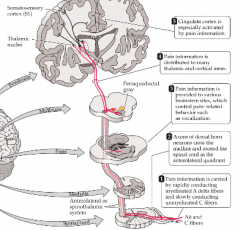
carries pain information to the brain. Fast (A delta) & slow (C fibers) conducting fibers carry information to the brain: two stages of pain sensation. Axons cross midline at point of entry in the spinal cord |
|
|
Pain is in the brain |
Expectation changes subjective experience of pain, and activation in cingulate cortex (Rainville et al., 1997) |
|
|
Endogenous opiates – Endorphins – are released from PAG in response to pain |
Opiate receptors are found throughout the brain: Limbic system (emotion and motivated behavior), hippocampus (learning and memory), thalamus (sensory relay nucleus). Endogenous ligands(endorphins) have been identified; mediate pain relief; Exogenous ligands (morphine, heroin) bind to opiate receptors |
|
|
Placebo effect |
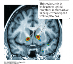
Can be blocked by naloxone – antagonist of endorphin receptors |
|
|
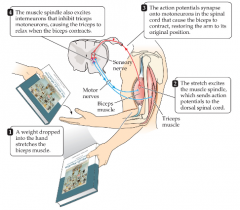
Reflex Action (the simplest circuit) |
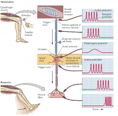
Sensory input triggers motor response. Speed of transmission: 100 m/s, total time ~40ms |
|
|
Sir Charles Sherrington |
(1932) Nobel prize for work on reflex action as well as concept of neural threshold and summation properties of the synapse. Reflexes, motor plans |
|
|
Reflexes |
simple, stereotypic, unlearned responses to external stimuli; typically involve a single body part |
|
|
Motor plans |
complex, sequential movements; typically purposeful and involving multiple body parts |
|
|
Construction of ____________ limit type of possible movements |
skeleton and joints
|
|
|
Movement results from _______of muscle Muscle types differ by _____ and _____. |
from stimulation differ by location and task Smooth muscle – smooth appearance, found in digestive system & circulatory system, not under voluntary control Striated muscle – striped appearance, skeletal muscles & cardiac muscle; skeletal muscles under voluntary control |
|
|
Muscular dystrophy |
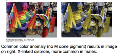
progressive weakening, degeneration of skeletal muscles; single-gene, X-linked disorder, affects young boys |
|
|
Pattern of attachment of muscle to bone determines range of action & pattern |
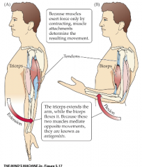
Stimulation of muscles results in specific movements Muscles act by contracting Limbs controlled by antagonistic sets of muscles: flexors, extensors Muscles contract when stimulated by motoneurons in spinal cord, cranial nerve ganglia |
|
|
Alpha motor neurons in the ventral horn receive many inputs, axons synapse on muscle fibers |
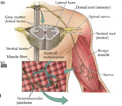
Alpha motor neurons receive local inputs as well as descending inputs from brain Axons branch widely at muscle; Stronger stimulation (more active inputs to muscle) results in stronger contraction |
|
|
ALS - Lou Gehrig's disease |
Degeneration of large motor neurons; progressively affects voluntary muscle control, eventually affects respiration |
|
|
Neuromuscular junction (NMJ) |
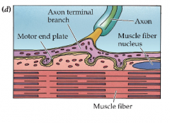
ACH (acetylcholine) is released at NMJ; depolarizes muscle fiber, triggers action potentials in muscle fibers. Muscle fibers have many nicotinic cholinergic receptors. |
|
|
Myasthenia Gravis |
is a type of vulnerable synapse. M.G is an autoimmune disease, attacks nicotinic receptors in muscle fibers. Toxins affect all stages of transmission of ACH (acetylcholine) |
|
|
Proprioception: Its two types of receptors are: |
sensory feedback from muscles, tendons, joints Proprioceptors provide information about where our limbs are, our posture, and what the state of each muscle is; local feedback for motor system The two types of receptors are muscle spindle and Golgi tendon organ. |
|
|
Muscle spindle |
senses passive stretch of muscle to aid in posture and control; |
|
|
Golgi tendon organ |
detects muscle tension, guards against excessive force on muscle by inhibiting alpha motor neurons |
|
|
Sensory-motor feedback control |
|
|
|
Motor commands arise in... |
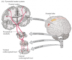
Motor commands arise in primary motor cortex, M1 |
|
|
Somatosensory & Motor Homunculus |
|
|
|
Cells in M1 change ___________ for specific movements. |
Cells in M1 change firing rates for specific movements |
|
|
Mirror neurons in premotor cortex |
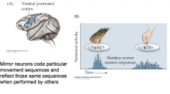
Mirror neurons code particular movement sequences and reflect those same sequences when performed by others |
|
|
Additional cortical motor areas aid in _______ and ______ of motor sequences |
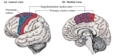
coordination and initiation Additional cortical motor areas like : Supplementary Motor Area (SMA), Premotor cortex |
|
|
Supplementary Motor Area |
important for movement initiation, movement sequencing |
|
|
Premotor cortex |
important for coordination of movement (hands, legs); may code motor behaviors; contains population of "mirror" neurons |
|
|
Motor control hierarchy - motor cortex modulated via other cortical areas |
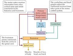
Descending motor control is necessary for planned movements. |
|
|
Motor loop between M1, basal ganglia, thalamus, and SMA modulates... _____ |
movements. Circuit enhances planned movements, allows intended movements to be executed |
|
|
Disorders in this motor loop of m1, basal ganglia, thalamus and SMA results in which two diseases? |
Parkinson’s disease Degeneration of brainstem dopaminergic neurons. (inability to initiate movements; show tremors) results from degeneration of dopamine neurons in substantia nigra. Huntington’s disease (inability to inhibit movement; cognitive impairment) genetic disorder characterized by degeneration of basal ganglia nuclei, GABAergic cells. |
|
|
Parkinson’s disease |
Degeneration of brainstem dopaminergic neurons. |
|
|
Auditory stimulus: sound waves The auditory organ (ear) has which three parts? |
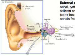
External, middle, inner ear |
|
|
External ear |
Pinna, ear canal, tympanic membrane – collects and focuses sound for better localization; enhances certain frequencies |
|
|
Middle ear does.... |
compresses and amplifies sound. Mechanical transfer of sound waves from tympanic membrane (eardrum) to inner ear; amplifies ~ 30X. |
|
|
Ossicles |
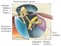
chain of 3 small bones moved by movement of eardrum, create movement of ‘Oval window’, moves fluid in inner ear. In the middle ear, muscles (tensor tympani; stapedius) damp ossicles to prevent damage to receptors from loud & self-made sounds. |
|
|
Inner ear (cochlea) contains... |
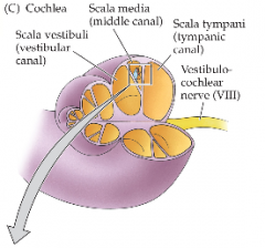
contains auditory receptors. Cochlea is coiled structure ~4mm long; 3-4 cm uncoiled 3 Fluid-filled chambers Scala media contains Organ of Corti: receptor cells (hair cells), basilar membrane, tectorial membrane Mechanical transduction process |
|
|
The two types of hair cells in the ear's scala media's organ of corti are: |
Inner and outer hair cells. Inner is the least numerous but most responsible for sound reception. Outer hair cells are the most numerous but don't do anything for sound reception. |
|
|
Exposure to very loud sound damages... |
hair cells |
|
|
Sound reception is a _____ process. |
Mechanical process. Hair cells transduce movements of the basilar membrane into electrical potentials Displacement of basilar membrane sets up shearing between hair cell stereocilia and tectorial membrane (overlies Organ of Corti) Shearing causes ion channels in stereocilia to open (tip links); K+ions enter cell, depolarizing hair cell membrane Depolarization opens Ca++ channels in base of hair cell, resulting in release of neurotransmitter (IHC – glutamate; OHC – ACH) |
|
|
Hermann von Helmholtz |
(~1850) Electrical conduction: measured speed of electrical conduction in frog nerve: 40m/s Found it was not as fast as wire conducts; so must be an active biological process Resonance theory of hearing: Auditory system decomposed sounds into basic frequency components which were represented in different places along the basilar membrane. |
|
|
Georg von Békésy |
(1890 – 1972) Nobel Prize, 1961, research on the function of the basilar membrane; demonstrated organization hypothesized by Helmholtz Dissected cochlea of cadavers Observed a tonotopy of the basilar membrane; high frequencies created maximal displacement near base (closest to oval window), and low frequencies created maximal displacement near apex (far end) Basis for “place theory” of pitch perception |
|
|
Basilar membrane _______________ at ____________ by sound frequency |
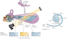
displaced maximally at different places |
|
|
Auditory nerve fibers are organized by... |
‘best frequency’ Auditory neurons encode frequency and intensity receptive fields are frequency tuned; intensity coded by number of neurons firing and duration of firing Pattern of firing across all auditory nerve fibers reflects frequency content of input and sound intensity (quality & quantity) Axons leaving the cochlea form VIIIth cranial nerve; goes to cochlear nucleus in brainstem |
|
|
Auditory pathways: cochlear nucleus are _______, higher areas are _______ |
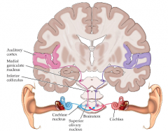
cochlear nucleus monaural, higher areas binaural Auditory information is processed at multiple levels of the brainstem before reaching inferior colliculus and thalamus Each stage of processing has a “tonotopic” map Stage after cochlear nucleus: information from two ears are combined (“binaural”) |
|
|
Coding of auditory space is done through: |
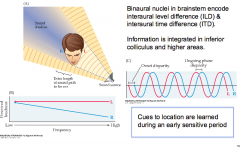
interaural time differences, and intensity differences Binaural nuclei in brainstem encode interaural level difference (ILD) & interaural time difference (ITD). Information is integrated in inferior colliculus and higher areas. Cues to location are learned during an early sensitive period |
|
|
Auditory-visual space mapping is.... |
calibrated during development . Plugging one ear during early life causes systematic mislocalization; young birds adapt; adult birds do not adapt Shifting visual input causes re-mapping of registration between auditory and visual space maps, only in young birds |
|
|
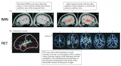
Tonotopic representation preserved in cortex in different species |
Auditory cortical neurons reflect, interpret biologically relevant sounds (speech; species typical calls; predator sounds, stimulus sweeps) No apparent map of auditory space, except laterality; basis exists for computation |
|
|
Comparison of auditory responses to noise or speech |
|
|
|
Three main types of deafness: |
Conduction deafness: sound stimuli do not reach cochlea (e.g. ossicles fused, or tymapnic membrane ruptured) Sensorineural deafness: disorders of inner ear or auditory nerve. Could reselt from damage to hair cells (loud stimuli, toxins like some antibiotics), nerve degeneration, genetic disorders Central deafness: damage to cortical areas involved in analysis of sound and speech |
|
|
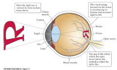
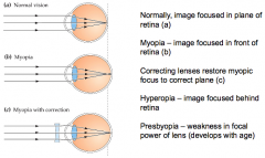
Structure of the human eye, collector of light energy |
|
|
|
Cornea and lens focus incoming light |
|
|
|
Retina has many cell layers; Visual receptor cells at the _____ of eye |
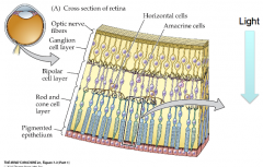
at the back of eye |
|
|
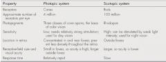
Visual receptors (photoreceptors): the two types are |
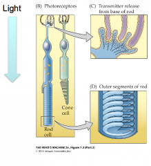
Rods and cones Chemical transduction process Photopigment molecules in discs of rods and cones. Incoming light of the right wavelength activates pigment molecules in discs. Light activation hyperpolarizes receptor cells; release less neurotransmitter at the synapse |
|
|
Light capture ___________ photoreceptors (Na+ channels close) |
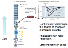
hyperpolarizes them. Light intensity determines the degree of change in membrane potential Photopigment in rods: Rhodopsin. Different opsins in cones. |
|
|
Sensitivity range result from _______ ____ and_________ |
receptor type and threshold. Different receptor types have pigments sensitive to different wavelengths of light. |
|
|
Sensitivity of different receptor types determines range of ________________. |
light sensitivity |
|
|
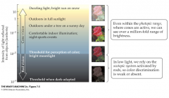
Rods and cones extend the range of visual sensitivity |
|
|
|
Photoreceptor distribution is _____ across the ______. Cone peaks in the ______. |
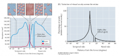
Photoreceptor distribution is uneven across the retina. Cone peaks in the fove. High density of cone photoreceptors in fovea is the basis for high acuity in fovea. |
|
|
Fovea |
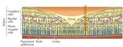
area of retina where acuity is highest, direct light access to receptors. |
|
|
Blind spot |
anglion cell axons leave eye and form optic nerve |
|
|
Where does information from the retina go? |
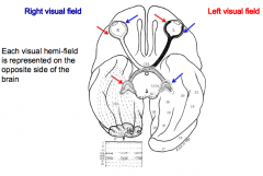
Ganglion cell axons form optic nerve; nasal ½ cross at optic chiasm Each visual hemi-field is represented on the opposite side of the brain |
|
|
Representation of the visual fields |
|
|
|
Location and contrast based on “receptive field” properties What is receptive field? |
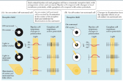
Receptive field: an area of sensory space within which stimulation excites the neuron [Each retinal bipolar cell and ganglion cells has a concentric receptive field, with antagonistic center and surround. Bipolar cells respond with changes in local membrane potentials, while ganglion cells respond with action potentials. ] |
|
|
Ganglion cell axons project to the thalamus ______ |
Lateral geniculate nucleus (LGN) |
|
|
LGN receptive fields are similar to ______ cell inputs |
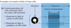
ganglion cell |
|
|
LGN cell axons project to ______ ______ ____ the ____ ________. |
LGN cell axons project to primary visual cortex (striate, V1, A17) – optic radiations |
|
|
V1 |
Primary visual “striate” cortex |
|
|
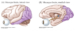
Visual areas occupy ~55% of neocortex in monkey, 30% in human |
|
|
|
Tatsuji Inouye |
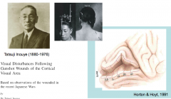
(1909) First to map cortical representation of visual space |
|
|
Non-uniform representation of visual field in primary visual cortex |
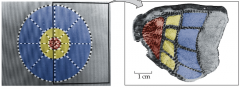
Area of highest acuity has the most receptors and the largest representation in the brain |
|
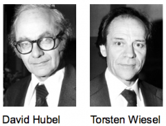
Hubel & Wiesel |
(1981) Visual cortical cells encode basic stimulus properties Binocular organization of visual cortex a substrate for stereopsis “feature detectors” in visual cortex respond preferentially to particular stimulus properties: orientation, motion direction, size, color |
|
|
Receptive fields of V1 neurons binocular, selective for stimulus properties |
Some V1 cells signal orientation, others signal direction of motion |
|
|
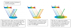
Object color depends on reflected light |
|
|
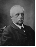
Hermann von Helmholtz |
(~1850) Electrical conduction Resonance Theory of hearing: Frequency components of sound resonate at different locations on basilar membrane Trichromatic theory of color vision: -Three separate kinds of color receptors will be found (blue, green, red peaks) -Each receptor type has a “labeled line” to the brain -All perceived colors can be understood based on activity of 3 receptor types |
|
|
Visual system encodes color in ... |
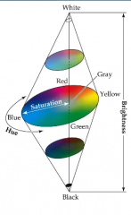
3 dimensions Hue – “color”, based on wavelength Saturation – Strong to flat (gray) Brightness – Lightness (light-dark |
|
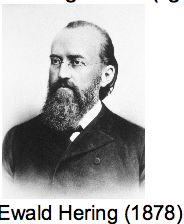
Ewald Hering |
(1878) Opponent-process theory All colors result from 4 unique hues (BYRG) 3 opponent processes: B/Y, R/G, Bk/Wh Explained much subjective experience of color |
|
|
Spectrally opponent cells aid in color discrimination |
Organization found at many levels of the visual system; begins with ganglion cells in retina |
|
|
Perceived color depends on... |
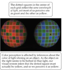
context. |
|
|
Color perception has basis in both trichromacy & opponent process. |
Relative activation of different photoreceptor types and balance of excitation in color opponent receptive fields determine inputs to cortical neurons that interpret the signals as perceived color. |
|
|
Color anomalies reinforce the importance of photopigments |
|
|
|
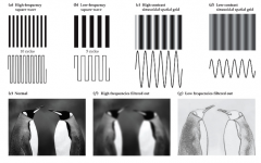
Visual system encodes size and contrast as well as color, luminance and position, |
|
|
|
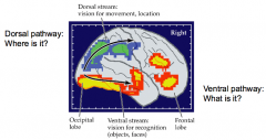
Two functionally distinct pathways beyond primary visual cortex |
|
|
|
Dorsal stream specialized for |
motion and spatial location |
|
|
Fusiform gyrus represents... |
faces. Prosopagnosia – inability to recognize faces; disorder linked to damage to fusiformgyrus |
|
|
Memory is... |
The retention over time of •learned (experience-dependent) behavior •newly acquired information/ knowledge, or the capacity to reactivate or reconstruct it From a neural-basis point of view: •The retention/maintenance of experience-dependent changes in a neural system’s response to the environment •The retention/maintenance of experience-dependent internal-representations Learning and memory must be investigated at multiple levels; modern neuroscience regards the representational level as a must |
|
|
Memory is a ________ that occurs __________. |
Memory is a process that occurs over time. |
|
|
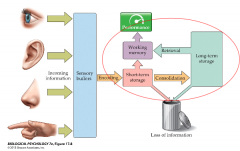
How are memories formed and stored? The threes steps: |
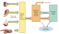
Encoding -> Consolidation -> Retrieval. |
|
|
Consolidation |
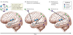
•How and where are memories stored for the long term? –HM could recall events prior to surgery without hippocampus •Systems Consolidation theory: Reorganization of memory trace in the brain |
|
|
Reconsolidation |
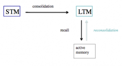
Retrieval of memories from LTM renders them susceptible to change |
|
|
Three types of models for reconsolidation |
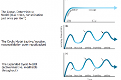
Linear Deterministic, Cyclic, Expanded Cyclic |
|
|
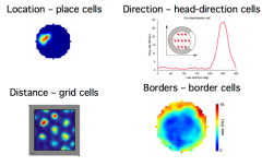
Spatially-tuned cells together provide for a sense of spaceand the knowledge to navigate space |
|
|
|
Kant said... |
If Kant is right then spatially-tuned cells should emerge from development without the need for experience (they are in fact the substrate for experience) |
|
|
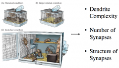
Experience-Dependent Neural Changesin STRUCTURE |
|
|
|
Experience changes... |
dendritic complexity i.e. dendrites GROW and ELABORATE |
|
|
Synapses are an the ends of DENDRITIC SPINES, which can be visualized in living brains Dendritic spines can enlarge and shrink if they are..... |
stimulated by neurotransmitter |
|
|
How do the cells “remember” where to fire?What changed in the brain to encode memory and allow it to persist for months and longer? |
|
|
|
Donald Hebb |
(1904-1985) How do neurons “remember” whether or not and when to be active? Due to activity-dependent synaptic plasticity. |
|
|
The Neural Basis of Learning & Memory |
Central tenets of neuroscience: •Learning involves changes in the strength of connections between neurons in the network: synaptic plasticity. The synaptic changes lead to modifications in the neural circuit properties, which in turn lead to new behavior •Synaptic plasticity is activity-dependent; it is induced at appropriate synapses during learning/memory formation, depending on activity in the pre- and post-synaptic neurons (and possibly on other factors) •Synaptic plasticity is both necessary and sufficient for the information storage underlying the type of learning mediated by the brain area in which that plasticity is observed. |
|
|
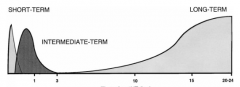
Temporal Domains of Memorydefined by BIOLOGY |
Short-term Memory (STM): Post-translational modifications •Alteration of existing proteins (e.g. phosphorylation) to cause conformational change, alter net charge, etc. •Intermediate-term Memory (ITM) –Translation •New Protein Synthesis •Long-term Memory (LTM) –Translation & Transcription •New Protein Synthesis and Synthesis of New Message (mRNA) |
|
|
Is there any advantage to having short-term memory sometimes convert to long-term memory? Is there any advantage to using proteins for memory storage? |
|