![]()
![]()
![]()
Use LEFT and RIGHT arrow keys to navigate between flashcards;
Use UP and DOWN arrow keys to flip the card;
H to show hint;
A reads text to speech;
73 Cards in this Set
- Front
- Back
|
Neuroethology |
is a multidisciplinary field combining neurobiology andethology (the study of behavior in natural conditions). |
|
|
most common animals used in neurobiology lab |
•Mouse(Mus musculus), zebrafish (Danio rerio), fruit fly (Drosophila melanogaster),and nematode (Caenorhabditis elegans) |
|
|
Microcephaly |
single-genemutation (abnormal spindle-like microcephaly-associated protein) |
|
|
TransgenicAnimals |
:insertion (“knock-in”) or deletion (“knock-out”) of genes in the genome |
|
|
Ramón y Cajal (Spanish, born in 1852): |
•Neuroanatomist who investigated the microscopic structure of thebrain. •Father of modern neuroscience. |
|
|
The'neuron doctrine' |
:relationship between nerve cells is not continuous but contiguous (neurons as'processing units'; neurons: Heinrich Wilhelm Gottfried von Waldeyer-Hartz) |
|
|
what are the two cell types in the nervous system? |
Neurons and glial cells (glia) |
|
|
neurons |
functional cells, fast and long-distance signaling● 100 billion in the human nervous system |
|
|
Glial cells (glia) |
support cells about 3-times the number of neurons |
|
|
name 4 parts of a neuron |
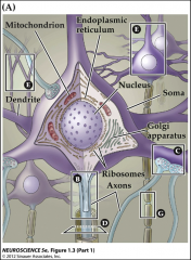
Dendrites – inputCell Body – integration, transcriptionAxon – conductionTerminal– transmission |
|
|
Memorize everything on slide 18-21 of lecture 1 |
|
|
|
what do glia cells do? |
•Maintainingthe ionic milieu•Controllinguptake and metabolism of neurotransmitters•Providingscaffolding for neuronal development•Aidingrecovery from neuronal injury |
|
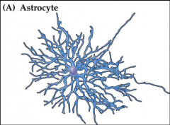
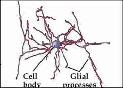
Astrocytes |
•Elaborateprocesses•Inbrain and spinal cord•maintainoptimal chemical environment for signaling•subsetsfunction as stem cells |
|
Oligodendrocytes |
•restrictedto the central nervous system•providea laminated, lipid-rich wrapping called myelin•theyare called Schwan cells in the peripheral nervous system•subsetsfunction as stem cells for oligodendrocytes and Schwann cells•Peripheralaxonensheathed bymyelin (red) except at node of Ranvier. Green labels ion channels in the node;bluelabels paranode. |
|
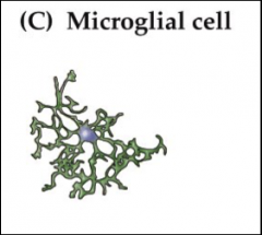
Microglialcell |
•mostlyderived from hematopoietic precursor cells•shareproperties with macrophages•functionas scavenger cells that remove cellular debris during cell turnover and injury•secretecytokines that modulate inflammation and influence cell survival and death•verymobile: migrate within brain to injured areas |
|
|
Stemcells |
proliferateand make othercell types |
|
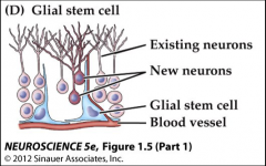
glial stem cells |
•foundthroughout adult brain and differentiated (adult) glia•somebecome differentiated neurons. These stem cells exist as astrocytes near ventricles in subventricular zone or near ventricular zone bloodvessels |
|
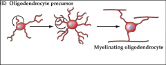
Oligodendrocytes precursors |
•scatteredthroughout white matter•becomemature oligodendroglialcells and some astrocytes |
|
|
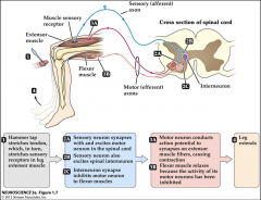
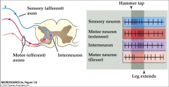
knee-jerk |
|
|
|
how to record the knee-jerk ExtracellularElectrical Recording |
|
|
|
how to record the knee-jerk IntracellularElectrical Recording |
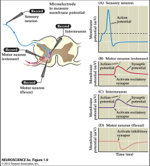
DetectsSmaller Graded Potentials in Neurons |
|
|
why is usinga “reporter gene” helpful |
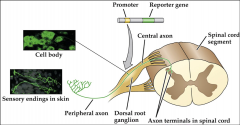
(GFP,green florescent protein) under the control of a cell-specific promoter gene toreveal only one cell type in the nervous system |
|
|
ReceptiveField (ref. lec. 2 slide 15) |
region in sensory space within which a specific stimuluselicits an action potential response(the receptive field represents all (sensory) inputs to aneuron. |
|
|
Sulcus(pl. sulci) |
depressionor groove in thecerebral cortex. |
|
|
Gyrus(pl. gyri) |
ridgessurrounding the sulci. |
|
|
Fissures |
largefurrows that divide the brain into lobes. |
|
|
X-Ray Computerized Tomography (CT) |
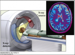
•basedon rotating x-ray beam and detectors•anatomicalimaging with a spatial resolution of millimeters |
|
|
Magnetic Resonance Imaging (MRI) |
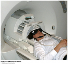
•basedon behavior of atoms in a strong magnet•typicallyimages watercontent for anatomy and deoxyhemoglogin for function (changesin blood flow)•sub-millimeterspatial resolution |
|
|
MRI plus Functional Magnetic Resonance Imaging (fMRI): |
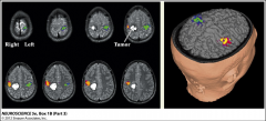
•forimaging neuronal activity using localized blood oxygenation level-dependent(BOLD) changes•exampleshows neuronal activity during hand movement |
|
|
Positron Emission Tomography (PET) |
•unstablepositron-emitting isotopes are detected using gamma ray detectors•Fluorodeoxyglucose (FDG,an analogueof glucose)is typically used as tracer.•The concentrationsof tracer imaged indicate tissue metabolic activity by virtue of the regional glucoseuptake•versatilefunctional measurements•2 to3 millimeter spatial resolution•temporalresolution in the seconds•oftencombined with X-Ray CT |
|
|
Magnetoencephalography (MEG) |
•measureselectrical signals due to the magnetic fields they produce•veryfast (millisecond temporal resolution)•littleanatomical detail•similarto electroencephalography (EEG) |
|
|
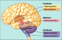
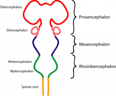
Major Subdivisions of the Central Nervous System |
|
|
|
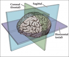
Anatomical terms of location: Planes |
|
|
|
Sagittalplane |
parallelto sagittal sulcus, divides body in left and right |
|
|
Coronal(frontal) plane |
dividesbody in back and front |
|
|
Horizontal(axial or transverse) |
•dividesbody in head and tail portions |
|
|
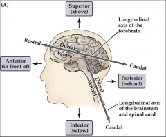
Anatomical terms of location: Axes |
|
|
|
•Dorsal: |
back |
|
|
ventral |
belly |
|
|
caudal |
tail |
|
|
cranial |
front |
|
|
Major Subdivisions of the Central Nervous System |
|
|
|
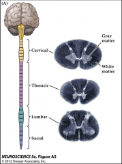
gray matter |
cellbodies and neuropil(dense tangle of axons and dendrites) |
|
|
white matter |
– myelinatedfiber tracts |
|
|
spinal cord pictures |
|
|
|
sinal cord pictures |
|
|
|
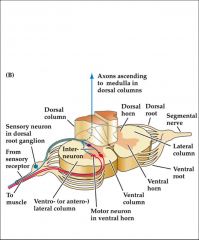
Ganglion(pl. ganglia) |
collectionof cell bodies |
|
|
Dorsalroot ganglia |
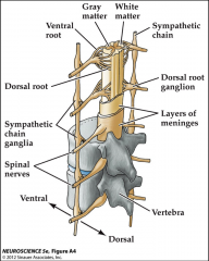
occuras chain along side of spinal cord, contain cell bodies of sensory neurons |
|
|
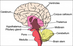
Medullaoblongata |
•resembles spinal cordin anatomy•control of breathingand cardiovascular system |
|
|
Pons – “bridge”: |
•massive enlargement•connects cerebrumwith cerebellum |
|
|
Midbrain: |
•control of eyemovement•essential auditoryrelay |
|
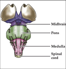
Brain stem general function |
•target or source ofcranial nerves•throughway forascending and descending tracts (brain <> spinal cord)•reticular formation(regulates level of consciousness)•Breathing,circulation, digestion, swallowing, etc. |
|
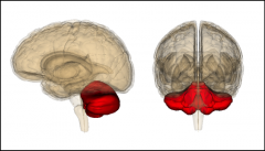
cerebellum general function |
•Coordinates andregulates motor activity•Equilibrium ,posture, motor learning (e.g. bicycling, etc.) |
|
|
granule cells |
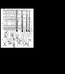
refer to let 3 slide 6 |
|
|
purkinje cells |
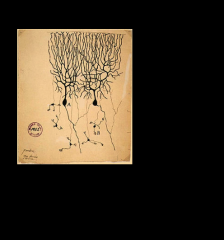
refer to let 3 slide 6 |
|
|
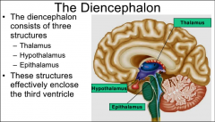
diencephalon |
|
|
|
Thalamus: |
•Major sensory relayarea |
|
|
Hypothalamus: |
•Integration ofautonomic nervous system•Reproduction,feeding, temperature regulation•Links nervous andendocrine systems (pituitary gland) |
|
|
Epithalamus: |
•Includes pineal gland•Connects limbicsystem with other parts of the brain•Circadian rhythms,emotion, motor pathways |
|
|
diagram of all |

|
|
|
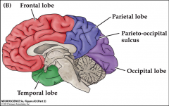
Cebrebral Cortex |
Functional Division:•Primarysensory and motor cortex:unprocessed information•Associationcortex: highly processedinformation |
|
|
frontal lobe |
planning, primarymotor cortex |
|
|
Parietal lobe |
attention, primarysomatosensory cortex |
|
|
Temporal lobe |
recognition, primaryauditory cortex |
|
|
Occipital lobe |
primary visual cortex |
|

Cortex |
•covers the entirecerebrum•Highly folded•gyrus is a crest, asulcus is a groove•Most cortex isneocortex with 6 cell layers•fewer cell layers in allocortex (olfactory systemand hippocampus) |
|
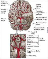
Circle of Willis |
mayhelp maintain perfusion of the brain when on the major arteries is occluded |
|
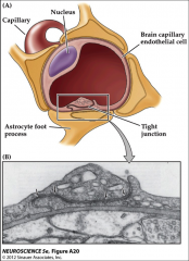
Blood-Brain Barrier |
•Protects the brainfrom toxins circulating in the bloodstream•Consists of tightjunctions between brain capillary endothelial cells and astrocyte footprocesses•Lipophilic moleculesreadily pass; hydrophillic molecules requiretransporters to pass••Some toxins can pass:•MPTP (MPPP: morphine)••Some bacteria gainaccess to the brain by attacking the endothelium:•Borrelia: Lyme disease•B streptococci:Meningitis•T.pallidum: Syphilis • |
|
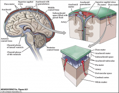
Meninges |
Three protective layers (membranes) of the central nervoussystem |
|
|
•Dura matter: |
“hard mother”, tough and thick, outermost |
|
|
Arachnoid mater |
“spider-like” processes, middle |
|
|
Pia mater |
“tender mother”,delicate, closely adhering to the brain |
|
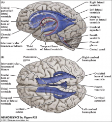
Ventricular System |
•Four, interconnected,fluid-filled spaces••Produces and isfilled with cerebrospinal fluid••Continuous with thecentral canal in the spinal cord |