Reading...
![]()
Play button
![]()
Play button
![]()
Use LEFT and RIGHT arrow keys to navigate between flashcards;
Use UP and DOWN arrow keys to flip the card;
H to show hint;
A reads text to speech;
300 Cards in this Set
- Front
- Back
|
In horses anesthetized with halothane or isoflurane, dobutamine administration may result in:
|
reflex bradycardia
|
|
|
What is the primary reason for fasting prior to anesthesia/
|
Minimize extent of lung collapse.
|
|
|
What are some of the effects of using atracurium during intraocular surgery in horses?
|
1. Paralysis of extraocular mm, keeps eyes in central position in the orbit.
(Does not affect pupil size, or lacramation). Also has no analgesia. |
|
|
Which cranial nerves are blocked with a properly placed Peterson eye block?
|
III, IV, V and VI
|
|
|
What is the one of the most common ocular disease in horses? Characterized by episodes of active inflammation followed by varying periods of quiescence.
|
Equine recurrent uveitis
|
|
|
What is the most common cause of blindness in horses worldwide?
|
Equine recurrent uveitis (due to the changes secondary to chronic inflammation)
|
|
|
What is the cause of Equine recurrent uveitis?
|
many potential initiating causes. is an immune mediated disease. common denominator is damage to the uveal tract, which may be initiated by trauma (both penetrating and blunt) or systemic diseases.
|
|
|
What are some of the specific conditions or agents which are implicated in equine recurrent uveitis?
|
leptospirosis, brucellosis, strangles (Streptococcus equi infection), onchocerciasis, equine influenza, tooth root abscess, hoof abscess.
|
|
|
What is the most widely investigated of the specific causes of equine recurrent uveitis?
|
Leptospira spp. in particular l interrogans serovar pomona.
|
|
|
What is the relationship between equine recurrent uveitis and lepto?
|
unknown. active uveitis does not occur often for months or even years after systemic disease. .
|
|
|
Why might active episodes of uveitis be seen after routine deworming?
|
dead or dying microfilaria that have aberrantly migrated to the eye. (onchocerciasis)
|
|
|
What is the pathogenesis of equine recurrent uveitis?
|
inflammation causes secondary side effects: releaseof inflammatory mediators like leukotrienes, prostaglandins, histamines causes increased permeability of the anterior uveal vessels, breakdown of the blood-aqueous barrier, iris sphincter spasm, and ciliary body muscle spasm. also, leakage of barier allows leakageof protein, fibrin, and cells into the aqueous, leading to blepharospasm, epipora, episcleral injection, corneal edema, aqueous flare, fibrin clots in the anterior of the chamber, and miosis.
|
|
|
Why is a complete eye exam so important to pre-purchase?
|
horses with chronic uveitis can have few or no anterior segment signes, but may manifest equine recurrent uveitis by retinal degeneration. fundoscopy cannot be overemphasized. such horses usually have normal PLR, and may not exhibit overt signs of visual compromise until late in the disease.
|
|
|
Once equine recurrent uveitis is identified, what treatment options are there?
|
topical and systemic anti-inflammatory meds are used ot minimize damage associated with intraocular inflammation.
|
|
|
For equine recurrent uveitis, which steroids work best?
|
Prednisone and dexamethasone are preferred to hydrocortisone, which penetrates the cornea poorly and is not sufficiently potent to be effective in anterior uveitis.
|
|
|
What medication might be used in anterior uveitis to paralyze the iris sphincter and ciliary body musculature?
|
Topical atropine (1% solution or ointment) This will dilate the pupil.
|
|
|
What medicine given systemically might be the single most effective treatment in anterior uveitis of horses?
|
flunixin meglumine (iv)
|
|
|
What are some potential problems associated with long term flunixin meglumine use in horses?
|
GI (ulcers) and hematologic complications. This is why in anterior uveitis fm is used as loading, then treatment is often switched to phenylbutazone or aspirin.
|
|
|
Flunixin is generally thought to be contraindicated in what species?
|
CATS
|
|
|
What are two surgical procedures that are sometimes used in the treatment of chronic recurrent uveitis in horses?
|
core vitrectomy and cyclosporine implant.
|
|
|
What viral respiratory infections are common to horses?
|
equine herpesvirus type 4 (EHV-4, rhinopneumonitis), equine influenza, and equine viral arteritis.
|
|
|
What are the clinical manifestations of equine viral respiratory diseases?
|
All are similar: pyrexia, serous nasal discharge, submandibular lymphadenopathy, anorexia, cough.
|
|
|
What is the newly recognized zoonotic disease of horses identified in Astralia?
|
Hendra virus. rapidly fatal in horses, close contact necessary for spread.
|
|
|
What is the most common opportunistic bacteria of the equine lung? What are other commonly isolated opportunistic pahtogens?
|
Streptococcus equi zooepidemicus. (although Actinobacillus equuli, Bordetella bronchiseptica, E. coli, Pasteurella and Pseudomonas are also frequently isolated)
|
|
|
vaccines are available for what respiratory infections?
|
equine influenza, viral rhinopneumonitis, equine viral arteritis and strangles.
|
|
|
What diagnostic tests are indicated in each of the following situations?
1. upper airway noise, inspiratory difficulty, poor exercise performance, and unilateral or bilateral nasal discharge 2. facial deformity, abn in sinus, gutteral pouch, and soft tissue structures (soft palate, epiglottis) 3. to obtain secretions for bacterial and fungal culture of the lower respiratory tract 4. cytologic evaluation of the lower respiratory tract for diffuse, noninfectious pulmonary disease 5. suspected strangles infection 6. assessing lower respiratory tract 7. identify pulmonary consolidation (pneumonia), peribronchial disease, pulmonary abscesation, interstitial disease, mediastinal masses) 8. evaluate fluid in the pleural space, peripheral pulmonary consolidation, and peripheral abscessation 9. accumulation of fluid in the pleural space 10. what do you do when all other diagnostics are tapped? |
1. endoscopic examination
2. rads of skull 3. transtracheal wash 4. bronchoalveolar lavage 5. nasal swab culture 6. thoracic rads (id abn in paranchyma, mediastinum and diaphragm) 7. thoracic rads 8. thoracic ultrasound 9. US guieded pleurocentesis 10. lung biopsy (very invasive) or FNA |
|
|
What are the two other names for equine herpesvirus infection?
|
equine viral rhinopneumonitis, equine abortion virus
|
|
|
What are the two types of herpes horses get? what are their similarities and differences?
|
EHV-1 and EHV-4
Both are ubiquitous in horse populations worldwide and produce acute febrile respiratory disease upon primary infection, characterized by rhinopharyngitis and tracheobronchitis. Horse is the natural reservoir, and latent and carrier states occur with both virus types. EHV-4: responsible for most outbreaks in weanlings. This strain rarely results in abortion. EHV-1: mares abort several weeks to months after exposure to clinical or subclinical infection. Neurologic disease is an infrequent sequela. |
|
|
What is the incubation period of EHV?
|
2-10 days.
|
|
|
When do mares with EHV exposure usually abort?
|
between 7 and 11 mo. of gestation.
|
|
|
What do EHV aborted fetuses look like/what happens to mom?
|
/fresh, minimally autolyzed. placenta expelled shortly after abortion. no evidence of damage to mare's repro tract, subsequent conception unimpared
|
|
|
What happens in the neurologic form of EHV? Which strain causes it? what are the clinical signs? who is affected?
|
clin signs: vary from mild incoordination and posterior paresis to severe posterior paresis/recumbency/loss of bladder/tail function etc. in severe cases, may progress to quadraplegia and death. more common in mares following abortion storms, but has been reported in all other equne signalments. EHV 1 responsible.
|
|
|
How does the pathogenesis of EHV-1 and EHV-4 differ?
|
Significantly. EHV-4 restricted to respiratory tract epithelium and associated ln. EHV-1 has predilection for vascular endothelium, especially nasal mucosa, lungs, adrenal, thyroid, and CNS. Gains access to peripheral tissues via cell-associated viremia.
|
|
|
What are the typical lesions associated with EHV-1 abortions?
|
interlobular lung edema, and pleural fluid; multifocal areas of hepatic necrosis, petechiation of myocardium, adrenal gland, and spleen, thymic necrosis. intranuclear inclusions can be foundin lung, liver, adrenal, and lymphoreticular tissues.
|
|
|
How do you diagnose EHV infection?
|
Clinically cannot be differentiated from equine influenza, equine viral arteritis or other equine respiratory infections. Dx: virus isolation from samples obtained via nasopharyngeal swab and citrated blood sample in early course of infection and by serologic testin of acute and convalescent sera.
|
|
|
What is the treatment for EHV?
|
no specific tx. rest and nursing care indicated. antipyretics if fever >104F. antibiotics upon aquisition of secondary infection.
|
|
|
What is the prognosis for EHV?
|
most foals affected prenatally succumb shortly after birth despite the best care. mare's repro tract is unharmed. neuro dz depends on severity and duration. if pt remains ambulatory, or are recumbent for only 2-3 days, prognosis is favorable.
|
|
|
What type of virus is equine viral arteritis?
|
RNA togavirus.
|
|
|
What type of virus is hendra virus?
|
Equine morbilliviurs.
|
|
|
The majority of horses with pleuropneumonia are in what signalment?
|
athletic horses less than 5 years old.
|
|
|
What is the most serious cause of pneumonia in foals 1-5 mo.?
|
Rhodococcus equi (not most common however)
|
|
|
Describe rhodococcus equi.
|
G+ facultative intracellular pathogen that is nearly ubiquitous in soil. Only certain types (Vap A, B, C) are pathogenic.
|
|
|
When would you use sodium chloride as a replacement fluid?
|
Saline is chosen if the sodium concentration is <125 mEq/L and there is no edema, there is a metabolic alkalosis, or the potassium is >5.9 mEq/L. Otherwise, a BES is used. (BES=balanced electrolyte solution)
|
|
|
if HYPP is suspected, and no blood values are available, what solution should be used for fluid therapy?
|
saline, dextrose, and/or sodium bicarbonate should be used.
|
|
|
What synthetic colloid is most frequently used in horses?
|
hydroxyethylstarch
|
|
|
What gauge needles are typically used in equine medicine?
|
14 gauge is common for adult horses. 10 or 12 can be used, but are more thrombogenic.
|
|
|
How should a horse be faced in the trailer for transportation for for/hind limb injuries?
|
If a regular straight-load trailer is used, the horse should face backward for a forelimb injury, and forward for a hindlimb injury, to help cushion sudden stops.
|
|
|
What is the problem with chest wounds in horses? How do you treat pneumothorax?
|
Because of the incomplete mediastinum in horses, a unilateral chest wound can lead to bilateral pneumothorax. An open pneumothorax is managed by providing a temporary seal over the chest wound. The wound is bandaged and an airtight layer of material (eg, conforming plastic sheets) is applied. The chest is then evacuated by inserting a 14-gauge catheter, using aseptic technique, in the dorsal aspect of the 12th intercostal space and aspirating the air out of the chest. Use of a 3-way stopcock facilitates this procedure.
|
|
|
Discuss basisphenoid fractures: what can they result in? How do you diagnose? Treat?
|
Basisphenoid fractures can result in acute optic nerve damage and cerebral signs. Temporary or permanent blindness may result. The diagnosis is made by radiography; treatment is supportive care and is focused on minimizing secondary brain damage.
|
|
|
Discuss rectus and longs capitis mm rupture: what usually causes this? Where are these mm located What are the clinical signs? How do you diagnose?
|
Rectus and longus capitis muscle rupture occurs most commonly from falling over backward. The muscles attach to the base of the cranium; with injury, hematoma and even avulsion fractures of the muscular insertion may result. Because of the location of these muscles within the guttural pouch septum, hematomas may rupture into one of the guttural pouches, resulting in epistaxis that may require blood transfusion. When epistaxis of guttural pouch origin, in conjunction with a large hematoma in the guttural pouch septum, is identified, diagnosis is made by endoscopy. Radiographs are useful to demonstrate an avulsion fracture accompanied by a soft-tissue opacity overlying the guttural pouches.
|
|
|
The basics of choke. Where are horses usually obstructed? predisposing factors?
|
Intraluminal esophageal obstruction is common in horses and is generally caused by impaction of feed material. The most frequent sites of impaction are the proximal esophagus and just cranial to the thoracic inlet. Predisposing factors include bolting of food, improper chewing of food (poor dentition), recent sedation, poor feed quality, and dehydration.
|
|
|
How do you diagnose choke?
|
Clinical signs of choke include nasal discharge containing saliva and feed material, hypersalivation, coughing, and frequent attempts to swallow. Esophageal obstruction is identified by palpation of the neck, passage of a nasogastric tube, or endoscopy. In refractory cases, radiography and contrast radiography may be used, particularly if a foreign body, stricture, or diverticulum is suspected.
|
|
|
What is the first step in relieving choke?
|
Once the presence of an obstruction has been confirmed, the horse should be muzzled to prevent packing of feed at the obstruction site. Many obstructions resolve with sedation and consequent relaxation of the esophageal musculature. An a2 agonist such as xylazine or detomidine provides good relaxation. Recently, oxytocin (0.11 mg/kg, IV) has been demonstrated to provide good esophageal relaxation and has been used successfully to resolve esophageal obstructions. Once an esophageal relaxant has been given, the obstruction often resolves within ~1 hr. If the horse is dehydrated, IV fluids may also help resolve the obstruction.
|
|
|
If the first step in relieving choke is not successful, what is step 2?
|
If the obstruction has not resolved after ~1 hr, a nasogastric tube is passed, and after adequate sedation (to lower the head), gentle lavage with water or 0.9% saline is used to flush the esophagus. Mineral oil should never be used due to the risks associated with aspiration. An esophageal lavage tube—essentially a nasogastric tube with a cuff—is useful to help resolve the obstruction. Alternatively, an endotracheal tube can be passed through the nasal passages and into the esophagus, and a smaller nasogastric tube is used for lavage. These procedures can be repeated intermittently and are facilitated by general anesthesia. However, if unsuccessful after a few hours, further tests may be required to exclude the presence of a foreign body.
|
|
|
What is the follow up treatment after relief of choke?
|
After the obstruction has been relieved, endoscopy can be used to assess the esophageal mucosa. Circumferential ulceration can lead to stricture formation with recurrence of the obstruction. Horses that have choked are at risk for recurrence in the 2-4 wk after the initial event even without visible esophageal damage. Feeding a slurried, pelleted diet or grass can prevent recurrence. When the esophagus has been damaged, narrowing maximizes at 30 days. Before attempts are made to resolve a potential stricture, the horse should be managed medically with dietary modification for 60 days. Broad-spectrum antibiotics are administered to prevent or treat aspiration pneumonia, along with anti-inflammatory drugs. Sucralfate has been advocated to facilitate healing of ulcers.
|
|
|
How do you grade rectal tears?
|
Grade I involves the mucosa and submucosa only; grade II involves the muscularis, with a mucosal-submucosal hernia; and grade III involves the mucosa, submucosa, and muscularis, leaving the serosal layer intact. In the case of a grade III tear located in the retroperitoneal area, there is no serosa, so the tear is complete and extends perirectally. Grade IIIa tears leave the visceral peritoneum intact; grade IIIb tears are located in the mesorectum. Grade IV involves the mucosa, submucosa, muscularis, and serosa. There is potential for fecal contamination of the abdomen.
|
|
|
Where are most rectal tears located?
|
Most tears resulting from rectal palpation are located dorsally within the peritoneal cavity and extend into the mesocolon.
|
|
|
How do you know a horse has a torn rectum? How do you decide how bad it is?
|
A rectal tear is suspected when there is sudden loss of resistance during palpation, and when a copious amount of fresh blood is present on the rectal sleeve. Blood-tinged mucus usually indicates mucosal irritation only. If a tear is suspected, the severity should be immediately assessed and measures taken to initiate treatment or referral.
The horse should be sedated during assessment and an epidural performed if there is any straining. Propantheline bromide can be given to decrease peristalsis. A speculum should not be used, as it can worsen the tear. Digital palpation (preferably bare handed) is carefully performed. A thin flap of tissue indicates a tear through only the mucosa. If a large cavity with a thin membrane is noted, then a grade III tear is present. If intestine can be palpated, the tear is a grade IV. |
|
|
How are grade I and II rectal tears managed?
|
Grade I and II tears can be managed medically with antibiotics and a laxative diet (oil, grass) and analgesics (flunixin meglumine) to facilitate defecation.
|
|
|
How do you manage grade III and IV tears?
|
rade III and IV tears should be referred to a surgical facility. However, it is essential to prevent fecal contamination during transportation. Rectal packing is highly recommended to achieve this goal. The horse is sedated, and an epidural is performed, using a combination of xylazine and mepivacaine. A tampon composed of a 6.5-cm stockinet filled with cotton is inserted until located at least 10 cm cranial to the tear, and the anus is occluded with a purse-string suture or towel clamp. It is important to insert the stockinet before filling it completely, to avoid further enlargement of the tear. The horse should be given systemic broad-spectrum antibiotics, flunixin meglumine, and appropriate tetanus prophylaxis. Prevention of fecal contamination of grade III and IV tears during referral can determine whether the outcome will be successful.
|
|
|
who is at greatest risk for postcastration evisceration?
|
Postcastration evisceration is always a risk following open castrations, but the risk is increased in Standardbreds and Belgians (due to their larger inguinal rings) or after castration of an adult stallion.
|
|
|
what are typical respiratory rates at birth, and just after for foals?
|
as 80 breaths/min, but should decrease to 30-40 breaths/min within a few hours
|
|
|
Concerning general anesthesia in horses: apneustic breathing is common after administration of what drug?
|
Ketamine
|
|
|
What is Biot's breathing?
|
Cluster breathing under anesthesia, not pathogenic per se.
|
|
|
True of false: Hypoxemia is difficult to predict under anesthesia clinically.
|
True. This is what monitors are for. Hypoxemia is NOT usually accompanied by cyanosis.
|
|
|
True or false: Eucapnia is more common with horses anesthetized with isoflurane over halothane.
|
Eucapnia is not likely with either: both depress respiratory centers.
|
|
|
Concerning foals: what is the most common cause of uroperitoneum? What is an unlikely way to get uroperitoneum? Is there a higher incidence in males/females? Why?
|
In foals, this most commonly results from tearing of the bladder during parturition or rupture of the urachus secondary to umbilical abscessation. Ureteral or urethral tears are rare. Some studies indicate a higher incidence of bladder rupture in males than in females, possibly because the narrower pelvis and the longer, narrower urethra of colts is a predisposing factor. Urachal rupture occurs in both males and females. Traumatic bladder rupture is thought to be caused by uterine contractions on a full bladder as the foal passes through the birth canal. Although most ruptured bladders at birth are thought to be traumatic, the presence of smooth edges and absence of hemorrhage around the tear in some foals suggest a congenital origin (developmental defect of the bladder wall).
|
|
|
Where do foal bladders usually rupture?
|
Most bladder tears are located on the dorsum of the bladder.
|
|
|
Clinical signs: foal uroperitoneum
|
Foals generally appear normal at birth but progressively become lethargic, tachycardic, and tachypneic over 24-48 hr. Signs may not appear for a longer period in foals with a ruptured urachus. As the condition progresses, the abdomen becomes noticeably distended, and ballottement may produce a fluid wave. Most foals attempt to urinate often, with small amounts of urine being produced. This stranguria is often misinterpreted as straining to defecate. Other foals may be anuric or urinate normally.
|
|
|
What do lab values of foals with uroperitoneum usually reflect?
|
Hyperkalemia (and associated cardiac abnormalities), hyponatremia, hypochloremia, neutrophilic leukocytosis. Serum BUN and creatinine may be normal or elevated.
|
|
|
What is the treatment for uroperitoneum? Prognosis? Perioperative considerations?
|
Surgery is necessary to correct the defect and, in uncomplicated cases, is very successful. The foal should be stabilized before surgery. Potassium >5 mEq/L should be lowered preoperatively by administration of insulin at 0.1 mg/kg, IV, in 500 mL of normal saline plus 5% dextrose, over 30-40 min. Sodium bicarbonate administration is also helpful in driving potassium into cells. Peritoneal dialysis can be considered if the above treatment is unsuccessful.
|
|
|
What are the predominant bacteria involved in foal septicemia?
|
The predominant bacteria involved in neonatal foal septicemia are the gram-negative organisms Escherichia coli , Klebsiella spp , Enterobacter spp , Actinobacillus spp , and Pseudomonas spp . About 50% of infections also involve gram-positive bacteria, with Streptococcus spp being the most common isolates. Anaerobic pathogens are involved in 30% of cases. The routes of entry for these bacteria include the placenta, umbilicus, lungs, and GI tract.
|
|
|
Bacteria infection accounts for approximately what percent of foal deaths?
|
33%
|
|
|
What might lab work reveal in a septic foal?
|
Septic foals are often neutropenic with a high ratio of band to segmented neutrophils. The neutrophils may exhibit toxic changes, which are highly suggestive of sepsis. Foals <24 hr old are often hypoglycemic. Fibrinogen levels >600 mg/dL in a foal <24 hr old is indicative of an in utero infection. Other chemistry abnormalities that may be evident include azotemia due to inadequate renal perfusion and increased bilirubin secondary to endotoxin damage to the liver. A high anion gap (>20 mEq/L), hypoxemia, hypercapnia, and a mixed respiratory and metabolic acidosis may be found on arterial blood gas analysis.
|
|
|
What are some ddx for foal sepsis?
|
Differential diagnoses include hypoxic ischemic encephalopathy ( Hypoxic Ischemic Encephalopathy: Introduction), hypoglycemia, hypothermia, neonatal isoerythrolysis ( Hemolytic Anemia), white muscle disease ( Nutritional Myopathy of Calves and Lambs), prematurity, neonatal pneumonia, and uroperitoneum ( Uroperitoneum in Foals).
|
|
|
What treatment is indicated in foal sepsis?
|
Broad spectrum antibiotics, serum transfer to increase IgG levels, nutritional support. Other support may include hyperimmune serum for endotoxemia, NSAIDs and other therapies. Fluids, oxygen and gastric protectants may also be used. Joints may be lavaged in joint sepsis.
|
|
|
What is the prognosis in foal sepsis?
|
in referral centers, 50-65% survivial with potential to become a healthy adult.
|
|
|
What is HIE?
|
Hypoxic ischemic encephalopathy. (lack of oxygen to brain, causing varying degrees of abnormalities in foals). Happens during parturition.
|
|
|
What is the prognosis for HIE?
|
Good without concurrent sepsis. Sepsis is often involved due to failure of passive transfer. In uncomplicated cases supportive care leads to 75% of patients making a full recovery.
|
|
|
What is NI? What species are affected?
|
Neonatal isoerythrolysis (NI) is an immune-mediated hemolytic disease seen in newborn horses, mules, cattle, pigs, cats, and, rarely, in dogs.
|
|
|
What causes NI?
|
NI is caused by ingestion of maternal colostrum containing antibodies to one of the neonate’s blood group antigens.
|
|
|
In NI, where/why does colostrum contain antibodies that attack fetal blood?
|
The maternal antibodies develop to specific foreign blood group antigens during previous pregnancies, unmatched transfusions, and from Babesia and Anaplasma vaccinations in cattle. Cats are unique in that blood type B cats have naturally occurring anti-A antibodies without prior exposure, and their kittens that are type A develop hemolysis after nursing. In horses, the antigens usually involved are A, C, and Q;
|
|
|
In horses, which speices/breeds are usually affected by NI?
|
NI is most commonly seen in Thoroughbreds and mules.
|
|
|
What are the clinical signs of NI?
|
Neonates with NI are normal at birth but develop severe hemolytic anemia within 2-3 days and become weak and icteric.
|
|
|
How do you diagnose NI?
|
diagnosis is confirmed by screening maternal serum, plasma, or colostrum against the paternal or neonatal RBC.
|
|
|
How do you treat NI?
|
Treatment consists of stopping any colostrum while giving supportive care with transfusions. If necessary, neonates can be transfused with triple-washed maternal RBC.
|
|
|
How do you prevent NI?
|
NI can be avoided by withholding maternal colostrum and giving colostrum from a maternal source free of the antibodies. The newborn’s RBC can be mixed with maternal serum to look for agglutination before the newborn is allowed to receive maternal colostrum.
|
|
|
What does NI stand for?
|
neonatal isoerythrolysis
|
|
|
What are equine uroliths usually made of?
|
Most equine uroliths are composed of calcium carbonate, in various hydrated forms, with struvite uroliths occasionally noted.
|
|
|
What does CEM stand for?
|
Contagious Equine Metritis.
|
|
|
What causes CEM? Describe the pathogen/name it/what other name does the disease have? what are the differences in strains? how do you diagnose?
|
CEM is caused by the gram-negative, microaerophilic coccobacillus Taylorella equigenitalis , also known as the contagious equine metritis organism (CEMO). Important strain differences exist; some strains are resistant to streptomycin (a fact that helps isolate this fastidious, slow-growing organism from contaminants), while others are streptomycin-sensitive. It is best cultured on chocolate Eugon agar at 37°C in an atmosphere of 5-10% CO2 in air. T equigenitalis is asaccharolytic but is positive for catalase, cytochrome oxidase, and phosphatase and unreactive to other conventional biochemical tests.
|
|
|
How is CEM transmitted? Where does the organism live?
|
CEM is transmitted primarily at mating, but infected fomites (instruments and equipment) also play a role. Undetected infected mares and stallions are the source of new outbreaks. Infected stallions show no signs and harbor the organism in the smegma of the prepuce and the surface of the penis, especially in the urethral fossa. The transmission rate is exceptionally high; virtually every mare mated by an infected stallion becomes infected.
|
|
|
What is EIA? What is the causitive agent?
|
Equine infectious anemia (EIA) affects Equidae and is caused by an equid-specific lentivirus in the retrovirus family, equine infectious anemia virus (EIAV).
|
|
|
Permanent equine dental formula:
|
I3, C1, P3-4, M3
I3, C1, P3, M3 |
|
|
What teeth are usually absent in mares? (vestigal)
|
Canine teeth.
|
|
|
What is one problem with canine teeth?
|
They are prone to supragingival calculus.
|
|
|
What is different about ruminant canines (vs equine)
|
They function as incisors.
|
|
|
Do horse canines make occlusal contact?
|
No
|
|
|
What are some of the potential complications of tooth extraction?
|
a. palantine artery hemorrhage
b. persistent sinusitis c. persistent fistula d. mandibular fracture |
|
|
What is the "bar" in a horses mouth?
|
Maxillary interdigital space.
|
|
|
In horses, what is the last permanent mandibular cheek tooth to erupt?
|
3rd premolar
|
|
|
What breed is a "ramped jaw" most common to?
|
Arabian
|
|
|
In horses a ramped jaw most commonly involves what teeth?
|
Ramped jaw refers to the angle of M3
|
|
|
Acurate aging of a horse by dentition can be done up to what age?
|
6 years. Research shows after all permanent teeth have erupted, accuracy is not what it was once thought.
|
|
|
What irregularities are usually encountered in parrot mouth horses?
|
no incisor occlusal contact
rostral hook on mandibular P2 caudal hook on mandibular M3 worn rostral surface of mandibular P2 |
|
|
What is the horse deciduous formula?
|
3030;3030
|
|
|
When do a horses deciduous teeth erupt?
|
they are erupted at birth or within a few weeks therafter. Di3 does not erupt until the 6th or 9th month.
|
|
|
How many baby teeth does a horse have?
How many adult teeth? |
24
36(most mares)-40(most stallions) |
|
|
When do canines usually erupt?
|
3.5-6 years(if ever)
|
|
|
what is the first premolar? when does it erupt?
|
wold, 6 mo to 3 yrs. (if ever)
|
|
|
when does m3 erupt
|
3-4 years.
|
|
|
leaving a full-mouth speculum can damage a horses...
|
masseter muscle (severe myositis)
|
|
|
In dental treatment of horses, the palantine artery is most frequently traumatized during:
|
use of an elevator to remove wolf teeth
|
|
|
What are some likely causes of tail rubbing in horse?
|
behavioral vice, food allergy, oxyuris equi infection, culicoides hypersensitivity.
|
|
|
What are straw itch mites, what lesions do they cause?
|
mites that eat the larvae of grain insects. horses become contaminated from eating hay from overhead racks. lesions are nonpruritic and consist of crusted papules on the dorsum.
|
|
|
What is leukoderma?
|
depigmentation of the skin.
|
|
|
what might be some rule outs for crusting dermatosis, matting of hair, hair loss along the trunk and distal legs of horses?
|
pemphigus foliaceus
generalized seborrhea dermatophilosis generalized granulomatous dermatitis |
|
|
What is the most common skin tumor in horses?
|
sarcoids
|
|
|
How do you distinguish dematophytosis from habronemiasis?
|
Dermatophytosis begins as papular eruptions that develop into a circular area of hair loss with crusting. Dermatophytosis in horses is not proliferative.
|
|
|
What are the vectors that deposit the agent for habronemiasis?
|
house fly (Musca domestica), and the stable fly (Stomoxys calcitrans)
|
|
|
What are the causitive agents for havronemiasis?
|
Larva of Habronema muscae, Habronema microstoma, and Draschia megastoma
|
|
|
Where do the lesions of cutaneous habronemiasis develop?
|
Moist areas of the body such as the penis, eye margins, prepuce or wounds
|
|
|
What are ddx for habronemiasis?
|
Squamous cell carcinoma, fibroblastic sarcoid, exhuberant granulation tissue, phycomycosis and dermatophytosis.
|
|
|
When do canines usually erupt?
|
3.5-6 years(if ever)
|
|
|
what is the first premolar? when does it erupt?
|
wold, 6 mo to 3 yrs. (if ever)
|
|
|
when does m3 erupt
|
3-4 years.
|
|
|
leaving a full-mouth speculum can damage a horses...
|
masseter muscle (severe myositis)
|
|
|
In dental treatment of horses, the palantine artery is most frequently traumatized during:
|
use of an elevator to remove wolf teeth
|
|
|
What are some likely causes of tail rubbing in horse?
|
behavioral vice, food allergy, oxyuris equi infection, culicoides hypersensitivity.
|
|
|
What are straw itch mites, what lesions do they cause?
|
mites that eat the larvae of grain insects. horses become contaminated from eating hay from overhead racks. lesions are nonpruritic and consist of crusted papules on the dorsum.
|
|
|
What is leukoderma?
|
depigmentation of the skin.
|
|
|
what might be some rule outs for crusting dermatosis, matting of hair, hair loss along the trunk and distal legs of horses?
|
pemphigus foliaceus
generalized siborrhea dermatophilosis generalized granulomatous dermatitis |
|
|
What is the most common skin tumor in horses?
|
sarcoids
|
|
|
skip
|
skip
|
|
|
What are the vectors that deposit the agents for habronemiasis?
|
house fly (Musca domestica), and the stable fly (Stomoxys calcitrans)
|
|
|
What is the etilogy for habronemiasis?
|
Larva of Habronema muscae, Habronema microstoma, and Draschia megastoma
|
|
|
Where do the lesions of cutaneous habronemiasis develop?
|
Moist areas of the body such as the penis, eye margins, prepuce or wounds
|
|
|
What are ddx for habronemiasis?
|
Squamous cell carcinoma, fibroblastic sarcoid, exhuberant granulation tissue, phycomycosis.
|
|
|
How can habronemiasis be definitively diagnosed?
|
skin biopsy
|
|
|
What is the treatment of choice for equine mite and lice infections?
|
Ivermectin
|
|
|
What are the clinical signs of culicoides hypersensitivity? How common is it?
|
One of the most common fly bite dermatoses of horses. Characterized by pruritis, lesions on head, tail, ventrum.
|
|
|
Where does scabies usually begin?
|
On the head
|
|
|
Where does Chorioptes infestation usually begin?
|
Legs
|
|
|
Where does Psoroptes infestation usually begin?
|
trunk
|
|
|
When are lice infestations common?
|
winter
|
|
|
What are the most commonly involved biting and sucking lice?
|
Biting: Damalinia equi
Sucking: Haematopinus asini |
|
|
What are common causes of scratches in draft horses?
|
lice and Chorioptes, but poor hygiene and stable management, bacterial infection and dermatophyte infections can be primary causes as well as autoimmune diseases such as pemphigus foliaceus and vasculitis.
|
|
|
What is digoxin incompatible with?
|
dobutamine HCL, acids and alkalies
|
|
|
What are the effects of digitalis glycosides?
|
In a failing heart: increased myocardial contractility (inotropism) with increased CO; increased diuresis and reduction of secondary edema secondary to a decrease in sympathetic tone; reduced heart size, rate, blood volume and pulmonary and venous pressures, and (usually) no net change in myocardial oxygen demand.
|
|
|
What are the electrocardiac effects of digitalis glycosides?
|
decreased conduction velocity through the AV node, and prolonged effective refractory period
|
|
|
What might and ECG reflect after administration of digitalis glycosides?
|
may increase PR interval, decrease QT interval and cause ST segment depression
|
|
|
How can the actions of digitalis glycosides be explained?
|
Exact mechanism not fully described, but their ability to increase the availability of Ca++ to myocardial fibers and to inhibit Na+-K+-ATPase with resultant increased intracellular Na+ and reduced K+ probably explain the actions seen.
|
|
|
What are the indications for digitalis glycoside use?
|
CHF, atrial fib or flutter, and supraventricular tachycardias.
|
|
|
When are digitalis glycosides contraindicated? In cats/dogs?
|
in cats with hypertrophic cardiomyopathy, collies (increased sensitivity to CNS effects), and in ventricular fibrillation or digitalis intoxication.
|
|
|
Where is the principal elimination of digitalis glycosides?
|
Kidneys-use in caution with renal dz.
|
|
|
What is quinidine?
|
Antiarrhythmic agent used in small animals and horses. Class IA
|
|
|
Can reticulocytes be used as a measure of regenerative response for anemia in the horse? Why or why not?
|
No. Not found in significant numbers. Methods used: repeat HCTs, bone marrow eval, RDW.
|
|
|
What is the average erythrocyte life span in the horse?
|
145 d.
|
|
|
What happens when horses ingest Acer rubrum?
|
Acer rubrum is Red maple. Causes oxidative injury to RBC leading to intravascular hemolysis. Characteristic changes include anemia, Heinz body and eccentrocyte formation, methemoglobinemia, and hemoglobinemia.
|
|
|
Identify features of equine erythrocytes.
|
5.7u in diameter, lack significant central pallor, and often have rouleaux formation.
|
|
|
When is rouleaux formation absent in horses?
|
Often in anemic and cachexic horses.
|
|
|
What do equine eosinophils look like?
|
Granules are lare and round.
|
|
|
When is a foal no longer a "foal"?
|
At weaning, they become colts and fillies.
|
|
|
When does a colt become a stallion?
|
3 yrs
|
|
|
When do fillies become mares?
|
3 yrs
|
|
|
What are are weanlings?
|
6 mo to 1 yr
|
|
|
what age are yearlings?
|
1-2 yrs
|
|
|
What are the differences between temporary and permanent teeth? (Describe the difference)
|
Temporary: more defined neck where crown and root meet the gumline, is whiter in color, and more rounded. It is not as long as a permanent incisor.
|
|
|
When do the deciduous teeth pertinent to aging a foal come in?
|
Deciduous Teeth:
Central incisor: present at birth or 1st wk Intermediate incisors: 4-6 wks Corner incisors: 6-9 mo. Rule of Thumb: 8 days, 8 wks, 8mo |
|
|
When are a horses deciduous incisors replaced?
|
Central: 2 1/2 yrs
Intermediate: 3 1/2 yrs Corner: 4 1/2 yrs |
|
|
When does a horse have a "full mouth"?
|
5 yrs
|
|
|
How do you know a 6 yr old horse?
|
Cups disappear from the bottom incisors.
|
|
|
How do you know a 9 yo horse?
|
Cups absent from upper central incisors.
|
|
|
How do you know a 10 yo horse?
(think on occlusal surface). |
Cups absent from upper intermediate incisors.
|
|
|
How do you know a horse is 10 or 12?
(occlusal surface) |
Cups absent from corner incisors. "smooth mouthed"
|
|
|
What is the Galvayne's groove?
|
longitudinal depression at the gum line on the surface of the upper corner incisor. appears at 9-10 years fo age.
|
|
|
How do you estimate a horses age by the Galvayne's groove?
|
Appears: 9-10
Halfway down tooth: 15 yrs Extends full tooth: 20 yrs Starts to disappear at gumline: 25 yrs Absent from upper 1/2 of tooth: 30 yrs |
|
|
Oleander: Extremely toxic to horses. Common ornamental bush.
Oleander is an evergreen shrub that typically grows from 5-25 feet tall. It prefers warmer climates, grows extensively in California and Florida and has become naturalized in Texas and Mexico. It has beautiful flowers (white, pink, purple or yellow) and is commonly grown as a highway divider. Oleander is extremely toxic to all species of animals, including humans. Inhalation of smoke, ingestion of food stirred with a stick, and ingestion of honey made from the flowers have all been responsible for deaths in humans, but the most common report of ingestion by animals is from discarded clippings. Greater than 50mg/kg bw may be lethal to horses with ruminants at a lower risk. The green leaves are not readily eaten because of their disagreeable taste. The toxic principles are numerous cardiac glycosides similar to digitoxin. The typical clinical signs, (depression, anorexia, salivation, diarrhea and vomiting in small animals) are associated with the heart and gastrointestinal tract. Hyperkalemia can often be seen on serum chemistry. Necropsy lesions are not diagnostic, but if the animal survives several days, a flabby heart with pale mottling of the myocardium may be seen. Activated charcoal, atropine, propranolol and dipotassium edetate have been used in the treatment of oleander toxicosis. |

What do you need to know about this plant?
|
|
|
Japanese yew (aka: Taxus cuspidata) is one of the most popular of all landscaping shrubs. It stays a beautiful dark green year around and has little red berries as flowers. It grows approximately 2-5 meters tall. Unfortunately, it is one of the most toxic of all the plants in North America. This plant has a reputation since ancient times as magical and extremely toxic. As little as 0.1% bw may be lethal in a mature horse. It contains several toxins, but the alkaloids: taxine A and B are considered to be the most important ones with Taxine B much more toxic than Taxine A. Clinically, sudden death is the most prominent clinical sign. Neurologic and gastrointestinal signs have been reported in some animals that ingest a nonfatal dose or survive several days before death. The most prominent necropsy finding is leaves in the stomach or rumen. Often the animal will be found with twigs still in their mouth. There is no specific necropsy lesion and no specific antidote. Ingestion of the dried clippings that were thrown over the fence has caused many deaths in livestock. Cases have been reported in humans, most animals and birds. There are several other Taxus sp. which are also toxic but there have been reports of moose and deer which have ingested Yew without a problem. It has been suggested that the younger shoots are less toxic or these animals have developed a tolerance to the plant.
|

What do you need to know about this plant?
|
|
|
Toxic to horses. Pattern of poisoning unknown. Horse must ingest 600lbs in 1-2 mo to produce symptoms. After acquiring a taste for it, horses actively seek it. Clinical signs: unable to swallow or grab food, pushing head into water to drink, wrinkled skin around mouth and nostril, wooden expression on face, lips pulled over teeth like purse-string suture, tongue running in and out of mouth, yawning, stanind or walking around with head down, exhibiting depression, and not being able to close mouth when grazing. In advanced cases, horse starves to death. No tx.
|
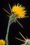
What do you need to know about this guy?
|
|
|
Sudangrass and hybrid sorghums are cyanogenic plants. Drought and freezing increase the hydrocyanic acid content of these plants, which cause death if ingested. Clinical signs include posterior ataxia, urine-stained hair and skin ulcerations in the perineal region, and frequent urination.
From Vin post: The main concern with sudan is sorghum cystitis, a syndrome that causes ataxia and cystitis from grazing only. Usually the hay is not a problem for this syndrome, although usually not a very good choice for horse hay in my opinion as I believe it could lead to colics. |

What are the concerns with this guy?
|
|
|
How does BCS work for horses?
|
Scale from 1-9. 9 is extremely fat. Areas to judge: along the neck, along the withers, ribs, and behind the shoulder, and other places.
|
|
|
What is one of the first signs of vit A deficiency? What are other signs?
|
impaired vision. also lacrimation because cornea is too dry. night blindness also occurs. skin lesions may develop that are difficult to heal. poor hoof growth, digestive disturbances, impaired conception, and respiratory illness may also be observed.
|
|
|
What should be known about vitamin D?
|
Rickets=deficiency, but rarely observed.
more common is OD: leads to calcium deposits in soft tissues such as the heart, mm, and arteries. Certain plants like day jasmine contain toxic levels of VitD-like compounds. Excess vit D stored in liver, so seldom necessary to supplement. (30 min a day outside adequate for horses to make their own vit D) |
|
|
Where do horses get vit C?
|
Synthesized in liver. Not essential to diet. Toxicity not reported.
|
|
|
What is the normal RR for a horse?
|
8-16 rpm
|
|
|
What is the normal body temp of a horse?
|
100 F or 38 C (varies from 99.5-101.5) for normal horses.
|
|
|
Classify fevers by temperature.
|
102 mild
104 moderate 106 high |
|
|
What is the normal heart rate for horses;
mature newborn yearlings |
Mature: 28-40 bpm
newborn: 80-120 foals: 60-80 yearlings: 40-60 |
|
|
Why do horses have cloudy urine?
|
Calcium carbonate crystals. (the longer the time between urination, the cloudier it is)
|
|
|
What are the 6 groups of roundworms that affect horses?
|
ascarids
small strongyles large strongyles (bloodworms) threadworms pinworms stomachworms |
|
|
Ascarids are commonly called what?
|
Large roundworms. (Parascaris equorum). Grow to be 12 to 15 inches, and are about the diameter of a lead pencil.
|
|
|
When do the usual clinical signs of a heavy ascarid infestation develop? (what age of horse?) What are the signs?
|
10-12 weeks (rough hair coat, poor appetite, pot belly)
|
|
|
When do horses usually develop a resistance to ascarids?
|
4-5 years old, but continuous deworming when the horse is young may interfere with the development of this immunity.
|
|
|
What parasite is the most severe threat to a horses health?
|
Strongyles. There are approximately 54 species that infect horses.
|
|
|
What are the stomach worms of horses?
|
Habronema muscae and Draschia megastoma.
|
|
|
What are the three types of mites that infest horses?
|
sarcoptic, psoroptic, and chorioptic
|
|
|
What are the differences between the 3 types of mites found on horses? How do you know which you are dealing with without a microscope?
|
Sarcoptic: burrow in skin, cause iirritation and itching. first lesions usually on head, neck, shoulders and flank, but later spread to entire animal.
Psoroptic mites do not burrow. Usually start in mane and tail. Puncture skin for food, and secrete poison into the wound. Cause itch, spread rapidly. Chorioptes: found below hocks and knees. Live on surface. Cause symptoms similar to psoroptes. horse may paw, kick, bite its lower limbs. |
|
|
What is the etiologic agent of Antrhax?
Describe it. |
bacteria: Bacillus anthracis. spore-forming, encapsulated, rod-shaped G+
|
|
|
How do horses get Anthrax?
|
ingestion or biting insects.
|
|
|
What tx is available for anthrax?
|
Tx not usually successful. Mortality high.
|
|
|
What are the clinical signs of Anthrax in horses?
|
Ingestion: colic, enteritis, septicemia.
Insects: fever, hot/painfl edematous swelling of throat, lower chest, abdomen, prepuce, and mammary glands |
|
|
Is there a way to prevent Anthrax?
|
There is a vaccine for horses.
|
|
|
What is the etiologic agent of Botulism?
|
Clostridium botulinum. Anaerobic bacteria. Toxins type A, B, C.
|
|
|
What are the symptoms of Botulism?
|
Flaccid paralysis of muscles via interference with the neuromuscular junction. Initially: loss of fx in tail, tongue. then inability to swallow, weak gait, decreased eyelid tone, decreased bowel sound, sweating, shaking forelegs, colic, bloat. horse becomes recumbant and dies of respiratory paralysis.
Foals: dz called "Shaker foal syndrome". Normally occurs between 2-8 weeks of age. |
|
|
How does a horse contract Botulism?
|
1. ingestion of toxin via feed/water (most common)(via contamination with dead bird/rodent bodies)
2. wound entrance 3. foals via umbilical stump |
|
|
Influenza is caused by what type of virus?
|
Myxovirus
|
|
|
Strangles is caused by what?
|
Streptococcus equi
|
|
|
Rhinopneumonitis is caused by what?
|
Herpes virus
|
|
|
Viral arteritis is caused by what?
|
Arterivirus
|
|
|
Etiologic agent of Potomac horse fever?
|
Ehlichia risticii
|
|
|
What causes Equine Protozoal Myeloencephalitis?
|
Sarcocystis neuona (a protozoa)
|
|
|
Rain rot is caused by what?
|
Dermatophilus congolensis
|
|
|
Strangles is also called what?
|
Distemper or shipping fever.
|
|
|
What are the places you can take the HR?
|
1. iner surface of groove under lower jaw (external maxillary artery)
2. the hack edge of the lower jaw (the cheek), four inches below the eye (facial artery) 3. under the tail, close to the body (medial coccygeal artery) 4. over heart with stethescope. 5. inside L elbow, up forward against the chest wall (heart) 6. inside of the foreleg (median artery) 7. behind the carpus (digital artery) 8. medial or lateral pastern (digital artery) |
|
|
What horse diseases can we vaccinate for?
|
Influenza, strangles, rhinopneumonitis, viral arteritis, equine encephalomyelitis, anthrax, Potomac horse fever, rabies rotavirus, salmonella
|
|
|
Describe Navicular disease.
|
Chronic degenerative condition of the navicular bursa and navicular bone that involves damage to the flexor surface of the bone and the overlying deep digital flexor tendon with osteophyte formation on the lateral and proximal borders of the bone.
|
|
|
What signalments get navicular disease?
|
One of the most common causes of chronic forlimb lamentss in horses but is essentially unknown in ponies and donkeys.
|
|
|
What is "ringbone"?
|
Periostitis or osteoarthritis of the phalanges that leads to exostoses.
|
|
|
What causes ringbone?
|
faulty conformation, improper shoeing, repeated concussion through work on hard ground are causitive; trauma and infection, especially wire-cut wounds are also incriminated. In light horses the strain of ligaments and tendinous insertions inthe pastern region are frequent causal facors. It may be a part of the OCD syndrome seen in young horses.
|
|
|
What is the progression of ringbone like?
|
Originally there is a lameness due to periosteitis,
|
|
|
WNV is in the genus?
|
Flavivirus.
|
|
|
Once horses show clinical signs of WNV, what is the fatality rate in the US?
|
Around 30%
|
|
|
How many of horses infected with WNV in the US become clinically infected?
|
10-39%
|
|
|
What are the clinical signs of WNE?
|
West Nile Encephalomyelitis
Presenting Complaints: neurologic abnormalities, colic, lamenes, anorexia and fever. Initial systemic signs: low grade fever, feed refusal, depression. |
|
|
What is the test of choice for detection of recent exposure to WNV?
|
IgM capture ELISA
|
|
|
What is the treatment for horses with WNV?
|
supportive-control pain and inflammation, prevent injury associated with ataxia or recumbency. Until EPM is ruled out, prophylactic antiprotozoal meds may be instituted.
|
|
|
Do vaccines for EEE, WEE provide cross protection for WNV?
|
No. Must vaccinate for WNV specifically.
|
|
|
What should OCD be referred to as when early in the disease? Why
|
instead of osteochondrosis, should be dyschondroplasia because early lesions are seen in cartilage
|
|
|
What factors contribute to the development of OCD?
|
rapid growth
overnutrition mineral imbalance biomechanics (trauma to cartilage) genetics implicated in some breeds (Standardbred and Swedish Warmblood) |
|
|
What is the most common sign of OCD?
|
nonpainful distention of an effected joint (eg, gonitis, bog spavin)
(OCD divided into a. seen in foals b. seen in older patients) first sign in a foal is often increased recumbency |
|
|
What mineral deficiency might be suspected in OCD?
|
copper deficiency
|
|
|
What is another name for "Wobblers"
|
Cervical stenotic myelopathy
|
|
|
What is the common signalment for Wobblers? What is the tx?
|
Familial basis in young, rapidly growing horses, particularly Thoroughbreds, with males more commonly affected. Overnutrition implicated, and clinical signs can be reversed if under 9 mo by reducing calories and restricting exercise.
|
|
|
What is the most serious cause of pneumonia in foals 1-5 months of age?
|
Rhodococcus equi pneumonia.
|
|
|
How do you characterize the equine breeding cycle?
|
Seasonally polyestrous.
|
|
|
When is the onset of the breeding season?
|
Spring, associated with an increase in daylight, temperature, and availability of food.
|
|
|
How many layers does a cross section of the reproductive organs have? Name the layers.
|
Serosa, muscularis, submucosa and mucosa.
|
|
|
Describe the serosal layer of the reproductive tract.
|
Single-cell layer of squamous cells
|
|
|
What type of epithelium lines the oviduct?
|
a mixture of ciliated and nonciliated simple columnar epithelium
|
|
|
Embryologically/anatomically, where does the repro tract develop?
|
Retroperitoneal
|
|
|
What is the broad ligament composed of?
|
double layered connective tissue sheets (peritoneum)
|
|
|
What is the name of the "broad ligament" that surrounds the ovary?
|
mesovarium
|
|
|
What type of uterus does a mare have?
|
bicornuate. poorly to moderately developed horns like the cow
|
|
|
When do fillies born in the spring normally reach puberty?
|
12 mo
|
|
|
How do you get a mare to cycle artificially?
|
Exposure to artificial light 16 hours per day causes serum melatonin to drop. If this continues for 8-10 weeks she will start to cycle.
|
|
|
Ovulatory season is longer in horses or ponies?
|
Horses. Ponies rarely ovulate in the winter. Jennies tend to have longer breeding seasons than mares.
|
|
|
When in the cycle does ovulation occur?
|
Typically one to two days before the end of estrus. (ie fourth or fifth day of estrus).
|
|
|
When is the earliest mares recognize fertilization?
|
Unfertilized ova do not usually pass into the uterus, but remain in the uterine tubes.
|
|
|
What age is optimal reproductive efficiency?
|
4 to 9 years
|
|
|
When are embyros usually collected for ET?
|
7 days after ovulation.
|
|
|
When twins occur, when must this be detected?
|
prior to day 16 so that when one embryo drifts away from the other, it can be crushed transrectally.
|
|
|
What are endometrial cups?
|
well circumscribed ulcer-like structures seen on the endometrium. they are fetal origin, arise from the chorionic girdle (a circumferential ring of specialized cells).
|
|
|
What do the endometrial cups make?
|
eCG
|
|
|
Do mares experience postpartum anestrus?
|
No
|
|
|
Use phenylbutazone with caution in:
contraindicated in: |
caution: foals, ponies, CHF, renal diasease or other drug allergies
contraindications: known hypersensitivity, history or preexisting hematologic or bone marrow abnormalities, preexisting GI ulcers, food producing animals |
|
|
Do NOT give phenylbutazone how? Why?
|
Do not give IM or SC; IA injections may cause seizures.
|
|
|
Adverse effects of phenylbutazone?
|
Adverse Effects: Horses: oral and GI erosions and ulcers, hypoalbuminemia, diarrhea, anorexia, and renal effects.
|
|
|
If first dose of flunixin meglumine is not successful at controlling pain, what do you do?
|
If first dose is ineffective for pain control, subsequent doses unlikely to be of benefit
|
|
|
Flunixin meglumine is a very potent inhibitor of what? Like other NSAIDS, it exhibits what behavior? How does it affect GI motility? What does it do in septic shock?
|
Flunixin is a very potent inhibitor of cyclooxygenase and, like other NSAIDs, it exhibits analgesic, anti-inflammatory and antipyretic activity. Flunixin does not appreciably alter GI motility in horses and may improve hemodynamics in animals with septic shock.
|
|
|
Butorphanol is how potent?
|
4 to 7 times morphine
|
|
|
Use butorphanol with caution/or contraindicated when?
|
Contraindicated or caution in patients with liver disease, hypothyroidism, or renal insufficiency, Addison's, head trauma, increased CSF pressure or other CNS dysfunction (e.g.,coma) and in geriatric or severely debilitated patients
|
|
|
What is the etiologic agent of tetanus?
|
Clostridium tetani
|
|
|
Describe clostridium tetani. Where does it live?
|
anaerobic bacteria, large gram-positive rods that have thick capsules and internal spores. soil and GI tract.
|
|
|
What is the pathogenesis of tetanus?
|
Bacteria multiply in vegetative form in tissues, causes autolysis, release neurotoxin, toxin absorbed by motor neurons in area and pas up to the spinal cord.
|
|
|
What are the clinical signs associated with tetanus?
|
spasmotdic, tonic contractions (b/c it interferes w/neurotransmitters), eventually respiratory failure and death.
|
|
|
How do you diagnose tetanus?
|
prescence of tetanus toxin in serum
|
|
|
How do you prevent tetanus? Describe what is used. Who gets prevention? Why?
|
Tetanus toxoid vaccine. (formalin deactivated tetanus toxin vaccine). should be given yearly after "puppy shots", also, brood mares 4-6 weeks before foaling to increase level of resistance in colostrum. (foal umbilical susceptible)
|
|
|
What are arboviruses? which ones are endemic to the US?
|
AR-thropod-BO-rne viruses; EEE, WEE, WNV.
|
|
|
Because arboviruses are transmitted by mosquitos, they are considered infectious, but not ____
|
contagious
|
|
|
What two members of the Alphavirus family do we need to vaccinate for?
|
WEE, EEV
|
|
|
What type of virus is WNV?
|
West Nile is a Alphavirus
|
|
|
For EEE, WEE and WNV, horses are considered what type of host?
|
dead end. (although the virus replicates in horses, they do not have enough viremia to transmit the virus back to mosquitos)
|
|
|
What are the case fatality rates for
EEE WEE WNV |
EEE : 90%
WEE: 20-40% WNV: 20-40% (majority in unvaccinated or incompletely vaccinated horses) |
|
|
How do you prevent EEE, WEE, WNV?
|
Vaccinate yearly (spring).
|
|
|
How do you diagnose the arboviruses?
|
serum
|
|
|
What are the clinical signs of WEE, EEE?
What are the R/O? |
quiet, depressed, neuro signs. (weakness, ataxia, stumbling, staggering, circline, muscle fasiculations, head pressing, blindness, seizures, recumbency or acute death). Fever, anorexia, dysphagia or other signs of abnormal mentation may be present.
R/O: rabies, neuro form of EHV-1, EPM, dn non-infectious causes like toxicities or spinal cord problems. |
|
|
What type of virus is Equine influenza?
|
orthomyxovirus. (RNA) two types: A/equine 1 and A/equine 2
|
|
|
What is the incubation of equine influenza? what are the clinical signs
|
1-3 day incubation. high fever, serous nasal discharge, submandibular L.N. enlarged, dry/harsh cough, depression, anorexia, and weakness
|
|
|
How long does damaged respiratory tract epithelium take to regenerate?
|
21 days. (up to 1 month)
|
|
|
Where do points usually develop in the horses mouth?
|
Upper buccal edge
Lower lingual surface |
|
|
What are some common oral neoplasias?
|
Gray horses: melanoma at commissure of the mouth
equine sarcoids, verrucous and fibrous forms |
|
|
What is omeprazole? What is it used for?
|
proton pump inhibitor used for GI ulcers and erosions
|
|
|
What is rantidine?
|
Like cimetidine, it is a histamine type 2 receptor antagonist, used to reduce acid output in the stomach. (doesn't work as well as omepramazole)
|
|
|
Where do gastric ulcers usually occur? Why?
|
proximal 1/3 of stomach (nonglandular portion), which is covered by stratified squamous epithelium and has no mucus or bicarbonate layer to protect it against acid induced injury.
|
|
|
What is the stomach capacity of an adult horse?
|
8-10 L
|
|
|
What type of muscle does the esophagus have? Discuss oxytocin and choke.
|
Proximal 2/3 striated, distal 1/3 smooth. (may not be as effective in distal portions) oxytocin relieves pressure in the striated muscle portion-mechanism unknown.
|
|
|
Contraindications, oxytocin in choke
|
pregnant, recently foaled, or if choke obstruction would cause additional damage due to increased motility of the esophagus (check w/endoscope prior to administration)
|
|
|
How long is a horses cecum? what capacity does it have?
|
4-5 foot (adult horse), holds 27-30L.
|
|
|
What do you need to know about acid/base and treating mastitis?
|
Nonionized drug crosses membranes readily, and "like is nonionized in like"
Mastitis: treated parenterally with antibiotics that are weak bases because milk is more acidic than plasma. Weak bases are highly unionized in plasma, and thus cross readily into the mammary gland where they become "trapped" in the more acidic environment of milk. |
|
|
Typical infections such as abscesses, metritis, and menngitis are typically acidic or basic when compared with plasma? How do you treat them?
|
These infections are typically acidic in relation to plasma, and therefore need to be treated parenterally with a weak base.
|
|
|
Basic drugs are what? Why do you use them?
|
Basic drugs include: aminoglycosides, macrolids, chlorampenicol/florfenicol Trimethoprim
They are used because they usually have a high volume of distribution. The acception is the aminoglycosides, which are very large, hydrophilic molecules that are highly ionized at physiologic pH and therefore do not cross biological membranes very well. They do not reach therapeutic concentrations in milk, abscesses or CSF. |
|
|
What are amphoteric drugs? Give examples.
|
They are drugs that have both acidic and basic groups as part of their chemical structures. They have a range of pHs where they are maximally unionized, so are lipid soluble in most cases. Fluoroquinolones and tetracycines are examples.
|
|
|
What is the implication of protein binding in drugs?
|
Bound drug is to large to pass through biological membranes, so only free drug is available for delivery to tissues. Degree of binding is only significant if more than 90%.
|
|
|
What antibiotic is more than 90% bound? How does that work?
|
Ceftiofur. Its efficacy is attributed to binding acute-phase proteins, which carry the drug to sites of inflammation. D/t high protein binding, does not cross into milk, and therefore there is no milk withdraw time in cattle.
|
|
|
Aminoglycosides are not effective in treating what?
|
Wounds that are in hyperosmoloar, anaerobic, acidic environments (such as abscess)
|
|
|
Sulfonamides are ineffective in what environment?
|
They act by replacing paraaminobenzoic acid (PABA) in the folic acid synthetic pathway of the bacteria and are ineffective in purulent material and necrotic tissue, which provide alternative sources of PABA.
|
|
|
Antimicrobial agents with activity against anaerobic bacteria, combinations of broad-spectrum agents and those agents that undergo extensive enterohepatic recirculation are often incriminated in what?
|
Disturbing the normal gastrointestinal flora and allowing the proliferation of Clostridium spp. and pathogens such as Salmonella.
|
|
|
What are some adverse reactions that may be seen with administration of Penicillins?
|
Fatal anaphylactic reactions and immune-mediated hemolytic anemia.
|
|
|
Fatal cardiac arryhthmias are associated with what drugs?
|
Concurrent IV administration of potentiated sulfonamides and detomidine.
|