Reading...
![]()
Play button
![]()
Play button
![]()
Use LEFT and RIGHT arrow keys to navigate between flashcards;
Use UP and DOWN arrow keys to flip the card;
H to show hint;
A reads text to speech;
23 Cards in this Set
- Front
- Back

|
|
|

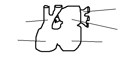
What causes the rough and smooth parts of the atria?
|
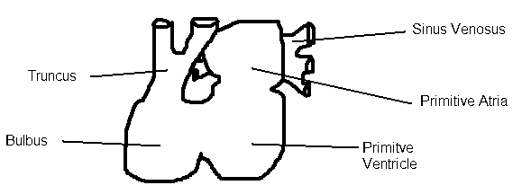
The rough comes from the primitive atria and the smooth comes from the sinus venosus
|
|
|
How are the atria separated from the ventricles?
|
Endocardial cushions from on the dorsal and ventral walls grow out and merge. The cushions function as AV valves.
|
|
What causes the rough and smooth parts of the atria?
|

The rough comes from the primitive atria and the smooth comes from the sinus venosus
|
|
|
How are the atria divided from the ventricles?
|
Endocardial cushions from on the dorsal and ventral walls grow out and merge. The cushions function as AV valves.
|
|
|
How is the left atria (& associated veins) created?
|
The L atria sends out a pulmonary vein bud. This grows out until it touches the pulmonary veins, which it begins to canabalize. The extent to which it canabalizes determines the # of pulmonary veins.
|
|
|
Where did the L atrial appendage come from?
|
The primitive atria. The rest of the L atrium is from the pulmonary veins that it ate.
|
|
|
What is the first major structure to form in the division of the atria?
|
The septum primum - descends from the roof of the Patria. As it descends a large opening called the foramen primum, forms betweens its free edge and the endocardial cushions. At the same time, programmed cell death causes another hole to form at the top of the septum - the foramen secundum. Once the second hole has opened the septum fuses completely and the foramen primum is fused shut. This allows for constant mixing of blood.
|
|
|
What is the second major structure to form in the division of the atria?
|
The septum secondum, a thicker membrane, descends from the ventrocranial wall to the right of the septum primum. The septum secondum covers the foramen secundum, however the septum itself has a hole (faramen ovale) that allows blood to continue to mix.
|
|
|
In the fetal heart, which atria has most blood return? Why? Which ventricle pumps the most blood? Which vessel carries the most blood? Why?
|
The R atria b/c of the foramen ovale. The R ventricle (67% of blood). The aorta carries most of the blood b/c the ductus arteriosus shunts the blood from the pulmonary trunk to the aorta.
|
|
|
How is the primitive ventricle divided?
|
The primordial interventricular septum is pused towards the fused endocardial cushions when the ventricles start to dilate and grow/fall downwards.
|
|
|
What form the membranous part of the IV septum?
|
The fushion of the bulbar cushions, the endocardial cushions to the muscular interventricular septum.
|
|
|
How do the aorta and pulmonary trunk form?
|
At the superior end (ie the Truncus) cushions emerge from the L & R (lateral) sides. These cushions spiral down so that at the bulbar portion they are anterior and posterior (180deg spiral). This creates a twisted separation of vessels with the anterior P trunk looping around the posterior aorta.
|
|
|
What causes a Vent. Septal Defect?
|
Failure of the membranous portion to form, leaving a hole through which the ventricles communicate. (creates a L --> R shunt)
|
|
|
How do you fix a VSD?
|
Suture a patch over the hole, but must be careful to avoid the bundle of His
|
|
|
What causes the R to left shunt of blood in the fetus?
|
The systemic resistance is low because the placenta has virtually no resistance. Also, the pulmonary resistance is high d/t the vasoconstriction of the pulmonary arterioles.
|
|
|
If pulmonary resistance is elevated blood is shunted in which direction?
|
from R to L; usually happens due to a defect. Once direction of blood flow is reversed - baby will turn blue
|
|
|
Describe "transposition of the great arteries" - what does this mean hemodynamically?
|
TGA is when the aorta is attached to the morphological right ventricle, and the Ptrunk is attached to the morphological L ventricle. Thus oxygen rich blood cycles around the pulmonary circuit, whereas oxygen-poor blood is stuck in systemic circuit. A patent ductus arteriousus can buy some time, but when this closes baby will need intervention FAST.
|
|
|
What can you do to treat TGA?
|
make a a big ASD to essentially create on big atrium where lots of mixing can occur. Let baby grow then repair defects.
|
|
|
What is aortic atresia? What is the hemodynamic complication?
|
The complete obstruction of the aorta (ie semilunar cusps are fused). This means there is no exit from the L ventricle, so heart relies on ASD to allow blood to mix and all blood leaves via P Trunk. A small amount can get into aorta via ductus arteriosus.
|
|
|
What can you do for a baby with aortic atresia?
|
Once the ductus arteriosus closes - baby in trouble as no access to systemic circuit. B/c L ventricle will have significant hypoplasia, it will be unable to function even if the defect is corrected - needs a transplant!
|
|
|
What does Situs Inversus mean?
|
The right atria and right lung are on the L side (and vice versa)
|
|
|
What is atrioventricular disconcordance?
|
R atrium + L ventricle
L atrium + R ventricle |