![]()
![]()
![]()
Use LEFT and RIGHT arrow keys to navigate between flashcards;
Use UP and DOWN arrow keys to flip the card;
H to show hint;
A reads text to speech;
6 Cards in this Set
- Front
- Back
|
How is the germ cell fate specified in nematodes? |
In nematodes the germ cell lineage is set up at the end of 4th cleavage and all germ cells are derived from the p4 Blasto mirror. This p for blessed Amir is derived from, 3 stem cell like divisions of the p1 Cell. At each of these divisions one daughter cell produces somatic cells and other divides again to produce a somatic cell progenitor and a p Cell. The egg contains P granules in the cytoplasm that become asymmetrically distributed before the first cleavage and are later restricted to p cell linage. In nematodes the pie-1 gene is involved in maintaining the stem cell property of the p blastomeres. It encodes a nuclear protein that is expressed maternally and his present only in the germline blastomeres. P granules and pie - 1 protein become asymmetrically distributed to germline cells during cleavage of the nematode eggs. Pi 1 represses new transcription of zygotic genes in the blastomeres. This repression protect themselves from the actions of transcription factors that promote development into somatic cells. In the absence of zygotic transcription maternal mRNAs express the germ cell components. |
|
|
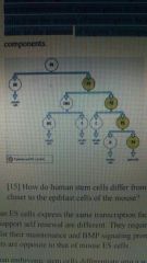
How do human stem cells differ from the mouse themselves? In what way are they closer to the epiblast cells of the mouse? |
Human ES cells express the same transcription factors as mouse ES cells, but signaling pathways that support self-renewal are different. They require FGF and activin/nodal signaling but not LIF for their maintenance and BMP signaling promotes differentiation in the cells. These effects are opposite to that of mouse ES cells. Human embryonic stem cells differentiate into a wide variety of cells but not all cells and are more advanced than most mouse stem cells. They are closer to Epiblast stem cells (epiSCs) which are pluripotent stem cells derived from the 5 day epiblast, a more advanced stage than the blastocyst in mice. They express transcription factors, Nanog, Sox2, Oct 4 and Klf4, similar to mouse epiSCs and can form teratomas. The human ES cells require FGF and activin/nodal signaling, but not LIF for the maintenance like mouse Epiblast stem cells (epiSCs). |
|
|
What does stem cell niche mean? |
The neighborhood where stem cells can remain undifferentiated. Include cells and molecules in the intermediate environment they inhabit and on which they depend for their survival and function |
|
|
What is the stem cell niche for a hematopoietic stem cell? |
In case of hematopoiesis stem cell niche is provided by the bone marrow stromal cells. Secreted signal proteins of the Wnt and BMP families, cell surface Notch ligands (which stimulate the notch pathway on cell-cell contact) retinoic acid and prostaglandin E2 have a role maintaining proliferation and self-renewal of hematopoietic stem cells in the stem cell niche of the bone marrow. |
|
|
Stem cell niche for neural stem cells |
Provided by adjacent glial cells in the dentate gyrus and subventricular regions. These produce signals that control neural stem cell behavior went through the beta-catenin pathway plays an important role in regulating adult neurogenesis. Notch signaling determines the number of stem cells and regulates the sonic hedgehog signaling. SHh promotes their survival |
|
|
When epidermis and mesoderm or mesoderm and endoderm tissues from an early stage embryo are disaggregated into single cells and mixed together they spontaneously reaggrerage as shown in the picture: the mesoderm migrates centrally with respect to the epidermis, adheringg to the inner epidermal surface. It also migrates centrally with respect to the gut or endoderm. Explain these results in terms of selective affinity |
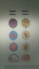
The final positions of the reaggregated cells reflect their embryonic positions. The mesoderm migrates centrally with respect to the epidermis, adhering to the inner epidermal surface. The mesoderm also migrates centrally with respect to the gut or endoderm. The inner surface of the ectoderm has a positive affinity for mesodermal cells and a negative affinity for the endoderm, while the mesoderm has positive affinities for both exctodermal and endodermal cells |