Reading...
![]()
Play button
![]()
Play button
![]()
Use LEFT and RIGHT arrow keys to navigate between flashcards;
Use UP and DOWN arrow keys to flip the card;
H to show hint;
A reads text to speech;
24 Cards in this Set
- Front
- Back
- 3rd side (hint)
|
Anatomy of the eye
|
Conjunctiva,sclera,cornea,pupil,lens,retina,aqueous humor and vitreous humor
|
|
|
|
path of light
|
path of light goes through the cornea to the pupil,lens,vitreos humor and retina
|
|
|
|
aqueous humor
|
separate cornea from the pupil
|
|
|
|
conjunctiva
|
white part of the eye
|
|
|
|
choroid cloat
|
contains blood vessels, pupil, lens
|
|
|
|
vitreous humor
|
gave eye shape and support the retina
|
|
|
|
Otitis Media
|
is an infection of the middle ear. caused by infections located in the nose and throat travel through eusthachian tube middle ear cause an inflammation
|
|
|
|
meniere's disease
|
is a disorder of the inner ear that causes spontaneous episodes of vertigo.result in abnormal volume or fluid in the inner ear.
|
|
|
|
cause of meniere's disease
|
a spinning sensation,hearing loss, ringing of the ear,feeling of pressure in the ear.one ear is affect
|
|
|
|
cause for otitis media
|
fluid and pus form in the middle of the ear cause fusion.eardrum could rupture.fluid back up in the affect esr
|
|
|
|
glaucoma
|
increase pressure in the eye and lead to blindness
|
|
|
|
open angle glaucoma(chronic disease)
|
most common
slowly no early signs and symptoms both eyes |
|
|
|
close angle glaucoma(acute disease)
|
less common
one eye happen suddenly |
|
|
|
signs and syptoms
|
early stage
eye pain difficulties adjusting to darkness unable to detect color sees halos around lights late stage nausea/vomiting headache fatigue blurred vision ranibow-halos |
|
|
|
cause of glaucoma
|
family history
fluid buildup poor blood flow damage to optic nerve |
|
|
|
treatment for glaucoma
|
medication and special eye drops
survey to drain the fluid |
|
|
|
what is cataracts
|
clouding of the lens that eye has impair vision.buildup of protein in the lens is when it occurs
|
|
|
|
signs and symptoms of cataracts
|
early
vision becomes slightly blurred colors seem faded glare increase halos double vision can become better w/ new glasses, better light, anti-glare sunglasses, magnifying lens advance visible white,milky spot marked vision loss entire becomes cloudy |
|
|
|
treatment for cataracts
|
surgery to removed clouded lens with artificial lens called intraocular lens
|
|
|
|
tympanic membrane
|
end of the canal is the eardrum
|
|
|
|
ossicles
|
middle ear made up three tiny bones
|
|
|
|
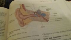
ear function to hear and balance
|
three parts outer,middle,inner ear
outer ear consists of visible external structure inner ear cochlea and three semicircular canals within the cochlea are tiny dendrites to hear |
|
|
|
semicircular canals
|
contain liquid and nerve endings. help us keep our balance
|
|
|
|
intracranial pressure
|
the skull exert a certain amount of pressure. due to cerebrospinal fluid, nervous tissue, blood flow through cerebral vessels
|
|