![]()
![]()
![]()
Use LEFT and RIGHT arrow keys to navigate between flashcards;
Use UP and DOWN arrow keys to flip the card;
H to show hint;
A reads text to speech;
30 Cards in this Set
- Front
- Back
|
Appendicitis
|
Definition: Appendicitis is an inflammation of the appendix, a finger-shaped pouch that projects from your colon on the lower right side of your abdomen (mayoclinic.org) Signs and Symptoms : Umbilical pain traveling RLQ and becoming severe, fever, RBT, malaise, N/V. RLQ=Right Lower Quadrant (abdominal) Abdominal Examination palpation, tenderness or pain may be felt (umbilical, RLQ) |
|
|
Constipation
|
Signs and Symptoms: Pain, inability to BM
Abdominal Examination palpation of large colon, masses may be felt. |
|
|
Acute Cholecystitis
|
Definition: Inflammation of the gallbladder. Your gallbladder is a small, pear-shaped organ on the right side of your abdomen, beneath your liver (mayoclinic.org) Localizedor diffuse RUQ pain Radiation to right scapula Vomiting Constipation Lowgrade fever |
|
|
Cholecystectomy |
Surgical removal of pt's gallbladder (Can be due to issues such as Acute Cholecystitis...) Laparoscopic cholecystectomy—The gallbladder is removed with instruments placed into small incisions in the abdomen (www.facs.org) |
|
|
Cholecystectomy continued... |
Post Surgery pt's commonly have abdominal distention and a referred shoulder pain. |
|
|
NasoGastric Tube (NG) |
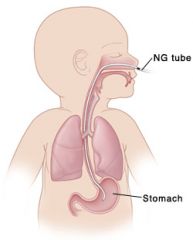
Uses: -On suction:Drain Gastric secretions. -Administer medications and nutrition. |
|
|
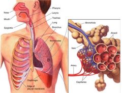
*Atelectasis |
Atelectasis refers to the incomplete expansion of the lungs (or partial incomplete expansion). It can be caused by airway obstruction, lung compression (such as occurs with pneumonothorax or pleural effusion) or increased recoil of the lungs due to loss of pulmonary surfactant. Atelectasis can be present at birth or attained later. |
|
|
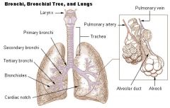
Atelectasis (online) |
Obstruction of bronchi / bronchioles and the collapse of the lung(s). -Common after receiving anesthesia, prolonged bedrest, mucous plug, particle in airway, pleural effusion (which is a buildup of fluids between the ribs and lungs) (remember bronchi lead to avioli) |
|
|
Atelectasis Continued... (medscape.com) |
Atelectasis is defined as diminished volume affecting all or part of a lung. Pulmonary atelectasis is one of the most commonly encountered abnormalities in chest radiographs. Recognizing an abnormality due to atelectasis on chest radiographs can be crucial to understanding the underlying pathology. Several types of atelectasis exist; each has a characteristic radiographic pattern and etiology. Atelectasis is divided physiologically into obstructive and nonobstructive causes. |
|
|
|
|
|
EKG |
EKG is a Cardiac monitor that monitors the electrical activity of the heart. (Some may call it a ECG but EKG is preferred) |
|
|
*Coronary Artery Disease (CAD) |
CAD describes heart disease caused by impaired coronary blood flow. Diseases of the coronary arteries can cause a spectrum os ischemic disorders ranging from angina to myocardial infractions.
|
|
|
*Atherosclerosis |
Most common cause of CAD, Stroke, peripheral artery disease. Atherosclerosis is a condition in which artery wall thickens as a result of the accumulation of fatty materials. (hypercholesterolemia is a major risk factor believed to cause Atherosclerosis. This can be due to lifestyle and/or genetics) |
|
|
*Chronic Obstructive Pulmonary Disease (COPD) |
COPD denotes a group of respiratory disorders characterized by chronic/reoccuring obstruction of airflow in the pulmonary airways. Airflow obstruction may also be accompanied by airway hyperreactivity and may be partially reversible. COPD is leading cause of death worldwide. Common cause of COPD is smoking. |
|
|
*Pleural Effusion |
Pleural Effusion refers to an abnormal collection of fluid in the pleural cavity . Pleural Effusion occurs when the rate of fluid formation exceeds the rate of removal. This fluid can be exudate, transudate, purulent (pus), chyle, sanguineous (bloody) |
|
|
*Pneumothorax |
Refers to the presence of air in the pleural space Causes partial or complete collapse of the affected lung. Open , Traumatic, Tension, spontaneous Pneumothorax. |
|
|
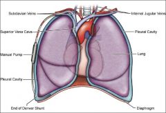
Pleural space: Surrounding Lungs |
|
|
|
-----> |
Acute Coronary Syndrome Angina Pectoris Complications of Myocardial Infarction Coronary Artery Vasospasm Isolated Coronary Artery Anomalies Myocardial Infarction Myocardial Rupture Pharmacologic Stress Testing Postinfarction Ventricular Septal Rupture Right Ventricular Infarction Saphenous Vein Graft Aneurysms Unstable Angina |
|
|
Exphixiation, anaphilaxix |
... |
|
|
Atrial Fibrillation (AFIB) |
Irregular Heart beat. Atrial fibrillation (AF or AFib) is the most common irregular heart rhythm that starts in the atria. Instead of the SA node (sinus node) directing the electrical rhythm, many different impulses rapidly fire at once, causing a very fast, chaotic rhythm in the atria. Because the electrical impulses are so fast and chaotic, the atria cannot contract and/or squeeze blood effectively into the ventricle. (http://my.clevelandclinic.org/) |
|
|
_____________________CANCERS_______________ |
______________CANCERS_________________________ |
|
|
Colorectal |
Cancer of the Colon or Rectum -Helpful site is American Cancer Society it tells all about risks and prevention |
|
|
cholecystectomy. |
removal of gallbladder |
|
|
Atelectasis |
partial or total collapse of lung/lobe (collapse of air sacs-alveoli) |
|
|
status asthmaticus |
multiple continuing asthma attacks |
|
|
bronchiectasis |
-airway damage, difficult to clear mucous -condition in which damage to the airways causes them to widen and become flabby and scarred. (http://www.nhlbi.nih.gov/) (can be caused by infection or condition causing damage to airways) |
|
|
bullectomy |
is the surgical removal of a bulla, which is an air pocket in the lung that is greater than one centimeter in diameter (across). Bullae tend to occur as a result of lung tissue destruction and diseases such as cancer and emphysema. (www.bmc.org/thoraciconcology/.) |
|
|
Bronchospasm |
Bronchospasm, which occurs in many pulmonary diseases, reduces the caliber of the small bronchi and may cause dyspnea, static secretions, and infection. Bronchospasm can sometimes be detected on auscultation with a stethoscope when wheezing or diminished breath sounds are heard. Increased mucus production, along with decreased mucociliary action, contributes to further reduction in the caliber of the bronchi and results in decreased airflow and decreased gas exchange. This is further aggravated by the loss of lung elasticity that occurs with COPD (GOLD, 2010). -manifestations: wheeze, decreased air flow, compromised gas exchange |
|
|
COPD |
Asthma Chronic Bronchitis Emphysema |
|
|
Asthma (COPD) |
Definition: S&S: Wheeze, dyspnea, cough Treatment: |