Reading...
![]()
Play button
![]()
Play button
![]()
Use LEFT and RIGHT arrow keys to navigate between flashcards;
Use UP and DOWN arrow keys to flip the card;
H to show hint;
A reads text to speech;
48 Cards in this Set
- Front
- Back
|
|
|
|
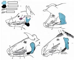
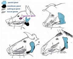
parotid salivary gland location
|
located superficially, ventral to ear
larger in herbivores innervated by cranial nerve IX (glossopharyngeal) |
|
|
parotid salivary gland ducts
|
serous in most species but not dog
travel across surface of masseter muscle in dog, opening in vestibule adjacent to upper fourth premolar tooth medial to central border of mandible in horse and ox- crosses ventral border of mandible laterally- enters vestibule as for dog |
|
|
mandibular gland
|
mixed serous and mucous
innervated by cranial nerve VII (facial) located at angle of jaw deeper and larger in herbivores duct opens at sublingual caruncle (close to frenulum) |
|
|
sublingual gland
|
mixed serous and mucous
innervated by cranial nerve VII (facial) monostomatic (single duct) and polystomatic (multiple ducts) parts in most speices duct runs along floor of oral cavity and open adjacent to frenulum |
|
|
zygomatic gland
|
CARNIVORES ONLY
innervated by cranial nerve VII (facial) present in dog and cats medial to zygomatic arch (in orbit) duct opens opposite last upper molar |
|
|
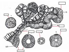
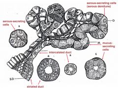
acini
|
clusters of cells arranged around a lumen
|
|
|
serous
|
cell pyramidal
round basal nuceli protein rich, watery basophilic perinuclear cytoplasm secretory glands in apical cytoplasm |
|
|
mucous
|
rich in carbohydrate, sticky, thick
cells swollen with mucous secretion flattened basal nucleus |
|
|
mixed glands
|
individual acini composed entirely of serous or mucous cells or both
|
|
|
intercalated duct
|
low cuboidal epithilium
|
|
|
striated duct
|
columnar epithilium with basal striations (alignment of mitochondria)
|
|
|
interlobular ducts
|
simple columnar,--> stratified columnar
|
|
|
innervation of salivary glands
|
secretions under autonomic (parasympathetic) control
|
|
|
|
|
|
pharynx
|
region common to digestive and respiratory tracts
lined by mucosa with mucous glands, and collagen and elastic fibers in lamina propria |
|
|
palatoglossal arch
|
demarcates pharynx from oral cavity
|
|
|
pharynx walls formed by
|
constrictor muscles
dilator muscles shortener muscles |
|
|
lymphiod tissue
|
assists with defense against infection
scattered and in tonsils |
|
|
pharynx divided by soft palate into
|
nasopharynx (dorsal to soft palate)
oropharynx (ventral to soft palate) |
|
|
pharynx innervation
|
contributions from cranial nerve IX (glossopharyngeal) and X (vagus)
|
|
|
deglutition steps
|
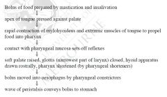
initiated voluntarily but taken over by reflexes once food reaches pharynx
|
|
|
esophagus or gullet fucntion
|
conduct food from the mouth to the stomach
|
|
|
esophagus structure
|
simple muscular tube that runs from the pharynx to the stomach in three segement: cervical, thoracic, and abdominal
|
|
|
Cervical segment of the esophagus
|
runs from pharynx to thoracic inlet (surrounded by 1st ribs, 1st sternabra, and 1st thoracic vertebraa)
begins dorsal to cricoid cartilage (immediatly rostral to beginning of trachea) follows trachea down neck, initially inclining to left medial to jugular groove returns to median position above trachea at about level of 1st rib |
|
|
throacic segment of the esophagus
|
runs within mediastinum in thorax
passes over base of heart crosses right side of arotic arch (beginning of aorta as it leaves heart) runs dorsal to bifurcation of trachea diverges slightly to left as it runs caudally in mediastinum ventral to aorta penetrates esophageal hiatus of diaphragm |
|
|
medistinum
|
formed from membranes that separate thorax into two pleural cavities and surround unpaired structures such as heart and esophagus
|
|
|
abdominal segment of esophagus
|
very short, especially in ruminants
]passes over dorsal border of liver to join the stomach dorsally at the cardia |
|
|
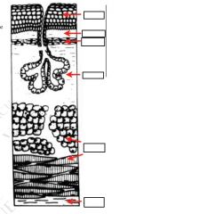
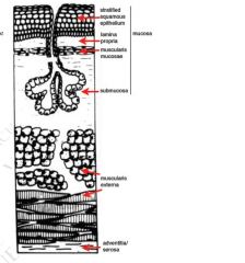
esophagus mucosa
|
innermost layer facing lumen
thrown into prominent longitudinal folds, thus great dilation possible composed of three layers |
|
|
epithilium of mucosa of esophagus
|
stratified squamous
keratinised or non-keratinised depending on species |
|
|
lamina propria of mucosa of esophagus
|
rich in collagen and elsatic fibers
leukocytes ducts of submucosal glands |
|
|
muscularis mucosa of the mucosa of the esophagus
|
thin layer of smooth muscle in mucosa at caudal end of esophagus
usually absent at cranial end (subject to specie variation) provides localised movement of mucosa |
|
|
submucosa of the esophagus
|
loose tissue, mainly collagen and some elastic fibers
rich in glands, produce mucus for lubrication contains glands at cranial end in most species but along full length of esophagus in dogs |
|
|
muscularis externa of the esophagus
|
muscle layer which moves food into stomach by contraction
usually thickest layer of wall two layers of muscle- difficult to define at cranial end, but at caudal end obvious inner circular and outer longitudinal layers |
|
|
cranial and caudal esophogeal sphincters
|
capable of maintaining intra-esophageal intra-luminal pressure higher than intra-gastric pressure
|
|
|
muscularis externa of the esophagus of dogs and ruminants
|
composed of skeletal muscle throughout
|
|
|
muscularis externa of the esophagus of species other than dogs and ruminants
|
skeletal muscle at cranial end, smooth at caudal 1/3
|
|
|
adventitia/serosa of the esophagus
|
outermost layer
|
|
|
adventitia of the esophagus
|
where esophagus lies outside thorax and abdomen
connective tissue merges with that of surrounding structures |
|
|
serosa of esophagus
|
where esophagus lies within thorax or abdomen
connective tissues covered by epithilial layer |
|
|
blood supply to esophagus
|
branches of common carotid, bronchoesophageal and left gastric arteries depending on region
|
|
|
nerve supply to esophagus
|
branches of sympathetic and vagus nerves
|
|
|
sites of possible obstruction of the esophagus
|
cranial and caudal esophagus
entrance to thoracic cavity-thoracic inlet over base of heart immediately anterior to diaphragmatic hiatus |
|
|
|