Reading...
![]()
Play button
![]()
Play button
![]()
Use LEFT and RIGHT arrow keys to navigate between flashcards;
Use UP and DOWN arrow keys to flip the card;
H to show hint;
A reads text to speech;
31 Cards in this Set
- Front
- Back
|
What is the main precursor to the cardiovascular system?
|
1. Mesoderm:
Some of it is outside the embryo (extraembryonic) and some of it is inside (intraembryonic) |
|
|
When do the heart precursors typically show up? Why does this timeline make sense?
|
At week 3. Note: It makes sense that a CVS is required at this point bc the embryo is growing - requiring a system to send nutrients to tissues and to efficiently remove waste products. Diffusion is not enough.
|
|
|
When do angioblasts typically appear?
|
About day 17
|
|
|
Where do "blood islands" begin to appear at week 3? On the embryonic disc? On the yolk sac? What's inside of them?
|
They appear on the outside of the yolk sac and are filled with angioblasts.
|
|
|
What tissue are angioblasts derived from?
|
Extraembryonic mesoderm (mesenchyme) (makes sense - especially considering where they're growing i.e. the outside of the embryo in the yolk sac)
|
|
|
When mesenchyme angioblast cells cluster, it is called what?
|
Blood island
|
|
|
How do we go from blood islands to primitive blood vessels?
|
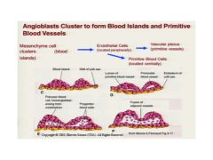
Some cells on the periphery of the island become flattened (endothelial cells) and form a lumen. Several lumens will coalesce to become primitive blood vessels. NOTE that some of the endothelial cells can break off and form primitive blood cells. Also note how "periphery" means the wall of the vessel.
|
|
|
In regard to angiogenesis, it first occurs in the __a__ and then happens in the __b__
|
a. Yolk Sac (umbilical vesicle)
b. Body Stalk |
|
|
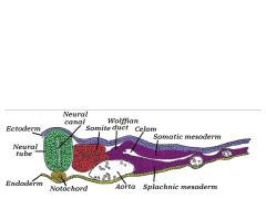
Don't forget where the splanchnic mesoderm lies - think about it - there's a pic on the reverse side
|
a
|
|
|
Review 9/20 10a about 16:00 describing how primitive heart forms. Hard to write questions for.
|
Review 9/20 10a about 16:00 describing how primitive heart forms. Hard to write questions for.
|
|
|
Note that the primitive heart (and throat) is initially cranial to the primitive brain (i.e. above it). However, after cranial-caudal folding, the heart becomes caudal located. (see 9/20 10a ~17:30 and image opposite side of card)
|
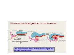
Note that the primitive heart (and throat) is initially cranial to the primitive brain (i.e. above it). However, after cranial-caudal folding, the heart becomes caudal located.
|
|
|
The primitive heart tube (PHT) is mostly the future what (three things)?
|
Left and Right atria and the L ventricle
|
|
|
If the PHT is mostly primitive left ventricle and atria, where does the other segments including atrium AV canal come from?
|
They are derived from the "secondary heart field" (a second area of mesoderm) located medial and dorsal to primary heart field.
|
|
|
At three weeks, the PHT is described as a "Tube within a Tube". Diagram that structure and label the three layers. What happens in the fourth week?
|
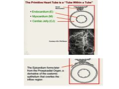
In the fourth week, a layer of epicardium forms around it.
|
|
|
How does the epicardium form?
|
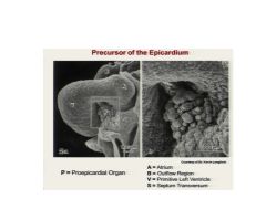
Just behind the heart is a cluster of cells called the proepicardial organ that delaminates and covers the myocardium. This epicardium will form both the visceral pericardium as well as the coronary vessels.
|
|
|
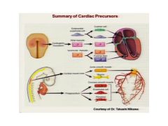
To summarize cardiac precursors:
1. Cardiogenic Mesoderm forms: a. b. c. 2. Proepicardium forms: a. b. c. |
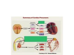
1. Cardiogenic Mesoderm:
a. Endothelial cell b. atrial myocyte c. ventricular myocyte 2. Proepicardium forms: a. coronary smooth muscle b. endothelial cell c. fibroblast (that forms visceral epicardium). |
|
|
What is heart looping? Growth of what drives it?
|
It's when the elongating heart tube begins to bend to the right. This is driven by addition of cells from the primary and secondary heart fields at the cranial and caudal ends.
|
|
|
See 9/20 10a at about 42:00 for overview of how looping arranges the heart
|
See 9/20 10a at about 42:00 for overview of how looping arranges the heart
|
|
|
Looping is a critical phase in development. Primarily because it:
|
...is required for proper alignment of the cardiac segments and septa.
|
|
|
Summary Q's for 9/20 10a
1. What is the hear primarily derived from? 2. Precursors organize at what end of the embryo. 3. What does folding accomplish? 4. The heart fields come together to form the what? 5. AS the tubular heart forms, it beings to loop to the...? 6. Blood flow change from a __a__ direction to a new direction that is __b__ 7. When does the heart begin to beat? (see 49:00) |
1. Splanchnic mesoderm
2. Cranial end 3. Forms primitive heart tube 4. Primitive heart tube. 5. Right 6. a. back and forth motion to b) two separate unidirectional, spiraling pathways. 7. 21-22 days. |
|
|
Partitioning of the heart primarily occurs between ? weeks.
|
5-8 weeks.
|
|
|
What are the four tissues used for cardiac partitioning?
|
1. Cardiac muscle
2. Cardiac mesenchyme (aka endocardial cushion tissue) 3. Extracardiac mesenchyme (mesoderm origin) 4. Neural crest. |
|
|
The left horn of the sinus venosus ultimately becomes what?
|
The coronary sinus
|
|
|
I stopped making note cards for 9/20 11a at around 13:00 min.
|
a
|
|
|
9/20 11a 15:40
Summary of RA Development 1. Coronary sinus derived from 2. Crista Terminalis? 3. Valve of the IVC? 4. Valve of CS? |
1. Lt horn of the Sinus Venosus
2. Rt valve of sinus venosus 3. Rt valve of sinus venosus 4. Rt valve of sinus venosus |
|
|
Where dose the terminal portion of the IVC come from?
SVC? Trabeculated Part & Auricle? |
1. Rt vitelline Vein
2. Rt common cardinal & proximal anterior cardinal v 3. Primitive Rt atrium |
|
|
For L atrium, the smooth part is derived from? The trabeculated part (auricle)
|
1. Resorption of the pulmonary veins.
2. Primitive L atrium |
|
|
Where does valve structures come from?
|
Cushion tissue.
|
|
|
Ventricle Development Derivations:
R Ventricle 1. Inlet portion: ? 2. Outlet portion: ? L Ventricle 1. Inlet portion: ? 2. Outlet portion: ? |
R Ventricle
1. Primitive R ventricle 2. Proximal outflow region L Ventricle 1. Primitive L ventricle 2. Proximal outflow region |
|
|
You may want to review/make note cards for the summary of RA development table on p11
|
Same with LA
|
|

Good review of precursors
|
a
|