Reading...
![]()
Play button
![]()
Play button
![]()
Use LEFT and RIGHT arrow keys to navigate between flashcards;
Use UP and DOWN arrow keys to flip the card;
H to show hint;
A reads text to speech;
61 Cards in this Set
- Front
- Back
|
What happens to the 1st right aortic arch?
|
Most of it disappears but it forms part of the maxillary artery.
|
|
|
What happens to the 1st left aortic arch?
|
Most of it disappears but it forms part of the maxillary artery.
|
|
|
What happens to the 2nd right aortic arch?
|
Most of it disappears but the rest forms the hyoid and stapedial arteries.
|
|
|
What happens to the 2nd left aortic arch?
|
Most of it disappears but the rest forms the hyoid and stapedial arteries.
|
|
|
What happens to the 3rd right aortic arch?
|
Ventral part- common carotid artery
Dorsal part - internal carotid |
|
|
What happens to the 3rd left aortic arch?
|
Ventral part- common carotid artery
Dorsal part - internal carotid |
|
|
What happens to the 4th right aortic arch?
|
Proximal part of right subclavian artery.
|
|
|
What happens to the 4th left aortic arch?
|
Part of arch of aorta.
|
|
|
What happens to the 5th right aortic arch?
|
Rarely recognizable, even in early embryo.
|
|
|
What happens to the 5th left aortic arch?
|
Rarely recognizable, even in early embryo.
|
|
|
What happens to the 6th right aortic arch?
|
Part of right pulmonary artery.
|
|
|
What happens to the 6th left aortic arch?
|
Ductus arteriosus and part of left pulmonary artery.
|
|
|
What are the 4 things that terminate in the sternal angle?
|
Azygos, pericardium, arch of aorta, bifurcation of trachea.
|
|
|
How many papillary muscles are in the right ventricle?
|
3
|
|
|
How many papillary muscles are in the left ventricle?
|
2
|
|
|
What structure is found only in the right atrium?
|
Crysta terminales.
|
|
|
Which valve has leaflets?
|
AV valves.
|
|
|
Which valves have cusps?
|
Aortic/Pulmonary valves.
|
|
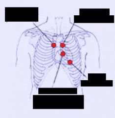
Identify each area of ausculation.
|
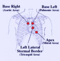
Answer
|
|
|
What does the left horn of the heart degenerate into?
|
Coronary sinus.
|
|
|
What does the right horn of the heart degenerate into?
|
Superior vena cava and all other vessels.
|
|
|
What forms the heart and most of the vessels?
|
Extraembryonic splanchnic mesoderm.
|
|
|
What are hemangioblasts?
|
Blood islands.
|
|
|
What forms the blood?
|
Hemocytoblasts.
|
|
|
What forms vessels?
|
Angioblasts.
|
|
|
What is angiogenesis?
|
Outgrowth or brenching of preformed vessels.
|
|
|
What is vasculogenesis?
|
Fusion of locally formed endothelial vesicles.
|
|
|
How is the coronary artery formed?
|
Angiogenesis
|
|
|
How are the vessels of the heart formed?
|
Vasculogenesis.
|
|
|
What is the intraembryonic vascular network development associated with?
|
Somite development (happens very quickly)
|
|
|
What are the 3 things that can happen to Vascular channels?
|
Large vessels, capillaries, disappear.
|
|
|
Can an artery become a vein in development and visa versa?
|
YES
|
|
|
Describe the formation of embryonic blood vessels.
|
Blood Island + Mesenchyme (vasculogenesis) -> Primary capillary plexus + Angiopoeiten (Angiogenesis)-> Lymphatic or completion of vascular wall + pericytes = blood vessel
|
|
|
How does the vessel attract mesenchyme?
|
PGEF
|
|
|
What are the 2 most important factors in embryonic?
|
Tif receptor, angiopoeitin-1
|
|
|
What are the 3 basic systems of the circulatory system? what happens to each?
|
Cardinal (most important, forms all major arteries), umbilical (disappears), omphalomesenteric (vitelline) (disappears)
|
|
|
What does the embryo's vein system develop out of? What is the result of this?
|
A very irregular network of capillaries. Venous system is not very uniform, and more variants of the venous system than on the arterial system.
|
|
|
Which arch becomes the aortic arch?
|
Left 4th aortic arch
|
|
|
Where does the brachiocephalic (B in BCS) come from?
|
The aortic sac.
|
|
|
What forms the right subclavian?
|
Proximal (right 4th aortic arch)
Middle (Portion of right dorsal aorta) Distal (Right 7th intersegmental artery) |
|
|
What makes up the right common carotid?
|
Entirely right 3rd aortic arch (also portions of internal and external carotic arteries)
|
|
|
Where does the left common carotid arise from?
|
Left 3rd aortic arch.
|
|
|
What forms the left subclavian artery?
|
Left 7th intersegmental artery.
|
|
|
Describe what happens in right aortic arch? How does it occur? When will they be asymptomatic? When will they by symptomatic?
|
The obliteration of the 4th left aortic arch artery or regression of the left dorsal aorta rather than the right one. Sometimes the ductus arteriosus will form the right arch to the right pulmonary artery and will be asymptomatic. However if the ductus arteriosus arises from the left pulmonary artery, it will have to cross the midline to reach the right aortic arch and this often occurs posterior to the trachea and esophagus, forming a vascular ring around these structures and compressing them.
|
|
|
What does a double aortic arch result from?
|
If the section of the right dorsal aorta inferior to the 7th intersegmental artery back to the point where it would meet with the left dorsal aorta (more inferior). This leads to persistence of both aorta and the formation of a ring which can compress the trachea and esophagus.
|
|
|
What is the cause of an interrupted aortic arch?
|
Obliteration of the 4th left aortic arch can possibly lead to an abrupt end to the aortic arch (there is no continuity between it and the descending aorta) instead the ductus arterious is patent and dialated leading to all of the blood being sent to the descending aorta have to come from the right ventricle.
|
|
|
What is the cause of an anomalous subclavian artery?
|
If the right 4th aortic arch artery regresses, the right subclavian will arise from the right 7th intersegmental artery, far left side of the aortic arch, passing posterior to the trachea and esophagus and can possibly compress these structures.
|
|
|
Why do the recurrent laryngeal nerves have different courses on either side of the body? Why is the right that way? Why is the left that way?
|
The differential development of the 6th aortic arch arteries explains why the recurrent laryngeal nerves have different courses on different sides of the body. The right 6th aortic arch degenerates and the recurrent laryngeal on this side moves superiorly to hook under the right subclavian. The left 6th aortic arch becomes the ductus arterios and and the left recurrent is hooked inferior to this.
|
|
|
What are the cardinal veins (anterior,posterior, commmon)? What do they drain? What happens to them later on?
|
Cardinal veins drain the embryo body and return the blood to the embryonic heart. The anterior cardinal veins drain the upper body and the posterior cardinal veins drain the lower body and both sets drain into common cardinal veins. Most regress or remodeled completely but they persist inthe veisn of the upper thorax and lower nexk, the upper limbs, lower limbs and sacral veins, and the most inferior portion of the IVC.
|
|
|
What are the subcardinal veins? What do the form? How about the left and righ?
|
These veins form in close proximity to the posterior cardinal veins and form an anastamosis with each other, the left one regresses and the right one becomes a significant portion of the IVC.
|
|
|
Where do the supracardinal veins form? What do they form?
|
Close proximity to subcarndinal and cardinal veins. They form a subsupracardinal anastamosis.
|
|
|
What do the supracardinal veisn become?
|
The azygos system, large portion of IVC, and their subsupracardinal anastamosis becomes the renal veins.
|
|
|
What does the left brachiocephalic vein form from? What does its shunt form?
|
An anastomotic shunt between the anterior cardinal veins, shunting blood from the left to the right anterior cardinal vein. The left anterior cardinal vein degenerates. The shunt leads to an enlargement of the right horn of the sinus venosus.
|
|
|
What happens the the left anterior cardinal vein?
|
It degenerates.
|
|
|
What does the ductus arteriosus form from?
|
Left 6th aortic arch artery, eventually attaching from the left pulmonary artery.
|
|
|
Where/how does the ductus venosus form?
|
The ductus venosus forms in the liver as the right umbilical vein degenerates and the left umbilical vein enlarges.
|
|
|
What could you contract in the fetal liver that would slow flow of blood to the heart?
|
sinus venosus.
|
|
|
What is the cause of double SVC? What does this lead to?
|
Persistence of the left anterior cardinal vein leads to a small left brachiocephalic and an extra SVC that opens into the right atrium through the coronary sinus.
|
|
|
What is the cause of left SVC? What does this lead to?
|
Persistence of the left anterior cardinal vein and regression of the right anterior cardinal vien. This leads to an odd left SVC that drains into the right atrium through the coronary sinus.
|
|
|
What causes absence of hepatic segment of IVC?
|
Normally forms most superior aspects of the IVC. IF it is absent, the IVC will not connect with the right atrium properly. This leads to the lower body being drained by the azygos and hemiazygos veins. THe hepatic veins also open seperately into the right atrium (normally connect to hepatic segment of IVC)
|
|
|
What is the cause of double IVC?
|
Failure of an anastamosis of the veins of the trunk to develop can lead to the persistence of the left supracardinal vein, forming a double IVC inferior to the renal veins.
|