Reading...
![]()
Play button
![]()
Play button
![]()
Use LEFT and RIGHT arrow keys to navigate between flashcards;
Use UP and DOWN arrow keys to flip the card;
H to show hint;
A reads text to speech;
35 Cards in this Set
- Front
- Back
|
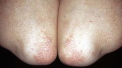
Psoriasis
Multiple small red plaques are visible on the elbows. |
|
|
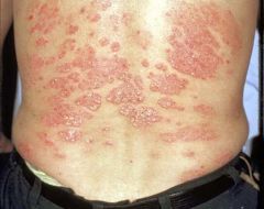
Psoriasis
Multiple red scaley areas are present over the lower back. The white scaley areas correspond to the microscopic finding of parakeratosis. |
|
|
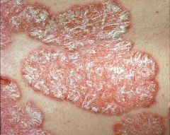
Psoriasis
Close-up view of a cutaneous plaque. Redness reflects vascularity. White indicates parakaratosis. |
|
|
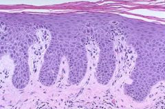
Acanthosis in psoriasis.
Microscopically, psoriasis is characterized by hyperplasia of the epidermis, downward elongation of the rete ridges with thinning of overlying stratum granulosum, with parakeratosis above this. Capillaries within dermal papillae are brought close to the surface. In some biopsies, neutrophils are visible in the stratum corneum or epidermis. |
|
|
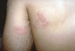
This is contact dermatitis, a form of type IV hypersensitivity reaction that is localized. This person contacted a plant in a tropical jungle to which he was already sensitized. A couple of days later, the skin is red from inflammation and slightly raised from local edema.
|
|
|
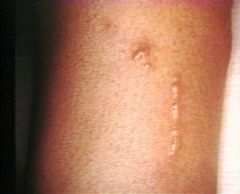
This shows a poison-ivy-like lesion of contact dermatitis in a linear distribution. An edematous oozing eruption of vesicles (small collections of fluid), which forms where the leaf brushes the skin. Pruritis may be pronounced.
|
|
|
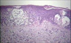
Spongiosis is intercellular edema in the epidermis, (visualized as the clear spaces between the epidermal cells).
|
|
|
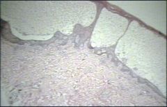
A blister
This is an epidermal vesicle, which has formed due to marked spongiosis. |
|
|
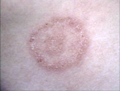
Dermatophyte "ring"
|
|
|
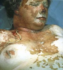
The skin of the face, neck, and chest is affected with sloughing of the epidermal layer due to a drug reaction. There are few dermatologic emergencies. This is one of them.
|
|
|
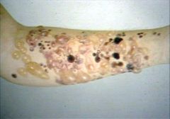
Bullous pemphigoid
Clear, tense bullae on an erythematous base. |
|
|
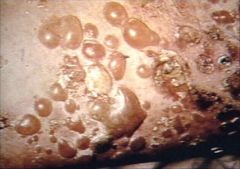
Bullous pemphigoid
Clear, tense bullae on an erythematous base. |
|
|
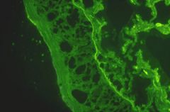
Bullous pemphigoid
Immunofluorescence studies can be performed on skin biopsies. Seen here is a bright green band of staining with antibody to IgG. The localization at the dermal-epidermal junction is typical, for immune complexes tend to be trapped along basement membranes. Diseases with this pattern may include systemic lupus erythematosus, discoid lupus erythematosus, and bullous pemphigoid. |
|
|
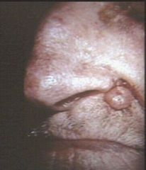
Nodular basal cell carcinoma
|
|
|
Nodular BCC
Pearly, opalescent. |
|
|
BCC w/ telangiectasia
|
|
|
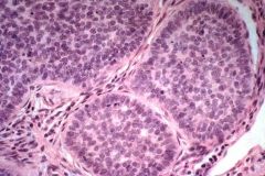
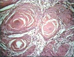
Palisading in BCC
The neoplastic masses of epithelial cells show palisading of the peripheral rim of cells, i.e the elongate cells are oriented perpendicular to the basement membrane. This characteristic helps pathologists recognize BCC. |
|
|
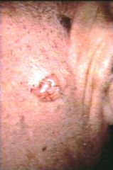
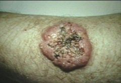
Invasive SCC
This is a nodular ulcerated lesion on the arm. |
|
|
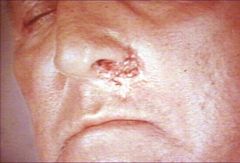
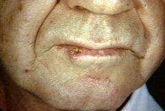
Invasive SCC
The lower lip has high exposure to the sun and is a frequent site for SCC. |
|
|
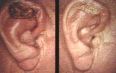
Squamous cell carcinoma
The lesion on the left appears to be ulcerated and covered by a dark crust. The lesion on the right appears to have a hyperkeratotic scale on the surface. |
|
|
Squamous cell carcinoma
This shows the invasive protion of a well differentiated SCC. The keratinization of nests of cells is prominent (“keratin pearls”). |
|
|
ommon epidermal lesions, especially in the elderly. Wart-like keratotic growths, often pigmented, predominantly growing above the level of the surrounding skin - imparting a "stuck on" appearance. They may be composed of basaloid cells, which often contain melanin, or basaloid keratinocytes which show keratin production with formation of keratin-filled channels ("horn pseudocysts").
|
Seborrheic keratoses
|
|
|
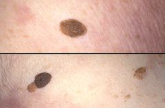
Seborrheic keratoses, skin
Seen here are examples of a very common lesion of older adults--a seborrheic keratosis (SK). They are rough-surfaced and brown coin-like plaques that vary from a few millimeters in size to several centimeters. |
|
|
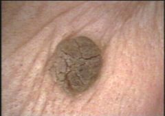
Pigmented Seborrheic Keratosis
This lesion is pigmented |
|
|
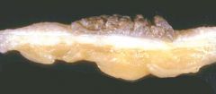
Seborrheic Keratosis
This resected seborrheic keratosis has been cut perpendicular to the skin surface. You can see that the lesion sits on top of the dermis and that it is pigmented. |
|
|
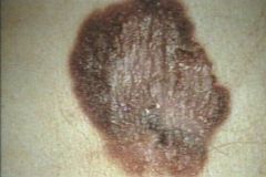
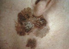
Superficial spreading melanoma
|
|
|
Superficial spreading melanoma
|
|
|
Superficial spreading melanoma
This skin lesion has an irregular border and variable pigmentation with nearly complete loss of brown color on the upper left margin and near the center. |
|
|
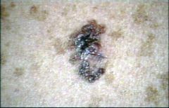
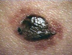
Pigmented nodular melanoma
|
|
|
Pigmented nodular melanoma
|
|
|
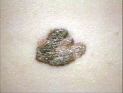
Lentigo Maligna Melanoma
Lentigo maligna occurs in the setting of sun damaged skin, usually on the face of elderly patients. However, with increased sun exposure, even early middle age patients develop lentigo maligna melanoma. |
|
|
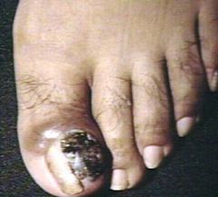
Acral-Lentiginous Melanoma
|
|
|
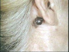
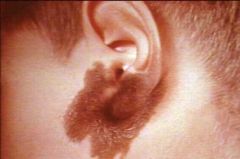
Congenital melanocytic nevus on the neck, face and ear of a child.
Precursor of malignant melanoma |
|
|
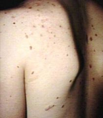
Dysplastic Nevi
Precursor to malignant melanoma |
|
|
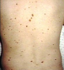
Dysplastic nevi
Precursor to malignant melanoma |