![]()
![]()
![]()
Use LEFT and RIGHT arrow keys to navigate between flashcards;
Use UP and DOWN arrow keys to flip the card;
H to show hint;
A reads text to speech;
132 Cards in this Set
- Front
- Back
|
Periodental disease |
Progressive condition that affects the supporting tissues of teeth |
|
|
Enamel
|
Outer covering of the crown composed of crystals of hydroxyapatite arranged in prisms
Formed by ameloblasts during tooth development Is acellular and considered nonliving Acts as an effective barrier to bacteria |
|
|
Dentin |
Makes up the bulk of the tooth Formed by odontoblasts located in the pulp chamber; arranged in series of tubules that transverse from the enamel to the pulp chamber As hard as bone but much softer than enamel |
|
|
Pulp |
Occupies interior cavity of tooth Rich with blood vessels, nerves, and lymphatics Composed of odontoblasts, fibroblasts, and other cells |
|
|
Cementoenamel junction (CEJ) |
Junction between crown and rooth |
|
|
Periodontium |
Tooth-supporting structure; collection of supporting structures surrounding the teeth |
|
|
Cementum |
Avascular and bonelike Attached to alveolar bone by periodontal ligament fibers Mineralized connective tissue |
|
|
Periodontal ligment |
Holds the tooth in the alveolus (socket) by attaching the tooth to the alveolar bone Composed of collagen with some elastic fibers, blood vessels, nerves, and lymphatics |
|
|
Alveolar bone |
Surrounds and supports teeth
|
|
|
Gingiva |
Soft tissue providing epithelial attachment |
|
|
Gingival sulcus |
Space between gingiva and tooth Normal depth in dogs is 1-3 mm and 0.5-1 mm in cats |
|
|
Crown |
Above gum line |
|
|
Root |
Below gum line |
|
|
Buccal |
Surface toward cheek |
|
|
Lingual |
Surface toward tongue |
|
|
Facial |
Surface that includes buccal and labial |
|
|
Labial |
Surface towards lips |
|
|
Palatal |
Surface towards the soft palate |
|
|
Mesial |
Surface toward the midline at front of the mouth |
|
|
Distal |
Surface toward the rear or back of the mouth |
|
|
Rostral |
Surface facing the nose of the animal |
|
|
Occlusal |
Chewing surface |
|
|
Furcation |
Space between 2 roots where they meet the crown |
|
|
Incisors |
Function to cut/nibble |
|
|
Canines |
Function to hold/tear |
|
|
Premolars |
Function to cut/shear/hold |
|
|
Molars |
Function to grind |
|
|
Carnassial teeth |
Largest cutting teeth Upper 4th premolars and lower 1st molars in dogs and cats |
|
|
28 |
Dogs deciduous |
|
|
42 |
Dogs permanent |
|
|
3142/3143 |
Dog dental formula |
|
|
26 |
Cat deciduous |
|
|
30 |
Cat permanent |
|
|
3131/3121 |
Cat dental formula |
|
|
24 |
Horse deciduous |
|
|
30-36, 40-42 |
Horse permanent (mare then male) |
|
|
3133/3133 |
Horse dental formula |
|
|
32 |
Swine deciduous |
|
|
44 |
Swine permanent |
|
|
3143/3143 |
Swine dental formula |
|
|
20 |
Ruminants deciduous |
|
|
32 |
Ruminants permanent |
|
|
0033/4033 |
Ruminant dental formula |
|
|
16 |
Hamsters, gerbils, rodents permanent |
|
|
1003/1003 |
Hamsters, gerbils, rodents dental formula |
|
|
20 |
Guinea pigs permanent |
|
|
1013/1013 |
Guinea pigs dental formula |
|
|
28 |
Rabbits permanent |
|
|
2033/1023 |
Rabbit dental formula |
|
|
103 |
Upper right 3rd permanent incisor # Triadan system |
|
|
308 |
Lower left 4th permanent premolar |
|
|
504 |
Upper right deciduous canine |
|
|
04 |
Canine tooth is always this number in Triadan system |
|
|
09 |
First molar always end this in the Triadan system |
|
|
Sickle scaler |
Curved or straight Triangular cross section and tapers to sharp pointed tip with 2 cutting edges on either side of face Removing supragingival calculus and for remvoing calculus from pits, fissures, developmental grooves, and interproximal areas |
|
|
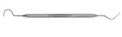
Curette |
U-shaped cross section with 1 or 2 cutting edges and round toe Used for subgingival calculus removal and root planning |
|
|
Gracey curette |
Face offset 20 degrees from terminal shank One cutting edge on edge tilted down Designed for specific teeth |
|
|
Universal curette |
Fx: scale and remove deposits and stain, remove calculus Characteristics: Face perpendicular to terminal shank Two cutting edges on either side of face Can be used in all areas |
|
|
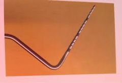
Periodental prode |
No sharp sides Used to measure depth of gingival sulcus and any other oral structures
|
|
|
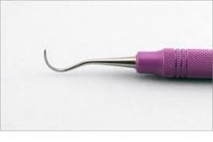
Shepherd's hook |
Sharp tip only Used to detect subgingival calculus and to detect enamel defects Assess tooth mobility Detect caries and fractured teeth |
|
|
Dental elevators |
Serve as wedge between root and bone to stretch and break periodontal ligament |
|
|
Periosteal elevators |
Used to elevate and reflect gingival, mucogingival, and palatal flaps Must be sharp so they will cut rather than tear |
|
|
Extraction forceps |
Used for tooth removal once tooth has been loosened and removal of heavy calculus |
|
|
Calculus-removing forceps |
Have hooked appearance to one of the jaws Specialized design allows for effective calculus removal |
|
|
Normal occlusion |
Scissor bite Upper incisors rostral to mandibular incisors Lower canines fit in diastema between upper maxillary canine and third incisor and should not touch
|
|
|
Mesticephalic |
Well-proportioned skull width and maxillary length |
|
|
Class I malocclusion (neutrocclusion) |
Maxillary and mandible correctly proportioned, but one or more teeth are misaligned |
|
|
Class II malocclusion (distoclusion) |
Teeth in maxilla occlude rostral to mandibular equivalents Maxillary prognathism and mandibular retrognathism More likely to occur in dolichocephalic breeds |
|
|
Maxillary prognathism |
Maxilla is forward |
|
|
Mandibular retrognathism |
Mandible is caudal |
|
|
Dolichocephalic |
Narrow skull and long maxilla (collies) |
|
|
Class III malocclusion (mesioclusion) |
Mandibular teeth occlude rostral to maxillary equivalent Maxillary retrognathism and mandibular prognathism Normal for brachycephalic breeds |
|
|
Maxillary retrognathism |
Maxilla is caudal |
|
|
Mandibular prognathism |
Mandible is forward |
|
|
Brachycephalic |
Wide skulls with short maxilla |
|
|
Level bite |
End-to-end bite of the incisors Genetically a degree of prognathism |
|
|
Wry mouth |
Nonspecifc term that refers to a variety of unilateral occlusal abnormalities; not recommended to use this term Genetically affects only one quadrant of the mandible or maxilla One segment of the jaw is disproportionately sized relative to the other half |
|
|
Oligodontia |
Only a few teeth present |
|
|
Anodontia |
Congenital absence of teeth |
|
|
Hypodontia |
One or a few teeth are missing |
|
|
Polydontia |
More teeth than normal Extra teeth are referred to as supernumerary (SN) |
|
|
Abnormal dental interlock |
Deciduous teeth that erupt in an abnormal pattern Upper deciduous canine teeth are pushed rostral to lower canine teeth Prevents normal forward growth of mandible Occurs in class II malocclusion and base-narrow malocclusion by impeding growth of mandible |
|
|
Retained deciduous teeth |
Should always be extracted if still present when permanent counterparts erupt Common in toy breeds |
|
|
Melanoma |
Malignant tumor Most common in dogs, rare in cats, poor prognosis Bone destruction evident around tumor Spreads early to regional lymph nodes and lungs |
|
|
Squamous cell carcinoma |
Malignant tumor Second most common tumor in dogs, most common in cats Grows fast, often ulcerated, spread slowly, invades bone Tumors in more rostral locations have better prognosis (surgical resection, slower to metastasize) |
|
|
Fibrosarcoma |
Malignant tumor Third more common tumor in dogs, second most common in cats Occurs at younger age than other oral malignancies Metastasizes slowly but is aggressively invasive, requiring wide surgical resection Guarded prognosis |
|
|
Osteosarcoma |
Malignant tumor May affect bones of mandible or maxilla Oral ___ spreads slower than appendicular version, so it is more responsive to surgery |
|
|
Epulis |
Nonmalignant tumor General term for any gingival mass
|
|
|
Gingival hyperplasia |
Thickening and excess growth of gingiva as a result of chronic inflammation and patient's response to plaque Most commonly seen in boxers, border collies, Labrador retrievers, German shepherds Gingiva grows to engult crowns |
|
|
Chronic ulcerative paradental stomatitis (CUPS) |
Severe ulceration on buccal mucosa covering teeth Associated with heavy calculus and gingival recession Severe halitosis, very painful |
|
|
Soft tissue lesions |
Lesions caused when a tooth makes contact with mucosa Cheek chew lesions Mucosa entrapped between teeth during chewing, may ulcerate |
|
|
Eosinophilic ulcers |
Rodent ulcers that occur on lip of cats; benign |
|
|
Gemini |
Attempt to make 2 teeth from 1 enamel organ Two crowns with single root |
|
|
Fusion |
2 tooth buds grow together to form 1 larger tooth |
|
|
Dilaceration |
Sharp end, curve, or angulation in root or crown Irregular surface, often has accessory canals to pulp chamber leading to periapical necrosis |
|
|
Enamel dyplasia |
Enamel didn't form properly; insufficient amount or hardness |
|
|
Brachyodont |
Short crown-to-root ratio with a true root No potential for further tooth growth once matured |
|
|
Hypsodont |
Long crown, short root Either radicular or aradicular |
|
|
Radicular hypsodont |
Grows for most of the animal's life until root apex closes late in life Horses and ruminants |
|
|
Aradicular hypsodont |
Never forms true root and teeth grow continuously Rabbits, guinea pigs, chinchillas |
|
|
Lagomorphs (Rabbits) |
Do not have canine teeth |
|
|
a.0.5 to 1mm |
The normal depth of the gingival sulcus in a cat is: a.0.5 to 1mm b.1 to 2mm c.1 to 3mm d.4 to 6mm |
|
|
d.Squamous cell carcinoma |
The most common oral malignant tumor in cats is: a.Melanoma b.Osteosarcoma c.Fibrosarcoma d.Squamous cell carcinoma |
|
|
d.Mandibular molars |
The teeth that are radiographed using standard parallel technique are the: a.Maxillary incisors b.Mandibular canines c.Maxillary premolars d.Mandibular molars |
|
|
c. 208 |
The fourth premolar in the upper left quadrant as: a. 108 b. 204 c. 208 d. 308 |
|
|
c. Remove supragingival calculi |
Sickle scalers: a. Have one sharp side b. Remove subgingival calculi c. Remove supragingival calculi d. Are pulled toward the gum line |
|
|
a.Cementum to the alveolar bone |
The periodontal ligament connects the: a.Cementum to the alveolar bone b.Gingiva to the cementum c.Cementum to the enamel d.Dentin to the enamel |
|
|
a. Hypervitaminosis D |
A new theory regarding the cause of resorptive lesions in cats is: a. Hypervitaminosis D b. Hyperparathyroidism c. A low pH diet d. Calicivirus |
|
|
a. 1 |
Tooth resorptive lesions that show a normal periodontal ligament space on a dental radiograph are type: a.1 b.2 c.3 d.5 |
|
|
b.90 |
The terminal shank of a universal curette should be lined up on the following line on a sharpening guide: a.110 b.90 c.30 d.0 |
|
|
c. 6 feet |
Bacterial contamination from a power dental scaler will reach distances of: a. 4 feet b. 5 feet c. 6 feet d. 8 feet |
|
|
d.Buccal |
The surface of the tooth that faces the cheeks is termed: a.Lingual b.Coronal c.Palatal d.Buccal |
|
|
a.Carnassial teeth |
The upper fourth premolar and first molar teeth are termed: a.Carnassial teeth b.Dilacerated c.Diphyodont d.Deciduous |
|
|
b. Rotary |
Which power scaler should be avoided in dental scaling? a. Sonic b. Rotary c. Ultrasonic d. Piezoelectric |
|
|
a.AVDC |
Which of the following organizations has a standardized reference for many of the grading indexes used for the teeth and gingiva? a.AVDC b.CVMA c.AVMA d.NAVTA |
|
|
a.Elongation |
Which image distortion occurs when the x-ray beam is directed too perpendicular to the long axis of the tooth? a.Elongation b.Foreshortening c.Grayish overlay d.Superimposition of mesial roots |
|
|
c. Side |
To accurately appreciate the direction that a canine tooth curves into the skull when preparing to take a dental radiograph, you should be standing at the patient’s: a. Front b. Rear c. Side d. Ventral aspect
|
|
|
a.Class I |
Rostral cross bite is classified under the following class of malocclusion: a.Class I b.Class II c.Class III d.Wry bite |
|
|
b.Class II |
Which type of malocclusion is a Collie most likely to have? a.Class I b.Class II c.Class III d.Wry bite |
|
|
b. 6 weeks of age |
An adult dog may have enamel dysplasia on their permanent teeth if they had a systemic disease at: a. 10 days of age b. 6 weeks of age c. 5 months of age d. Day 50 of gestation |
|
|
c.PD3 |
It has been determined that a tooth has 37% attachment loss. Which corresponding periodontal index should be assigned? a.PD1 b.PD2 c.PD3 d.PD4 |
|
|
Ruggae |
Ridge in palate |
|
|
Glossitis |
Inflammation of the tongue |
|
|
Stomatitis |
Inflammation of the mouth |
|
|
Polyhyodont |
Permanent replacement of teeth |
|
|
Diphyodont |
2 sets of teeth |
|
|
Monophyodont |
One set of teeth |
|
|
Egg caruncle |
All egg layers Not actual tooth Structure epidermal, horny, keratinized On tip of snout To penetrate egg shell |
|
|
Egg tooth |
Lizards and snakes Actual tooth Upper jaw To penetrate egg |
|
|
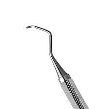
Explorer |
Fx: To examine teeth for decay (caries), calculus, furcation, or canals and other anomalies Characteristics: Pointed tips: sharp, thin and flexible Variations: Orbin, Shepherds, Pigtail |
|
|
Perioprobe |
Fx: incremental marks on tip to measure periodontal pockets Characteristics: Designed with different increments |
|
|
Vestibular |
Surface of tooth facing the vestibule or lips |