![]()
![]()
![]()
Use LEFT and RIGHT arrow keys to navigate between flashcards;
Use UP and DOWN arrow keys to flip the card;
H to show hint;
A reads text to speech;
100 Cards in this Set
- Front
- Back
|
which of the following is NOT associated with low-Q transducer? a. wide bandwidth b. multifrequency selection c. long pulse length d. diagnostic pulsed-wave ultrasound e. loss of most energy in the first few vibrations |
c. long pulse length transducers used for diagnostic ultrasound are low-Q transducers. low-Q transducers have a wide bandwidth, which allows multifrequency selection. they have short pulses for good axial resolution. because the pulse is short, most of the energy within it is lost after the first few vibrations. |
|
the image shows clear distinction between the gray scale of various structures in the fetal brain. the ability to distinguish similar structures with varying gray scale is termed: a. axial resolution b. contrast resolution c. lateral resolution d. temporal resolution e. none of the above |
b. contrast resolution |
|
|
if the number of cycles in the transmitted pulsed wave is increased : a. axial resolution is degraded b. lateral resolution is degraded c. spatial pulse length becomes shorter d. rate of attenuation is increased e. penetration is decreased |
a. axial resolution is degraded axial resolution is proportional to pulse length. axial resolution is degraded as pulse length increases. pulse length is increased by increasing the number cycles in the pulse. |
|
|
if the frame rate is too slow for adequate temporal resolution, what action could you take to improve frame rate? a. reduce the number of transmit focal zones b. increase the scan line density c. increase the scanning depth d. decrease the dynamic range e. reduce the transmit frequency |
a. reduce the number of transmit focal zones a pulse/listen cycle is required for each transmit zone. it follows then, that as the number of zones are increased, the time required to create one frame is decreased. |
|
|
you have been requested to image a superficial mass and to adjust your equipment to optimize spatial resolution. spatial resolution consists of: a. contrast and temporal resolution b. temporal and axial resolution c. axial and contrast resolution d. contrast and lateral resolution e. lateral and axial resolution |
e. lateral and axial resolution |
|
|
what is the purpose of the radiofrequency shield that is placed around the crystal and backing material of the ultrasound probe? a. it helps to transmit radio waves into the body. b. it aids transmission of the electric signal to the crystal. c. it reduces the incoming electrical signal strength from the body. d. it reduces electromagnetic interference e. it improves axial resolution |
d. it reduces electromagnetic interference electromagnetic interference contributes to the noise level and reduces detection of weak echoes. the use of a radiofrequency shield around the crystal and backing material reduces this noise and enhances sensitivity to weak signals |
|
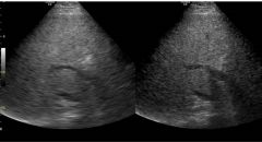
the image on the left demonstrates poor lateral resolution compared to the image on the right. how were the system controls adjusted to optimize the image on the right? a. increase dynamic range b. decrease transducer frequency c. change gray map d. increase line density e. reduce frame averaging |
d. increase line density the image on the left shows lateral smearing of the echoes. this is the result of poor lateral resolution. by increasing line density, lateral resolution is improved in the image on the right |
|
|
what type of resolution is affected most by pulse duration? a. lateral resolution b. contrast resolution c. temporal resolution d. axial resolution e. elevational resolution |
d. axial resolution we usually think of axial resolution as being determined by spatial pulse length. spatial pulse length and pulse duration are directly related. pulse duration ( also called temporal pulse length) is the length of time required to complete one pulse. the shorter the pulse duration, the better the axial resolution. |
|
|
with a standard one dimensional linear array transducer, what type of resolution is affected most by electronic focusing and dynamic aperture? a. lateral resolution b. contrast resolution c. temporal resolution d. axial resolution e. elevational resolution |
a. lateral resolution lateral resolution is improved at depth by increasing transducer width or aperture size in array. essentially, the larger the aperture (the active portion of the array), the smaller the beam width at the focal point. |
|
|
what type of resolution is most affected by the mechanical focus on a linear array transducer? a. lateral resolution b. contrast resolution c. temporal resolution d. axial resolution e. elevational resolution |
e. elevational resolution elevational resolution is determined by the slice thickness. (slice thickness is also known as the z-axis or elevational plane). elevational resolution is a measure of the beam width perpendicular to the imaging plane. on linear, curved linear, and phased array probes, elevational resolution is determined by the mechanical focus that is placed along the width of the array. the mechanical focus narrows the beam at one fixed point in the z-axis. |
|
|
the spectrum of frequencies emitted by a pulsed-wave transducer is known as: a. Reynolds number b. center frequency c. F-number d. resonance frequency e. bandwidth |
e. bandwidth |
|
|
what is the optimal thickness for the matching layer? a. equal to the thickness of the crystal b. one-fourth of a wavelength c. one-half of the pulse length d. twice the transducer e. one-half the width of the backing material |
b. one-fourth of a wavelength |
|
|
a method used to improve frame rates with multizone electronic focusing is termed: a. apodization b. parallel processing c. F-number focusing d. low Q transducers e. none of the above |
b. parallel processing with parallel processing (also called coprocessing), frame rates can be improved simultaneously acquiring data for multiple acoustic scan lines. this is accomplished by firing one set of elements and forming more than one acoustic scan line signal with the signals by the active elements. |
|
|
which of the following best describes the F-number? a. focal length divided by the aperture b. aperture multiplied by the number of elements in the transducer c. number of transducer elements divided by the Fraunhofer zone d. beam width 1cm from the transducer surface e. focal length multiplied by the number of elements in the transducer |
a. focal length divided by the aperture the F-number is the ratio of the focal length to the size of the aperture. |
|
|
if you wish to use an array transducer that allows you to control transmit focal depth, what type would you select? a. linear array b. convex array c. phased array d. annular array e. any of the above |
e. any of the above all array transducer types permit operator control of transmit focal depth |
|
|
if you wish to perform a sonogram with the largest possible field of view in both near field and at depth, what type of array transducer would be most preferable? a. linear array b. curved array c. phased array d. sector e. any of the above |
b. curved array |
|
|
a commonly used material in modern transducer elements is: a. tungsten powder and epoxy resin b. quartz c. lead zirconate titanate d. rubber e. none of the above |
c. lead zirconate titanate |
|
|
you are using a linear array transducer to evaluate the thyroid gland. for standard B-mode imaging, the transducer most likely produces pulses of how many cycles? a. one to three b. three to six c. six to nine d. nine to twelve e. twelve to fifteen |
a. one to three transducers used for diagnostic ultrasound are damped to improve axial resolution. damping reduces the pulse duration and spatial pulse length. these transducers typically have pulse lengths of one to three cycles. longer pulse trains carry more energy and provide greater penetration but give very poor axial resolution. |
|
|
what is the purpose of applying multiple matching layers to the transducer face? a. the gel gradually erodes the matching layer, and using multiple layers lengthens the life of the transducer assembly b. because the impedance of tissue is quite variable from one patient to the next, multiple matching layers improve the likelihood of providing a close impedance match to each c. each individual coating of the matching layer contributes to the damping of the sound pulse, which results in improved axial resolution d. multiple matching layers result in increased transducer bandwidth e. each coating of the matching layer provides focusing at a specific depth. with multiple layers, multiple points of focus are possible |
d. multiple matching layers result in increased transducer bandwidth the purpose of the matching layer is to reduce the impedance mismatch between the crystal and the tissue. this will improve sound transmission into the body and increase the transducer bandwidth. the optimal thickness for the matching layer is one-fourth of the wavelength of the center frequency of the transducer. use of multiple matching layers will increase the transducer bandwidth with the result of improving axial resolution. |
|
|
for a nonfocused transducer, the region between the transducer element and the point at which the sound beam is the narrowest is called: a. far field b. Fraunhofer zone c. near zone length d. focus distance e. both A and B |
c. near zone length the near zone length is also called the Fresnel zone or the near field. the Fraunhofer zone is the term used to describe the far field. |
|
|
axial resolution is chiefly determined by: a. beam width b. transducer diameter c. pulse duration d. line density e. frame rate |
c. pulse duration |
|
|
what is the main advantage of using an intracavity probe compared to a standard transducer? a. the intracavitary probe has superior focusing capabilities because of the increased radius of the curved probe face b. the intracavitary probe uses a lower frequency that provides superior penetration and closer inspection of the pelvic structures c. the intracavitary probe does not demonstrate side lobes or grating lobes because of the close spacing and curvature of the elements d. since the intracavitary probe is closer to the area of interest, a higher frequency transducer can be used, resulting in superior spatial resolution e. bioeffects are greatly reduced with the intracavitay probe because of its frequency range and power output |
d. since the intracavitary probe is closer to the area of interest, a higher frequency transducer can be used, resulting in superior spatial resolution |
|
|
what defines the best axial resolution that can be obtained with a given transducer? a. spatial pulse length x (times) 2 b. spatial pulse length x (times) 4 c. spatial pulse length divided by 2 d. spatial pulse length divided by 4 e. equal to the spatial pulse length |
c. spatial pulse length divided by 2
|
|
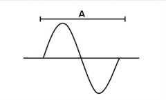
the following illustration depicts acoustic pressure vs. distance in a medium in which a sound wave is present. what sound parameter is measured by the length of the bar labeled A in the following illustration? a. amplitude b. wavelength c. pressure d. pulse repetition frequency e. duty factor |
b. wavelength |
|
|
what term describes the ability of an imagine device to separate closely spaced objects?
a. resolution b. penetration c. duty factor d. interference e. reflection |
a. resolution |
|
|
what happens to pressure when pressure is applied to a piezoelectric crystal? a. it emits and electrical signal b. it increases temperature c. it emits radio waves d. it emits photons e. it becomes phosphorescent |
a. it emits an electrical signal |
|
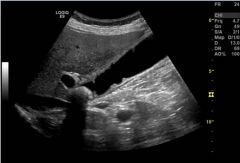
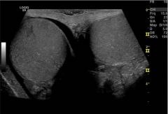
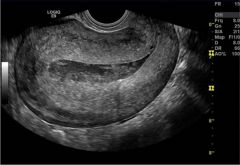
what type of transducer was used to obtain this image? a. linear array b. sector c. phased array d. micro convex e. curved array |
e. curved array |
|
|
the interpreting physician has asked you to use a stand-off pad to evaluate a superficial mass. what is the main advantage the stand-off pad provides to improve the image? a. the stand-off pas has an acoustic impedance closer to that of the crystal which results in improved transmission of sound into the body b. the stand-off pad has a propagation speed equal to twice that of soft tissue, which decreases the wavelength of the sound beam c. the stand-off pad increases the distance between the transducer and the mass, making it more likely that the mass will be located close to the elevational focus of the sound beam d. the stand-off pad creates a soft interface between the crystal an the tissue, increasing the constructive interference and reducing scattering. e. the stand-off pad eliminates artifacts associated with reverberation, side lobes, and grating lobes. |
c. the stand-off pad increases the distance between the transducer and the mass, making it more likely that the mass will be located close to the elevational focus of the sound beam
|
|
|
what array transducer fires all of the elements for each acoustic scan line, using small time delays to steer the beam? a. linear segmental array b. convex array c. annular array d. linear sequential array e. phased array |
e. phased array the phased array transducer fires all of its elements to create each acoustic scan line. small time delays are employed to focus the beam and beam steering. |
|

the shape corresponding to the image created by a phased array transducer most closely corresponds to which of the following? a. A b. B c. C d. D e. E |
d. D |
|

a linear array will produce which of the following image shapes? a. A or B b. B or C c. C or D d. D or E e. A, C or E |
e. A, C or E shape A is the most typical of linear array. shape C occurs when the linear array is steered in both directions resulting in a trapezoidal display. shape E occurs when the linear array is steered in one direction |
|
|
if you are using a transducer with poor elevation resolution, which of the following problems are you most likely to encounter? a. inability to resolve two structures that lie close together and parallel to the path of the beam b. inability to resolve two structures that lie close together, perpendicular to the path of the beam, and parallel to the long axis of the transducer c. inability to penetrate through dense tissue d. inability to clearly demonstrate small cystic structures e. inability to demonstrate rapidly moving structures accurately |
d. inability to clearly demonstrate small cystic structures eleavational resolution is determined by beam slice thickness. if the slice is thick, small cystic structures may not appear cystic. they will be displayed with the internal echoes reflected from the surrounding tissue. this occurs because the beam is wider than the cystic structure, and tissue on either side reflects the sound beam from the same depth as the cyst. since the echoes arrive back at the transducer at a round-trip time that is consistent with both the tissue and cyst, they are assigned the same depth location on the image. if the beam slice is narrower, it will not cover the cystic structure and the surrounding tissue at the same time. since the fluid portion of eh cyst will not send back reflections, it will then appear anechoic. |
|
|
what can you do to achieve a higher frame rate? a. decrease the number of focal zones b. decrease the line density c. decrease sector width d. all of the above e. none of the above |
d. all of the above (A, B, C) a. decrease the number of focal zones b. decrease the line density c. decrease sector width |
|
|
what can you do to improve contrast resolution in the ultrasound image? a. increase sector width b. decrease the number of focal zones c. use a 2D or matrix array transducer d. use a phased array transducer e. use a curved array transducer |
c. use a 2D or matrix array transducer
a 2d or matrix array transducer has both rows and columns of elements. this allows electronic focusing in the out-of-plane dimension with the result that the slice thickness of the sound beam is thinner compared to traditional 1D transducers. contrast resolution is improved with thin slices due to decreased volume averaging. |
|
|
increasing which of the following will result in improved axial resolution? a. pulse duration b. pulse length c. frequency d. period e. focusing |
c. frequency axial resolution is improved by reducing pulse length or pulse duration. this can be accomplished by reducing the number of cycles in a pulse (damping) or by reducing the size of the wavelength (increased frequency). |
|
|
which represents the best measure of resolution for modern day ultrasound scanners? a. contrast b. axial c. elevational d. temporal e. lateral |
b. axial axial resolution is the best measure of resolution for today's instruments. the worst measure of resolution is usually elevational. lateral and elevational resolutions vary considerably with depth. axial resolution does not. |
|
|
which of the following transducers produces side lobes? a. mechanical sector b. linear sequential array c. phased array d. convex array e. all of the above |
e. all of the above (A, B, C, D) a. mechanical sector b. linear sequential array c. phased array d. convex array side lobes are secondary beams of energy that are displaced from the main beam and produced by all probes. side lobes can be made as much as 60 to 100dB less intense than the main beam through broad bandwidth excitation of the transducer and through apodization of array transducers. at higher intensities, they produce noise and artifacts in the image |
|
|
which transducer type is best suited to vascular imaging? a. mechanical sector b. linear array c. phased array d. convex array e. annular array |
b. linear array |
|
|
which of the following transducer types is best suited for cardiac imaging? a. mechanical sector b. linear array c. phased array d. convex array e. annular array |
c. phased array |
|
|
which of the following can NOT be evaluated with a tissue equivalent phantom? a. axial resolution b. lateral resolution c. contrast resolution d. slice thickness e. temporal resolution |
e. temporal resolution
temporal resolution is related to the amount of time it takes to complete a single frame of information. this cannot be assessed with a tissue phantom in which there are no moving structures. |
|
|
increasing the frequency on a multifrequency transducer from 3.5MHz to 5.0MHz will: a. increase the sound propagation speed b. increase the pulse length c. increase penetration d. decrease the pulse repetition frequency e. decrease the wavelength |
e. decrease the wavelength
|
|
|
you notice a loss of detail in the lateral dimension of the ultrasound image. what can you do to improve this? a. decrease the number of focal zones b. increase the scan line density c. decrease the transducer frequency d. increase the frame averaging e. increase the dynamic range |
b. increase the scan line density
a loss of detail in the lateral dimension is related to poor lateral resolution. this can be improved by increasing the scan line density, increasing the number of focal zones, or increasing the transducer frequency. the frame averaging and dynamic range will NOT affect lateral resolution. |
|
|
you are performing a sonogram on a patient with a highly attenuating tissue. which of the following solutions would be most likely to improve penetration through the tissue? a. decrease dynamic range b. decrease near field TGC c. choose a lower frequency transducer d. increase the scan line density e. change the gray scale map |
c. choose a lower frequency transducer |
|
the image was obtained with a linear array transducer. the trapezoidal field of view increases the field of view. how is the trapezoidal shape created? a. electronic beam steering b. side lobes c. grating lobes d. mechanically steering the elements e. mirror |
a. electronic beam steering a trapezoidal image can be obtained with a linear array transducer by electronically steering the beam |
|
in the image to the left, how many transmit focal zones are being used? a. the number of transmit focal zones is not indicated b. 1 c. 2 d. 3 e. 4 |
d. 3 the number of transmit focal zones is shown by an indicator on the screen. the symbol used varies somewhat by manufacturer, but is clearly visible with one symbol per focal zone. |
|

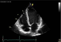
what type of probe was used to take this image? a. curved array b. phased array c. linear array d. mechanical sector e. tightly curved array |
b. phased array |
|
what best describes the method used to create the triangular shaped image format in this image? a. mechanical steering b. electronic steering with elements fired in small groups c. electronic steering with most elements fired for each pulse echo sequence. small time differences steer the beam d. curved lens e. mirror |
c. electronic steering with most elements fired for each pulse echo sequence. small time differences steer the beam
phased array transducers (sometimes called linear phased array or sector transducers) form the beam by firing most, if not all, elements at once with small time differences. the time delays are changed with each subsequent firing so that the beam direction is varied. the result is that the beam is electronically steered to create the sector image format. |
|
|
you can reduce beam width to improve spatial resolution in the image by which of the following? a. focusing b. decreasing aperture c. decreasing transducer diameter d. decreasing transducer width e. increasing backing material |
a. focusing |
|
|
which transducer frequency would have the thinnest crystal? a. one with a 15MHz center frequency b. one with a 10MHz center frequency c. one with a 7MHz center frequency d. one with a 5MHz center frequency e. all would have the same crystal thickness |
a. one with a 15MHz center frequency
the crystal (or element) must be thinner for higher frequencies |
|
|
what component of a pulsed wave transducer helps to reduce the spatial pulse length and improve axial resolution? a. matching layer b. lens c. element d. backing material e. radiofrequency shield |
d. backing material the backing material reduces the length of time the crystal rings. this, in turn, shortens the spatial pulse length and improves axial resolution. |
|
|
which transducer would have a lower line density in the far field compared to the near field? a. linear array b. curved array c. tightly curved array d. phased array e. B, C, and D |
e. B, C, and D
b. curved array c. tightly curved array d. phased array only the linear array transducer has the same line density for both the near and far field. all transducers producing a curved or sector image format have more spreading of the lines in the far field. this results in lower lateral resolution in the far field. |
|
|
what transducer component reduces reflection at the transducer/skin interface? a. matching layer b. lens c. element d. backing material e. radiofrequency shield |
a. matching layer |
|
|
axial resolution is improved with: a. increased bandwidth b. shorter pulse length c. shorter wavelength d. B and C e. all of the above |
e. all of the above
a. increased bandwidth b. shorter pulse length c. shorter wavelength |
|
|
what is a side lobe? a. uneven transducer element b. accessory pulse used for electronic steering c. sound beam emitted from the transducer that travels in a different direction from the main beam d. nonlinear propagation of the sound beam e. additional backing material on the transducer edge used to reduce lateral vibrations |
c. sound beam emitted from the transducer that travels in a different direction from the main beam
|
|
|
what is the purpose of curving the transducer elements or applying a lens over the elements? a. to provide focusing in the out-of-plane (elevational) direction b. to reduce the acoustic impedance mismatch between the crystal and skin c. to reduce crystal ringing and improve axial resolution d. to help reduce electronic interference e. all of the above |
a. to provide focusing in the out-of-plane
(elevational) direction |
|
what advantage is obtained by steering a linear array transducer to create a trapezoidal shape to display as demonstrated in this image? a. improved lateral resolution b. improved axial resolution c. improved contrast resolution d. improved elevational resolution e. improved field of view |
e. improved field of view
the steered beams allow for increased field of view along both edges of the displayed image. lateral resolution is slightly decreased in the far field because the scan lines are not farther apart, but axial, contrast, and elevational resolution are not affected. |
|
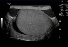
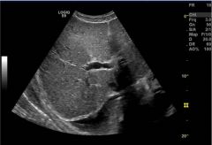
what type of transducer was used to create this image of the uterus? a. linear array b. phased array c. single element mechanical d. tightly curved array e. sector |
d. tightly curved array a tightly curved array has elements arranged in a linear fashion on a tightly curved radius. the tight curvature increases the field of view while keeping the probe size small. |
|
|
what disadvantage is related to the use of a tightly curved array transducer? a. axial resolution degrades with depth b. lateral resolution degrades with depth c. field of view is compromised compared to the other transducer types d. temporal resolution is compromised compared to the other transducer types. e. elevation resolution is compromised compared to the other transducer types |
b. lateral resolution degrades with depth lateral resolution degrades with depth because the scan lines become spaced farther apart in the far field compared to the near field. |
|
|
what advantage is related to the use of a tightly curved array transducer? a. axial resolution is improved b. lateral resolution is improved c. field of view is improved d. temporal resolution is improved e. elevation resolution is improved |
c. field of view is improved
|
|
|
what best describes the difference between the ultrasound frequencies used for doppler and for B-mode imaging with a given transducer? a. the frequency for doppler is usually lower than for imaging b. the frequency for doppler is usually higher than for imaging c. the transmit pulse for doppler has a greater bandwidth compared to the bandwidth for imaging d. the frequency for doppler and for B-mode imaging must be identical for a given transducer e. the frequency for doppler is usually double for B-mode imaging |
a. the frequency for doppler is usually lower than for imaging
in general, the transmitted frequency for doppler studies is lower than the imaging frequency for a given transducer. this relates to the different types of reflectors being evaluated in doppler (red blood cells) and imaging (tissue). since the scattered echoes from blood are much weaker than echoes from tissue, lower frequencies help overcome attenuation losses. |
|
|
in order to focus a sound beam relatively far away from the transducer, it is advantageous to: a. increase the thickness of the element b. increase the diameter of the element c. decrease the width of the element d. decrease the frequency of the element e. increase the curvature of the element |
b. increase the diameter of the element
|
|
|
which factor LEAST influences axial resolution? a. damping b. beam width c. frequency d. pulse duration e. pulse frequency |
b. beam width
beam width affects lateral resolution rather than axial resolution. all others are factors that influence axial resolution. |
|
|
spatial pulse length is equal to: a. the wavelength times the number of cycles in the pulse b. the wavelength minus the number of cycles in the pulse c. the wavelength times the axial resolution d. the wavelength times the beam width e. the wavelength divided by two |
a. the wavelength times the number of cycles in the pulse
it is more common for engineers to specify the pulse duration. then spatial pulse length is equal to the pulse duration times the speed of sound. |
|
|
what is the disadvantage of using multiple transmit focal zones? a. decreased lateral resolution b. decreased temporal resolution c. decreased elevation resolution d. decreased axial resolution e. all of the above |
b. decreased temporal resolution
each transmit focal position requires a separate pulse echo sequence (transducer firing). |
|
|
what is the advantage of using multiple transmit focal zones? a. increased lateral resolution b. increased temporal resolution c. increased elevation resolution d. increased axial resolution e. all of the above |
a. increased lateral resolution
|
|
|
what factor LEAST influences lateral resolution? a. frequency b. focus depth c. beam width d. transducer diameter e. damping |
e. damping
|
|
|
which transducer will exhibit the LEAST amount of volume averaging? a. mechanical single element b. 1.5D or multi-row array c. linear array d. phased array e. convex array |
b. 1.5D or multi-row array
it allows electronic focusing in the elevational (out of plane) direction. |
|
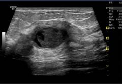
the ability to differentiate between two regions having similar echogenicity, such as in this liver image with focal fat, is termed: a. temporal resolution b. contrast resolution c. axial resolution d. lateral resolution e. elevation resolution |
b. contrast resolution
|
|
|
imaging frame rate decreases when you: a. decrease frequency b. increase dynamic range c. increase frame averaging d. increase sector width e. decrease the number of transmit focal zones |
d. increase sector width
by increasing the sector width, the user forces the system to fire more lines of sight (scan lines) for each imaging frame. this increases the length of time it takes to create each frame and causes a decrease in frame rate. |
|
|
the optimal transducer technology for cardiac scanning is: a. curved array b. tightly curved array c. linear array d. phased array e. mechanical sector |
d. phased array
a phased array transducer has a small surface area with a flat face. this allows for the best contact between the ribs. in addition, high frame rates can be achieved by electronically steering the beam. |
|
|
what system control determines the amount of amplification that occurs in the receiver? a. gain b. acoustic power output c. rectification d. pulse repetition frequency e. dynamic range |
a. gain
|
|
|
when you adjust the output power control, you affect the following system component: a. pulser b. beam former c. scan converter d. memory e. receiver |
a. pulser
|
|
|
what system control do you adjust to equalize the differences in echo amplitudes received from similar structures situated at different depths? a. dynamic range or compression b. rectification c. time gain compensation d. pulse repetition frequency e. rejection |
c. time gain compensation
|
|
|
electronic noise is reduced in the ultrasound system by this method: a. demodulation b. compensation c. rectification d. amplification e. rejection |
e. rejection
|
|
|
you have chosen to enlarge an ultrasound image with a read magnification. which of the following is NOT related to read magnification? a. preprocessing b. increased pixel size c. performance on a frozen image d. resolution loss e. B and D only |
a. preprocessing
read magnification is a postprocessing method of enlarging a portion of the ultrasound image. a preprocessing method of enlarging the image is termed write magnification. read magnification increases pixel size and results in some resolution loss in the enlarged image. |
|
|
what term below describes the rate at which the transmitter applies electronic voltage pulses to the transducer? a. period b. pulse repetition frequency c. depth gain compensation d. demodulation e. apodization |
b. pulse repetition frequency
the pulse repetition frequency (PRF) is the number of pulses emitted by the transducer per second. the number of pulses determined by the rate (per second) at which the crystal is stimulated by electronic voltage from the transmitter. |
|
|
pulsing the transmitted sound wave is necessary for real time imaging because: a. the transducer becomes too hot to handle if continuous sound waves are emitted b. the crystal in the transducer will break under the stress of the continuous emissions c. the depth of the interface from which the echo originated can be determined d. lateral resolution is improved by pulsed transmission e. temporal resolution is improved by pulsed transmission |
c. the depth of the interface from which the echo originated can be determined
pulsed wave (PW) ultrasound determines the depth of origin of reflected echoes by use of the range equation. since the speed of sound in tissue is known, the system can count the elapsed time after pulse transmission to when the echo is received and determine the depth from which the reflection originated. |
|
|
what receiver function is responsible for decreasing the difference between the smallest and largest received signal amplitudes? a. amplification b. compensation c. compression d. demodulation e. rejection |
c. compression
the range of echo strengths reflected from the tissue is too great for the display to handle. for this reason, the difference between the smallest and largest amplitudes of the received signals must be reduced. this is accomplished by a logarithmic compression, which amplifies weak signals more than strong signals. |
|
|
what receiver function listed below is NOT operator adjustable? a. amplification b. compensation c. demodulation d. rejection e. A and D |
c. demodulation the operator can control amplification, compensation and rejection. |
|
|
while performing a sonographic examination, you have performed both preprocessing and postprocessing functions. which of the following functions is postprocessing? a. white zoom b. frequency change c. gray scale map assignment d. scan line density e. acoustic power output |
c. gray scale map assignment
the easiest way to determine if a function is pre- or postprocessing is this: if it can't be performed on a frozen image, it is preprocessing; if it can be performed on a frozen image, it is postprocessing. |
|
|
which of the following would be most helpful to enhance the contrast difference between tissues having subtle variations in echogenicity? a. decreasing the acoustic power output b. decreasing the scan line density c. performing a read-zoom over the area of interest d. changing the gray scale map assignment e. increasing the overall receiver gain |
d. changing the gray scale map assignment the gray scale map assignment determines the shades of gray that are assigned to incoming signal amplitudes. because the maximum number of gray shades is predetermined, the distribution of brightness levels over the range of signal amplitudes must be set by some assignment. the is accomplished by various assignment curves in which different signal amplitudes are given their gray assignment. common curves are a linear or S-curve assignment. the contrast resolution in a certain range may be diminished or emphasized, depending on the map selection. |
|
|
what part of the sonographic instrument is responsible for apodization, beam steering, focusing, and aperture control? a. beam former b. receiver c. memory d. pulser e. scan converter |
a. beam former |
|
|
what control should you adjust to better compensate for the attenuation of sound as it propagates through tissue? a. dynamic range b. time gain compensation c. acoustic power output d. rejection e. focusing |
b. time gain compensation |
|
|
what control would you adjust to increase the intensity of the transmitted pulse? a. receiver gain b. depth of scanning c. output power d. time gain compensation e. dynamic range |
c. output power the output power control is used by the operator to adjust the magnitude of the voltage pulse applied to the crystal. as it increases, the intensity of the transmitted pulse increases. |
|
|
which control would you adjust to alter the dynamic range of the displayed echoes? a. compression b. transmit power c. scanning depth d. time gain compensation e. focusing |
a. compression the compression control reduces the difference between the largest and smallest reflected signal amplitudes, decreasing the dynamic range of the incoming signal to a level that the display can handle. |
|
|
the technique of frame averaging during real time acquisition is designed to: a. reduce random noise b. decrease pixel size c. redistribute the gray scale d. enhance spatial resolution e. increase frame rate |
a. reduce random noise frame averaging (also known as persistence) averages the data in pixels over successive frames. (the number of averaged frames is set by the user.) slight movements of the transducer or patient result in slight changes in the speckle pattern, so frame averaging has the effect of smoothing the image or reducing speckle. additionally, electronic noise is smoothed. |
|
|
you are performing a sonographic examination and select the tissue harmonics operating mode. what advantage will you obtain over conventional imaging? a. improved contrast resolution b. improved penetration c. improved temporal resolution d. improved signal-to-noise ratio e. increased bandwidth |
a. improved contrast resolution tissue harmonics is a new method of ultrasound imaging in which the harmonic of the transmitted frequency is used to create the image. typically, the second harmonic is used, which is twice the transmitted frequency. since the harmonic frequency of the transmitted pulse is generated within the body, it has numerous advantages over conventional imaging. improved contrast resolution is one of the main advantages. there are several reasons for the improvement. first, contrast resolution is always improved by increasing frequency, and harmonics always results in use of a higher frequency. secondly, harmonics reduces clutter and side lobe artifacts. the absence of these extraneous echoes improves contrast resolution compared to conventional scanning methods. additionally, effects of reverberations are reduced because the harmonic component is weak at shallow depths where reverberations often occur. |
|
|
you have decreased the scan line density. what technique will be employed to fill in the empty data between the scan lines? a. interpolation b. rejection c. compression d. autocorrelation e. demodulation |
a. interpolation interpolation is used to fill in the missing data between scan lines in both gray scale and color doppler. the missing data are estimated from the data present in nearby pixels on adjacent scan lines. |
|
|
while performing an ultrasound examination, you decide to adjust the system settings to improve the image. which of the following is NOT user-adjustable? a. frequency b. power c. gain d. compression e. echo arrival time |
e. echo arrival time echo arrival time is determined by the medium through which the sounds travels. |
|
|
what control should you adjust to optimize the image if you are scanning a structure that produces very bright echoes on the display? a. rejection b. TGC c. edge enhancement d. frame averaging e. frequency |
b. TGC |
|
|
to generate a sonographic image, what is the order in which the following system components are activated?
a. pulser, receiver, display, beam former, memory b. pulser, beam former, receiver, memory, display c. beam former, pulser, memory, display, receiver d. memory, beam former, pulser, receiver, display e. beam former, memory, pulser, receive, display |
b. pulser, beam former, receiver, memory,
display the action begins with the pulser (which contains the clock). the pulser produces the electric voltage pulses to the beam former and on the transducer. the returning echoes are sent to the receiver. the resultant image is stored in the memory and then displayed on the video monitor. |
|
|
what is a typical frame rate for B-mode real time imaging? a. 1-9kHz b. 10-50Hz c. 100-200Mhz d. 200-300Hz e. 0.05-0.9Hz |
b. 10-50Hz
although frame rates vary, typical real time frame rates range from about 10Hz to 50Hz. faster frame rates are found with cardiac imaging. frame rates are displayed in terms of Hz a unit of frequency equal to one cycle per second. if the frame rate is 30Hz, that means 30 frames are produced in one second. |
|
|
what control could you adjust to improve the signal-to-noise ratio on the image? a. dynamic range b. frame averaging c. gray-scale map d. edge enhancement e. receiver gain |
b. frame averaging |
|
|
the ratio of the largest to the smallest signal that a system can handle is termed: a. apodization b. compression c. threshold d. dynamic range e. pulse repetition frequency |
d. dynamic range |
|
|
the term duty factor is defined as: a. the fraction of time the transducer is actively transmitting sound b. the fraction of time the transducer is actively receiving sound c. the fraction of time between the transmitted and received sound pulse d. the fraction of time between transmitted sound pulses e. the fraction of time required for round-trip travel of a sound pulse to one cm in tissue |
a. the fraction of time the transducer is actively transmitting sound
this value is quite low in diagnostic ultrasound, typically less than 0.01 (1%). |
|
|
if you increase the pulse repetition frequency and leave all other controls unchanged, what will happen? a. lateral resolution will improve b. frame rate will increase c. frame rate will decrease d. axial resolution will improve e. frequency will increase |
b. frame rate will increase
the pulse repetition frequency (PRF) is the number of pulses emitted by the transducer in one second. if the PRF is increased and all other factors remain the same, the frame rate will increase because it will take less time to fire all the necessary pulses to create one frame. |
|
|
if you increase pulse repetition frequency to a level too great for the depth of field, the result will be: a. increased side lobes b. increased grating lobes c. range ambiguity d. decreased frame rate e. increased electronic noise |
c. range ambiguity
if the PRF is too high for the imaging depth, range ambiguity will result. the PRF is set so that a pulse can be received from the bottom of the field of view before the next pulse is transmitted. if another pulse is transmitted before all echoes from the first pulse are received, then the echoes would be misplaced axially on the image. why? because the system must assume that all received echoes have arrived from the most recently transmitted pulse. |
|
|
you have the ability to adjust the following controls during an abdominal ultrasound study. which control most closely affects patient exposure? a. receiver overall gain b. TGC c. dynamic range d. reject e. output power |
e. output power Gain and TGC (time gain compensation) amplify the reflected signal. they do not increase patient exposure. |
|
|
what effect will you see in the image if you increase the reject level? a. increased number of shades of gray b. decreased low-level echoes c. decreased frame rate d. decreased scanning depth e. decreased amplitude of the brightest shades of gray |
b. decreased low-level echoes
the reject control is used to eliminate weak echo signals from the display. as signal rejection increases, display of the weaker signals decreases. this results in fewer low-level gray echoes. |
|
|
if sound did not attenuate with increasing depth, what system control would you no longer need? a. dynamic range b. reject c. frame averaging d. TGC e. master gain |
d. TGC TGC (time gain compensation, also called depth gain compensation) is used to amplify echoes from deeper structures so that they appear as bright as similar structures located at more shallow depths. TGC is necessary because sound beam attenuation with depth results in reception of weaker signals from structures located deep in the image compared with those located deep in the image compared with those located more superficially. if the sound beam did not attenuate with depth (i.e., distance and travel time), TGC would not be needed. master gain is used to adjust the amplification of all the reflected signals, not just those at a specific depth. |