![]()
![]()
![]()
Use LEFT and RIGHT arrow keys to navigate between flashcards;
Use UP and DOWN arrow keys to flip the card;
H to show hint;
A reads text to speech;
22 Cards in this Set
- Front
- Back
|
Major Muscles and Muscle Groups: Torso- Back |
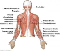
Trapezius, Latissimus Dorsi, Rhomboid Major, Erector Spinae |
|
|
Major Muscles and Muscle Groups: Torso- Front |
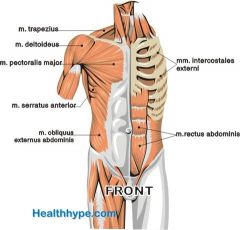
Pectoralis Major / minor (chest), Serratus Anterior, Rectus Abdominis, Internal / external obliques |
|
|
Major Muscles and Muscle Groups: Arms- Shoulder |
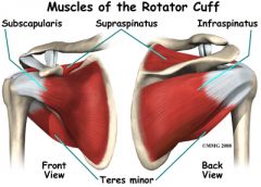
deltoids (anterior, medial, posterior), supraspinatus, infraspinatus teres major, teres minor |
|
|
Major Muscles and Muscle Groups: Arms |
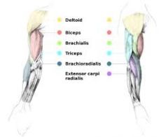
bicep brachii, brachialis, brachioradialis, triceps brachii |
|
|
Lower body- Hip / buttocks |
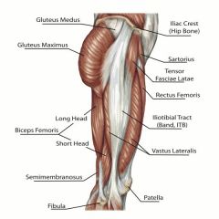
psoas major / minor, Iliacus, piriformus, gluteus |
|
|
Lower Body- Quads |
vastus intermedialus, rectus femoris, vastus lateralis, vastus medialis |
|
|
Lower Body- Hamstrings |
biceps femoris, semitendinosus, semimembranosus |
|
|
Muscle Cell Description |
Muscle cell is enclosed in epimysium. Within epimysium there are bundles of muscle fibers, calls Fasciculus, enclosed by perimysium. Within the Fasciculus, myofibrils are separated by endomysium. Myofibril's are enveloped in sarcolemma (muscle cell membrane). Muscle cells are also surrounded by sarcoplasm, which release calcium during contraction. Within the myofibril are myofiliments. |
|
|
Structure of Sarcomere: what is it and what is it comprised of? |
A sarcomere is the smallest contractile unit ofmuscle. It is comprised of actin andmyosin filaments. |
|
|
Describe the Z and M line and their relation to the sarcomere |
Z lines are at the ends of each sarcomere. M line is in the middle of the sarcomere. H Zone is where only the myosin filament is present |
|
|
Describe the I band and A band in relation to the sarcomere |
I band corresponds with the areas where only the Actin filament is. A band is the length of myosin filaments. |
|
|
What is the sliding filament theory? |
actin filaments at each end of the sarcomere slide inwards on myosin filaments, pulling z lines towards the center of the sarcomere and shortening / contracting muscle fiber. Both H - zone and I-Band shrink. |
|
|
phases of sliding filament theory |
resting: little calcium present (stored in sarcoplasmic reticulum). Actin and myosin have a weak bond interaction. Excitation-contraction coupling phase: before flexing, myosin and actin must attach. Sar. Reticulum releases calcium, binding with troponin, getting tropomyosin shifted away Contraction phase: cross-bridge flexion occurs from hydrolysis ofATP to ADP, this reaction catalyzed by myosin ATPase, another molecule of ATPmust replace ADP on cross-bridge to allow detachment and to recock forcontinued contraction Recharge phase: significant muscle shortening accomplished whencycle of contraction phase occurs over and over Relaxation phase: occurs when stimulation of motor nerve stops,calcium pumped back into sarcoplasmic reticulum |
|
|
Define different types of muscle action |
eccentric, concentric, and isometric |
|
|
How does the type of muscle action relate to the amount of force it is able to produce? |
Concentric – occurs when tensionin cross-bridges can overcome any resistance Isometric – occurs when tension incross-bridges is equal to resistance Eccentric – occurs when tension incross-bridges is less then external resistance |
|
|
What factors affect the production of force within a muscle? |
The number of cross-bridge heads attached at anyinstant, frequency of stimulation of motor units, number of motor unitsactivated, preloading – isometric forces first in order to generate concentricforces, cross-sectional area of the muscle, velocity of muscle shortening –more sarcoemeres in line allow for greater speed of movement but greater speedof movement produces less force while slower speed of movement produces greaterforce, angle of pennation – how the muscle fibers align with the tendon andmuscles with greater angle of pennation have more force capabilities, sarcomere and muscle length – peak forceusually at resting length where most cross bridge attachments occur, ,prestretching or stretch-shortening cycle, DOMS, muscle fiber type I or II |
|
|
What are the different types of muscle fibers? |
slow twitch (type I) and fast twitch fibers (type 2: 2A and 2X). Type 2 fatigues faster but has higher power output, while type 1 has higher endurance but lower power output. Type 2A fatigues slower than type 2x (higher aerobic-oxidative energy supply) |
|
|
What functions do muscle spindles perform, and where are they located? |
Muscle spindles are proprioceptive intrafusal muscle fibers that runparallel to extrafusal muscle. Theyprovide information concerning muscle length and rate of change. When muscle lengthens, spindles arestretched. The deformation activatessensory neuron, sends impulse to spinal cord, and synapses with motorneurons. Motor neurons send signal backindicating degree to which muscle must be activated. |
|
|
What functions do Golgitendon organs perform? Where are theylocated? |
GTO’s are proprioceptors located in tendons. Activated when tendon attached to activemuscle is stretched. Sensory neuron inGTO synapses with inhibitory neuron in spinal cord, which synapses withinnervating motor neuron. Motor neuronis inhibited resulting in reduction of tension in muscle. GTO’s inhibit muscle activation as aprotective mechanism. |
|
|
How do nerves stimulate muscle activity? |
action potential arrives atthe nerve terminal. Causes a release ofacetylcholine which diffuses across the neuromuscular junction, exciting thesarcolemma. With sufficient amount ofacetylcholine released, action potential is generated across sarcolemma and fiber contracts. |
|
|
How does nerve conduction velocity adapt to training? |
Twitch of a muscle can be increased by decreasing the time interval btwn.twitches. There is a greater summation of force until they actually fuse tobecome one, which is called tetanus. |
|
|
How do recruitment patterns adapt to training? |
increasing the frequency amount of motor neuron activation utilized to innervate the muscle leads to a more efficient movement pattern. Also, in trained individuals, athletes can typically skip over smallest to largest principle, and go straight to the muscle fiber needed the most from recruitment. |