Reading...
![]()
Play button
![]()
Play button
![]()
Use LEFT and RIGHT arrow keys to navigate between flashcards;
Use UP and DOWN arrow keys to flip the card;
H to show hint;
A reads text to speech;
38 Cards in this Set
- Front
- Back

|
|
|
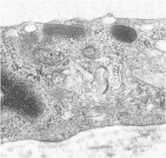
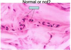
What are dark spots on top and what is this picture of?
|
picture is tunica intima and dark spots are called Weibel- Palade bodies which are cylindrical granules
|
|
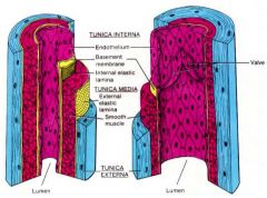
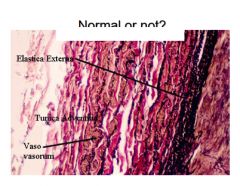
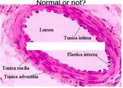
Label different layers briefly say important things
|
1. tunica intima- endothelium, basal lamina, type IV collagen, proteoglycans
2. tunica media- smooth muscles fibers, elastic and reticular fibers, external elastic lamina 3. tunica adventitia- longitudinaly arranged Collagen (type 1) and thickest layer in veins, vasa vasorum, thicker in veins |
|
Compare the blood vessels with Vein vs. Artery for ...
a. intima b. media c. adventitia |
a. intima (vein)- endothelium same for both
b. Media- artery many layers opposite veins c. Adventitia- thin in artery and vasa vasorum inn veins |
|

|
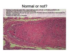
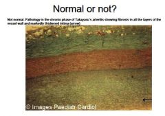
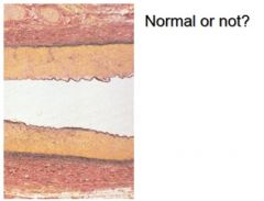
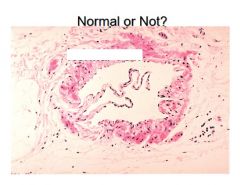
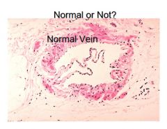
not Normal
|
|
|
|
|
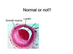
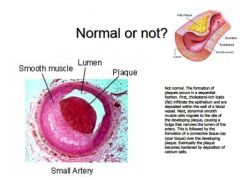
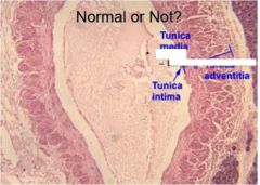
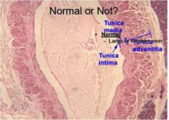
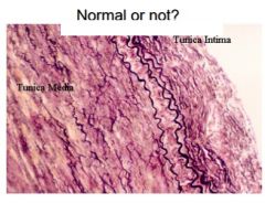
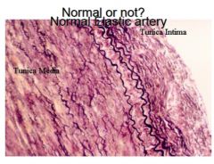
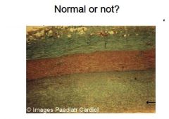
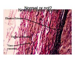
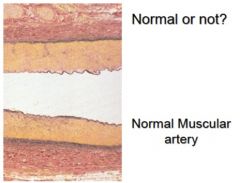
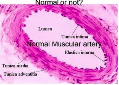
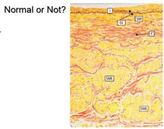
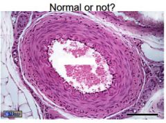
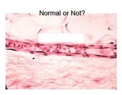
Normal or not?
|
not normal arterity
|
|
|
List the 3 main classes of arteries
|
1. large elastic or conducting arteries-
2. medium-sized muscular or distributing arteries- 3. small arteries and arterioles- |
|
|
Describe the following terms of tunic composition and function.
a. large artery |
1. TI-
subendothelial layer – INTERNAL ELASTIC LAMINA BETWEEN INTIMA & MEDIA • May be obscured by elastic lamina in T. media 2. T. media: 40-70 fenestrated elastic lamellae, Smooth muscle – Reticular (collagen II) fibers 3. T. adventitia: – Elastic & Collagen I fibers – Vasa vasorum |
|
|
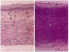
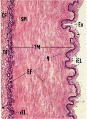
Describe the following terms of tunic composition and function.
a. muscular artery |
a. most arteries in body...
- Prominent internal elastic lamina 1. T media- 40 layers s.m., thick external elastic lamina (xEL) 2. T. Adventitia- mostly collagen fibers |
|
|
Describe the following terms of tunic composition and function.
a. small artery |
a. narrow lumen,
1. TI endothelium, no defined IEL 2. TM 1-5 continuous layers of sm 3. TA- thin |
|
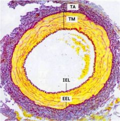
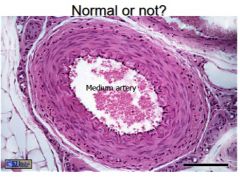
What is this (be specific)
|
medium sized artery
|
|
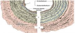
What is this? (be specific)
|
muscular artery, see prominent internal elastic lamina
|
|
Which is the vein and artery?
How can you tell? |
vein on right see squashed lumen
|
|
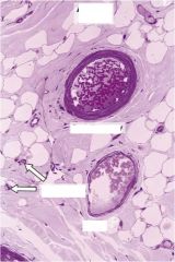
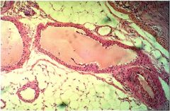
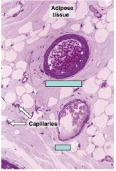
Identify the type of artery and veins here, also identify capillaries and adipose tissue
|
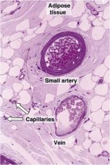
T. adventitia mroe developed in veins for small vessels
|
|

Which is the artery and which is the vein?
|
see larger squashed lumen in vein
|
|
Which is the artery and which is the vein?
|
see larger squashed lumen in vein
|
|
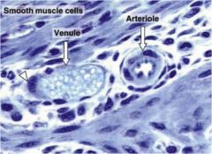
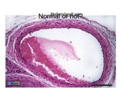
What type of vessel is this? how do u know?
|
medium sized veins due to best developed tunic, and no internal elastic lamina
|
|
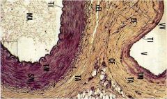
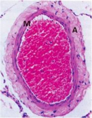
WHat is this and how can you tell which is which?
|
on the left is a vein and the right is an artery see the large adventitia on the vein and abnormal lumen (squashed) and the larger media with collagen on the left
|
|
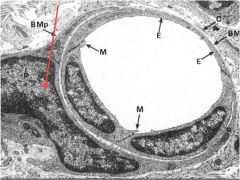
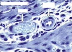
What is this pointing to? and what is EM of?
|
pericyte and EM of capillary
|
|
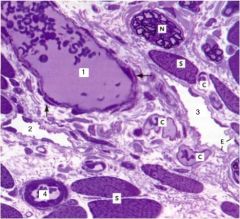
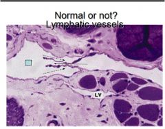
Label numbers also explain what two and three structure is like
|
Lymphatics:
Terminate in blood vascular system via 2 large trunks: thoracic duct & right lymphatic duct |
|
|
|
|

|
normal
|
|
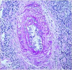
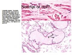
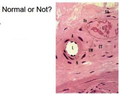
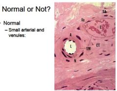
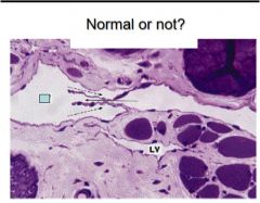
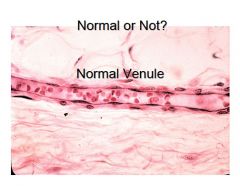
Normal or not? what is it?
|
small artery and vein normal
|
|
|
|
|
|
|
|
|
|
|
|
|
|
|
|
|
|
Normal capillary
|
|
|
|
|
|
normal large veins
|
|
|
normal
|
|

|

|
|
|
|
|
|
normal
|
|

|
Normal
|
|
|
|