Reading...
![]()
Play button
![]()
Play button
![]()
Use LEFT and RIGHT arrow keys to navigate between flashcards;
Use UP and DOWN arrow keys to flip the card;
H to show hint;
A reads text to speech;
65 Cards in this Set
- Front
- Back
- 3rd side (hint)
|
Describe the MECHANISMS OF EXCHANGE OF MATERIALS ACROSS THE CAPILLARY WALL
Besides open pores/ gaps... the are four more. list them. |
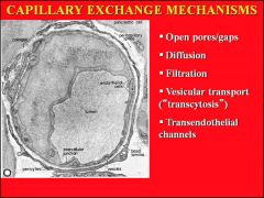
1. DIFFUSION
2. FILTRATION 3. VESICULAR TRANSPORT 4. TRANSENDOTHELIAL CELLS |
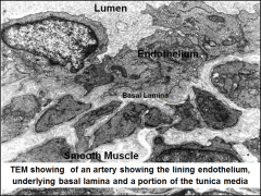
A. Diffusion: Gases and lipid soluble substances can pass across the plasmalemma.
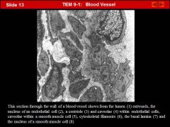
B. Filtration: Hydrostatic versus oncotic pressures causing the movement of water and small molecules between the "leaky tight junctions." C. Vesicular transport: Main mechanism: for movement of large molecules (> 9 nanometers, e.g. plasma proteins) across the endothelium by way of pinocytotic pits and vesicles. -Not energy dependent. D. Transendothelial channels: Formed by the fusion of vesicles and spanned by diaphragms. For transport of molecules < 9 nanometers in size. |
|
|
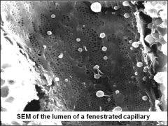
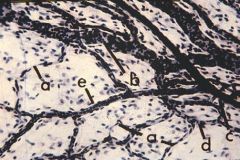
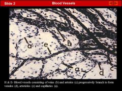
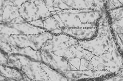
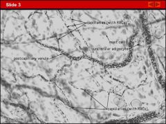
What are the three major types of capillaries?
|
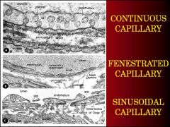
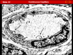
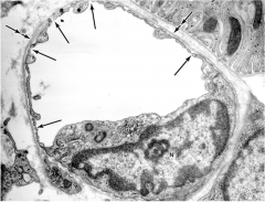
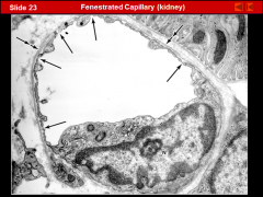
1. CONTINUOUS CAPILLARIES
2. FENESTRATED CAPILLARIES 3. SINUSOIDAL CAPILLAIRES |
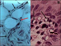
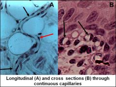
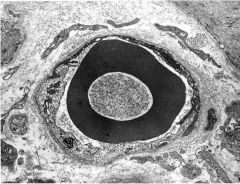
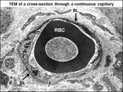
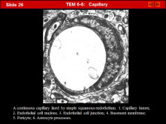
1. CONTINUOUS CAPILLARIES- The most common; found in muscle connective tissue, CNS, etc.
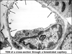
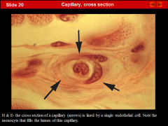
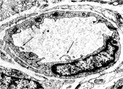
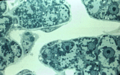
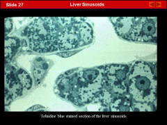
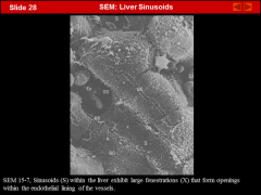
a. Luminal diameter of 7-9 microns. (Just large enough for a RBC to squeeze through) b. very THIN endothelial wall surrounded by a basal lamina. Endothelial NUCLEI BULGE into lumen. Scattered mitochondria, ribosomes, Golgi, microtubules and microfilaments. Most pronounced ultrastructural feature is numerous pinocytotic pits and vesicles (approximately 80 nanometers in diameter). c. Pericytes (perivascular cells): Function is not clear. May be able to differentiate into smooth muscle or phagocytic cells when the need arises. 2. FENESTRATED CAPILLAIRES a. Numerous PORES in lining endothelium. These pores are spanned by a thin diaphragm (e.g. found in intestines, kidneys, endocrine glands) b. DISCONTINOUS sinusoidal capillaries LACK DIAPHRAM with the open pores surrounded by a thick continuous basal lamina (e.g. renal glomerulus). 3. TRUE SINUSOIDS a. Usually large (up to 30-40 microns in diameter) with irregularly shaped lumens. b. There may be prominent intercellular spaces between endothelial cells and fenestrations in the endothelium. c. The basal lamina may be missing or discontinuous. d. Examples: Found in LIVER, SPLEEN, and BONE MARROW e. Fenestrated sinusoids: Large fenestrated capillaries. Examples: Found in ADENOHYPOHYSIS and ADRENAL GLAND |
|
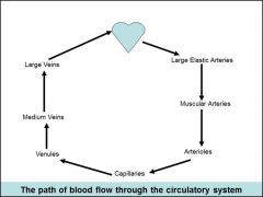
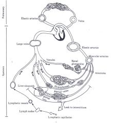
Generally, the CAPILLARY connects the arterial side to the venous side, BUT there are some important exceptions to this:
1. What do you call a system when the blood flows through not just one capillary bed, but also a SECOND capillary bed? 2. How about of when the path of blood flow SKIPS the capillary bed entirely? |
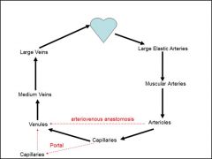
1.PORTAL SYSTEM
2. ARTERIOVENOUS ANASTOMOSES |
Deviations from this general circulatory plan:
PORTAL SYSTEMS of blood flow. a. Hepatic portal system: Hepatic portal vein deliveries materials picked up in capillary plexus of gut to the liver capillary plexus (i.e. liver sinusoids). b. Hypophyseal portal system: Neurosecretory material picked up in capillary plexus in the hypothalamus drain by way of portal vein down pituitary stalk to capillary plexus in adenohypophysis. ****(as seen in liver, basically superfilters stuff ) ARTERIOVENOUS ANASTOMOSES: Blood bypasses capillaries and passes from arterioles to venules. Examples found in dermis of skin of hands and feet. ****(as seen in circle of willis in brain... basically makes multiple back ups so your body isn't screwed in case one artery/vein goes missing) |
|
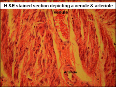
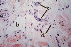
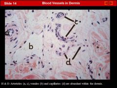
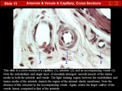
Think about all the blood vessels in the body (large & small... arteries/ veins) What are some features that are similar? What are some traits that are unique to a certain blood vessel? *take time to think about it*
What is one thing they ALL have in common? |
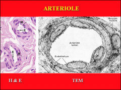
ALL BLOOD VESSELS ARE LINED BY ENDOTHELIAL CELLS. With variety in the layers of connective tissue.
|
|
|
|
In the capillaries, what force is causing the fluid that gets squished out by the HIGH HYDROSTATIC PRESSURE at the ARTERIOLE end of the capillary to get back into the circulatory system at the VENULE end of the capillary?...
Think about it... I know you know the answer... think about the physics **He showed a slide in class about it** |
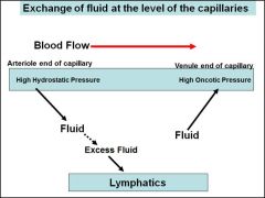
HIGH ONCOTIC PRESSUE in the VENULE end of capillary helps soak up the tissue fluid in area that leaked out. (I blacked out during MCP. something about... albumin... osmotic potential? plasma protiens?... )
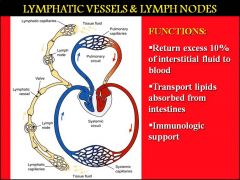
The LYMPH system helps pick up any excess fluid tissue (about 10%) that gets leftover. Pretty efficient little system! |
|
|
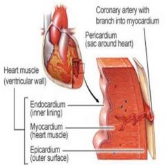
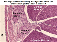
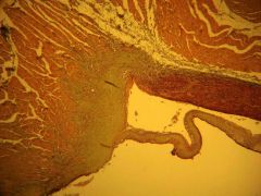
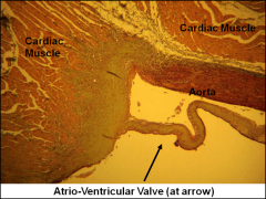
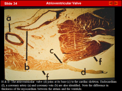
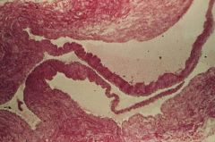
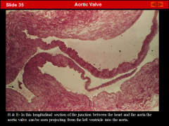
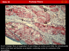
How is the heart almost like a unique modified blood vessel?
*hint: the heart has three layers... what are they homologous to in the blood vessel? |
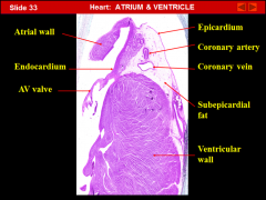
endocardium= tunica intima
myocardium= tunica media epicardium = tunica adventitia |
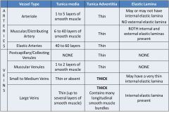
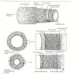
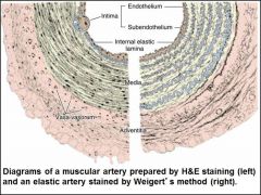
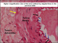
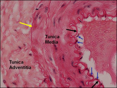
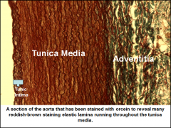
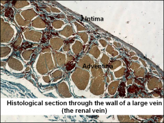
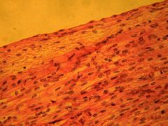
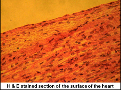
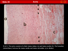
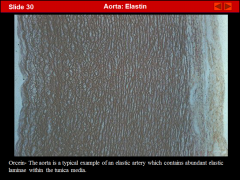
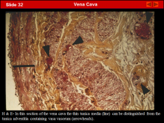
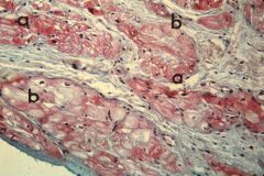
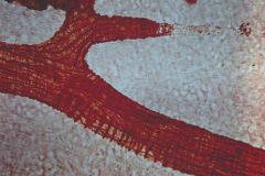
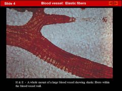
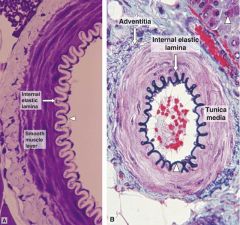
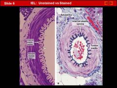
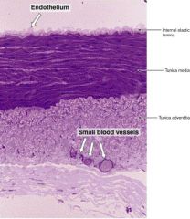
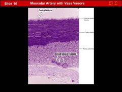
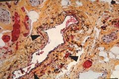
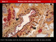
STRUCTURE OF LARGER VESSELS
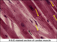
TUNICA INTIMA: Endothelium and subendothelial connective tissue TUNICA MEDIA: Smooth muscle TUNICA ADVENTA: Connective tissue -Vasa vasorum ELASTIC LAMINA: Internal and external. STRUCTURE OF THE HEART ENDOCARDIUM: Lines all INTERNAL l surfaces of the heart, and is continuous with the tunica intima of blood vessels. The endocardium consists of an endothelium and an underlying thin layer of connective tissue. Under the endocardium is a thicker layer of subendocardial connective tissue. MYOCARDIUM: Forms the MAIN MASS of the heart and consists of CARDIAC MUSCLE. The myocardium is thin in the atria, and much thicker in the ventricles. The muscle fibers within the myocardium are attached to the "fibrous skeleton II of the heart.” The fibrous skeleton consists of dense fibrous connective tissue. EPICARDIUM (visceral pericardium): Forms the outermost covering of the heart. a. a Subepicardial region consisting of loose connective tissue, blood vessels and nervous tissue lies under the epicardium. b. Parietal layer of pericardium: Consists of a serosal layer with mesothelial surface facing the epicardium. c. Pericardial cavity: A potential space between the visceral and parietal pericardium which in health contains up to 50 mL. of fluid distributed as a thin film between the apposed mesothelial surfaces. |
|
The LYMPH and CIRCULATORY system are both VASCULAR systems and are VERY similar.
What are some clues/ key differences to help you tell the difference between the two on a slide? |
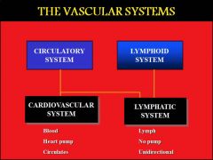
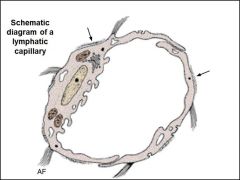
Lymph capillaries have:
* NO RBCs. Only lymphocytes! * THINNER WALLS (only a single layer of endothelium) *BASAL LAMINA is discontinuous or MISSING |
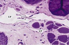
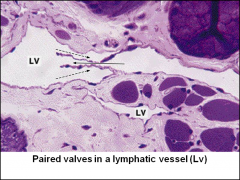
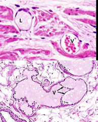
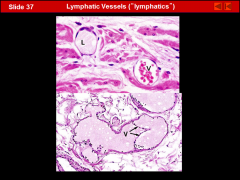
LYMPH VESSELS
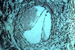
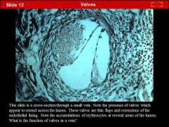
The lymphatic vessels represent a drainage system whereby fluid and plasma protein which has collected in the interstitium is returned to the blood. Lymph capillaries end blindly and the lymph empties into the blood via the thoracic duct and right lymphatic duct. They have the following characteristics: *Lymph capillaries consist only of a single layer of endothelium. *The basal lamina is discontinuous or missing. *No pericytes. *Fibers of connective tissue anchor endothelium to surrounding tissue. The larger lymph vessels have a structure similar to veins: * Many valves * Thinner walls than veins * NO RBCs. Only lymphocytes! |
|
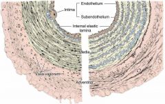
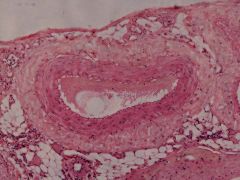
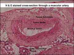
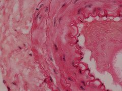
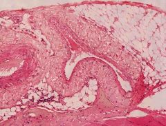
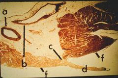
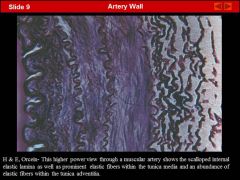
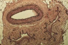
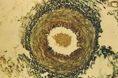
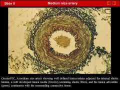
Which one is a MUSCULAR ARTERY? Which one is an ELASTIC ARTERY?
|
|

|
|
|
|
|
|
|
|
|
|

|
|
|
|
|
|
|
|
|

|
|
|
|
|
|
|
|
|
|
|
|
|
|
|

|
|
|
|
|
|
|
|
|
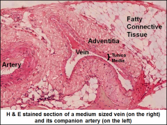
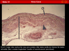
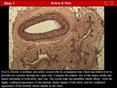
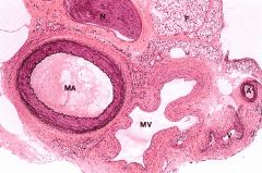
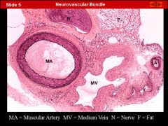
BASIC DIFFERENCES BETWEEN ARTERIES AND VEINS
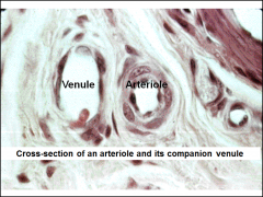
*Usually run along side of each other (i.e. artery has a "companion vein"). *Veins have less muscle, more connective tissue, less elastic tissue, larger lumens, and valves (in the extremities). |
BASIC DIFFERENCES BETWEEN ARTERIES AND VEINS
*Usually run along side of each other (i.e. artery has a "companion vein"). *Veins have less muscle, more connective tissue, less elastic tissue, larger lumens, and valves (in the extremities). |
|
|
|
|
|
|
|
|
|
|
|
|
|
|
|
|
|
|
|
|
|
|
|
|
|
|
|
|
|

|
|
|
|
|
|
|
|
|
|
|
|
|
** Be careful- he mentioned in class this slide might be mislabeled.
|
|
|

|
|
|
|
|
|
|
|
|
|
|
|
|
|
|
|
|
|
|
|
|
|
|
|
|
|
|
|
|
|
|
|
|
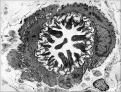
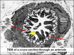
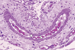
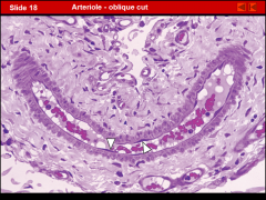
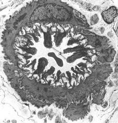
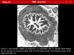
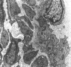
Remember: This is NOT the glomerulus!!
|
|
|
|
|
|
|
|
|
|
|
|
|
|
|
|
|
|
|
|
|
|
|
|
|
|
|
|
|
|
|
|
|
|
|
|
|
|
|
|
|
|
|
|
|
|
|
|
|
|
|
|
|
|
|
|
|
|
|

|
|
|
|
|
|
|
|
|
|
|
|
|
|
|
|
|
|
|
|
|
|
|
|
|
|
|
|
|
|
|
|
|
|
|
|
|
|
|
|
|
|
|
|
|
|
|