![]()
![]()
![]()
Use LEFT and RIGHT arrow keys to navigate between flashcards;
Use UP and DOWN arrow keys to flip the card;
H to show hint;
A reads text to speech;
13 Cards in this Set
- Front
- Back
|
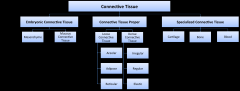
CT chart |
|
|
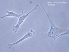
fibroblast - elongated and spindle-shaped (i.e. fusiform, tapered at the ends) |
|
|
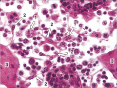
mast cells - large (20-30 µm) oval-shaped cells - round nucleus in the center - cytoplasm is filled with granules containing heparin, histamine, and other factors. - granules stain dark purple with toluidine blue |
|
|
plasma cells - derive from B-lymphocytes and secrete antibodies - "clock face": heterochromatin is positioned in clumps around the periphery of the nucleus |
|
|
macrophage - large (10-30 µm) phagocytic cells - irregularly-shaped with cytoplasmic projections extending from the cell surface - macrophages develop from monocytes, which circulate in the blood |
|
|
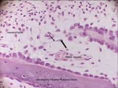
mesenchyme CT - pale tissue that fills most of the image - almost all of the visible cells are mesenchymal cells - no visible fibers in the matrix - pink tissue at the bottom and right of the image are areas of newly formed bone |
|
|
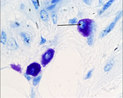
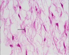
mucous CT - section of an umbilical cord - in addition to mesenchymal cells, there are also fibroblasts (one is indicated by an arrow) which produce collagen fibers - COLLAGEN FIBERS PRESENT |
|
|
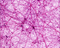
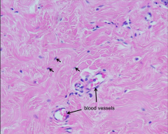
loose areolar CT (has elastic fibers) - image of mesentery (a fold of the peritoneum that attaches organs to the posterior wall of the abdomen) - many cells present - look for elastic fibers (very thin purple lines) and collagen fibers (thicker pink lines) |
|
|
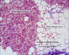
loose adipose CT - shows both brown (multilocular) fat and white (unilocular) fat - collagen fibers in blue (note that there are few collagen fibers in the matrix) |
|
|
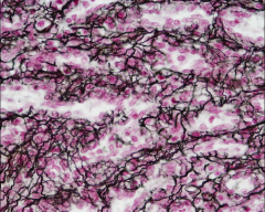
loose reticular CT - has reticular fibers (type III collagen) --> forms a net like mesh - using a silver stain, so the reticular fibers appear black - pink-purple cells scattered throughout are blood cells and lymphocytes - section is from a spleen |
|
|
dense irregular CT - section from the mammary gland - wavy pink material that fills the field is collagen fibers |
|
|
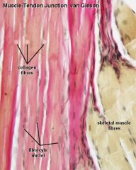
dense regular CT - image of a tendon - collagen fibers are red/pink, nuclei are grey, and cytoplasm is tan/yellow - collagen fibers of the tendon all run parallel to one another - Fibroblasts are visible as thin oval-shaped nuclei squished between bundles of type I collagen |
|
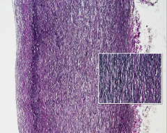
|
dense regular elastic CT - section through the aorta - parallel elastic fibers can be seen in dark purple |