![]()
![]()
![]()
Use LEFT and RIGHT arrow keys to navigate between flashcards;
Use UP and DOWN arrow keys to flip the card;
H to show hint;
A reads text to speech;
8 Cards in this Set
- Front
- Back
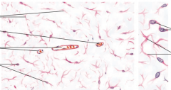
What type of tissue is this? Where can it be found? Identify important structures. |
Embryonal Connective Tissue Umbilical Cord **Identify fibroblasts (dark nuclei looking things that appear oval or cigar shaped) present and scattered bundles of collagen fibers (lighter squiggly lines). The relatively unstained semifluid gel called ground substance. (SLIDE 2) |
|
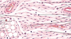
What type of tissue is this? Where can it be found? Identify important structures. |
Loose Areolar CT Mesentary spread **Larger ribbon like structures are small blood vessels or capillaries. The pale staining bundles are collagen fibers. The fine darkly stained individual lines are elastic fibers. Should be able to also identify mast cells (larger granulated cells) and eosinophiles (granulated bi-nucleated cells) as well as fibroblasts (most abundant) (SLIDE 3) |
|
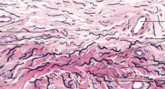
What type of tissue is this? Where can it be found? Identify important structures. |
Dense, Irregularly* Arranged Collagenous CT Skin **Note the dense* irregular* interweave of collagen bundles (located below the epithelial layer of the skin; epidermis) in the dermis. With dense irregular remember More fiber, less cells. (SLIDE 11) |
|
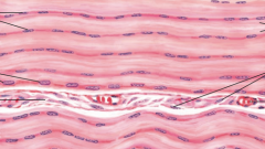
What type of tissue is this? Where can it be found? Identify important structures. |
Dense Regularly Arranged Collagenous CT Tendon **Dense means more fiber, less cells. Note the densely packed bundles of collagen fiber running parallel in an arranged* manner and the pale staining fibroblast nuclei squeezed between the bundles. Tendon found between muscles (SLIDE 4) |
|
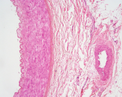
What type of tissue is in the wall of the artery/vein? Identify important structures. |
Elastic Fibers Artery and vein (elastic tissue stain) **the wall of the artery is composed of mainly smooth muscle with occasional elastic fibers (thin, wavy, dark stained). The bundles of elastic fiber (laminae) are present on the inner and outer layer. (SLIDE 28) |
|
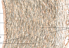
What structure is this? Identify the squiggly black lines. |
Aorta **in the wall of the aorta there are layers of elastic tissue rather than individual fibers. Called elastic laminae* (SLIDE 29) |
|
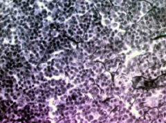
What type of tissue is this? Where can it be found? Identify important structures. |
Reticular Fibers aka Type III Collagen* Lymph Node **The Connective tissue matrix of lymph nodes consists of a delicate network of reticular fibers that stain black (black branchy lines) (DEMONSTRATION SLIDE**) |
|
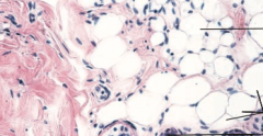
What type of connective tissue is this? |
Adipose Tissue Artery and vein The "bubble-like" tissue containing fat cells. The boundaries are the thin rings and cell nuclei. (SLIDE 28) |