Reading...
![]()
Play button
![]()
Play button
![]()
Use LEFT and RIGHT arrow keys to navigate between flashcards;
Use UP and DOWN arrow keys to flip the card;
H to show hint;
A reads text to speech;
66 Cards in this Set
- Front
- Back
|
vacuum disc
|
air visible in a degenerated disc in osteoarthritis
|
|
|
Classic findings for osteoarthritis on plain film (4)
|
osteophyte formation, joint space narrowing, subchondral cysts, and joint line sclerosis
|
|
|
umbrella osteophyte
|
forms around the entire periphery of the femur or humeral head
|
|
|
intramuscular calcification presenting as calcareous fascial sheets and subcutaneous calcification presenting as linear sheets of calcification
|
poly/dermatomyositis - inflammation leads to atrophy, edema, and calcification in muscle
|
|
|
What causes scapholunate advanced collapse (SLAC)?
|
disruption/damage of the scapholunate ligament - CPPD can cause (pseudogout)
|
|
|
femoral notching sign
|
scalloping of the anterior aspect of the femur by the patella; occurs with loss of the patellofemoral joint space
|
|
|
Causes of chondrocalcinosis (calcification of cartilage)
|
DJD, CPPD (pseudo-gout), Gout, Acromegaly, Hemochromatosis, Hyperparathyroidism, Ochronosis, Wilson’s disease, Oxalosis
|
|
|
Causes of sclerotic bone lesions
|
Sclerotic bone metastases are most frequently due to prostate cancer in males, breast cancer in females, and Hodgkin’s lymphoma
|
|
|
Codman's triangle
|
aggressive form of periosteal reaction; periosteum separates from cortex of bone
|
|
|
What is a periosteal reaction? What causes it?
|
Periosteal reaction occurs in response to acute or chronic insult to cortical long bones and has a long differential
|
|
|
Hair-on-end periosteal reaction
|
Spiculated, perpendicular periosteal reaction, termed “hair-on-end,” as in this patient, is aggressive and highly suggestive of Ewing’s sarcoma (diaphyseal)
|
|
|
Non-common cause of hair-on-end periosteal reaction
|
Chronic osteomyelitis (not Ewing's)
|
|
|
DDx for Hypertrophic osteoarthropathy (thick, fluffy, symmetric periosteal reactions along shafts of multiple long bones, sparing the epiphysis)
|
pulmonary: primary intrathoracic neoplasms e.g. non-small cell lung cancer and mesothelioma, as well as suppurative lung lesions e.g. lung abscess, bronchiectasis, empyema, and cystic fibrosis
Non-pulmonary: biliary cirrhosis, ulcerative cholitis, and Crohn’s disease |
|
|
What is Caffey Disease?
|
a rare inflammatory disease of infancy with soft-tissue swelling and cortical hyperostosis of the mandible and facial bones. It may also involve the scapula, clavicle, ulna, and/or ribs. The pattern of thick periosteal reaction is nonaggressive
|
|
|
Chondroblastoma
|
benign cartilage producing lesions that typically occur in the epiphyses of skeletally immature patients.
-->The lesions are lytic and may have a sclerotic margin; -->laminated AND disorganized periosteal reaction |
|
|
"onionskin" periosteal reaction
|
laminated proliferation of periosteum: sarcomas, osteomyelitis, and chondroblastomas
|
|
|
central nidus with surrounding SOLID periosteal reaction - classic for what?
|
osteoid osteoma, which is a benign bone-forming tumor affecting children and adolescents
|
|
|
solid periosteal reaction with marginal erosions
|
psoriatic or reactive arthritis
|
|
|
sunburst periosteal reaction?
|
osteosarcoma
-->often secondary to Paget's disease |
|
|
increased density, size, and coarsened trabeculae of the entire visualized bone
|
Paget's
|
|
|
Sail sign
|
separation of anterior fat pad from the bone; indicates fluid in the joint space, very suspicious of fracture
-->appearance at all of the posterior fat pad is also indicative of fluid |
|
|
rugger jersey spine
|
alternating sclerotic and lucent bands in the vertebrae; central horizontal stripe of lucency
-->sign of hyperparathyroidism |
|
|
H-shaped vertebrae
|
The central area of the vertebral bodies are not as tall as the periphery of the vertebral bodies
-->sickle cell anemia |
|
|
Triad of renal osteodystrophy
|
diffuse osteosclerosis
Secondary hyperparathyroidism Osteomalacia |
|
|
Where is osteosarcoma, who gets it, what does it look like
|
metaphysis of long bones, esp femur and tibia
15-25 yr olds or >55 with Paget Sclerotic lesion with densities in surrounding soft tissue |
|
|
triad of paget's disease
|
increased bone density, increased bone size, coarsened trabeculae
|
|
|
How do you recognize a super scan on bone scintigraphy
|
The kidneys do not light up but everything else does!
|
|
|
ground glass bone?
|
diffuse - osteomalacia
focal - fibrous lesions |
|
|
Sail sign
|
fat pad separating away from the bone - indicative of fluid in the joint space and highly suspicious of fracture
|
|
|
tumor in the metaphysis?
|
osteosarcoma
|
|
|
tumor in the diaphysis, very young child?
|
Ewing's
|
|
|
tumor in the epiphysis
|
Giant cell
|
|
|
dense matrix with "clouds and clumps"
|
osteoid lesion
|
|
|
dense matrix with rings and C's
|
chondroid
|
|
|
what is the only non-aggressive type of periosteal reaction?
|
solid and continous
|
|
|
What are the terms used to describe the border of a bone lesion (3)?
|
geographic - non-aggressive
moth-eaten permeative |
|
|
What is the first question to ask yourself when faced with a bone lesion?
|
aggressive or non-aggressive?
|
|
|
Colles' fracture?
|
distal radius after FOOSH in older people
|
|
|
H-shaped vertebra
|
sickle cell anemia - due to chronic ischemia; will always see a SCLEROTIC vertebral body
|
|
|
fish-mouth or codfish vertebra
|
compression fractures! Will always see a LUCENT vertebral body
|
|
|
Three possible bone findings in multiple myeloma
|
1. punched out lytic lesions
2. diffuse osteopenia 3. osteosclerosis (very rare - slow growing type) |
|
|
bone scan in multiple myeloma
|
may look normal - osteoblasts have no time to respond and hence are not overactive
|
|
|
Four possibilities if a bone is bent or bowed
|
osteomalacia, osteogenesis imperfecta, Paget's, fibrous dysplasia
|
|
|
Common site of bone resporption in secondary hyperparathyroidism?
|
distal clavicles - wide AC joint
|
|
|
subperiosteal reabsorption of the 2nd and 3rd proximal and middle phalanges on the radial side
|
hyperparathyroidism
|
|
|
what do infarcts look like in bone?
|
sclerotic!
|
|
|
bone tumor with pain at night relieved by NSAIDS
|
osteoid osteoma
|
|
|
Boxer's Fracture
|
4th or 5th metacarpal head
|
|
|
Galeazzi Fracture
|
radial mid diaphyseal fracture AND distal radial ulnar joint dislocation
|
|
|
Monteggia Fracture
|
fracture of ulna and anterior radial head dislocation
|
|
|
Osgood Schlatter DIsease
|
painful tibial tuberosities - due to microavulsions in a growing young person
|
|
|
What is an exostoses?
|
It is a spur like defect in bone at the metaphysis with a cartilage cap; benign; caused by growth plate cartilage misplaced in the metaphysis - danger is nerve compression
|
|
|
Appearance of AVN
|
# Early AVN may present with a crescentic lucency below the cortex.
# Moderate AVN usually shows sclerosis of the head. # Advanced AVN shows inhomogeneous sclerosis and flattening of the femoral head as in this patient. |
|
|
periarticular erosions are common in what type of arthritis?
|
Gout
|
|
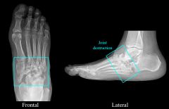
What is this?
|
neuropathic arthritis
|
|
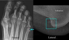
Findings and diagnosis
|
Reactive Arthritis
# There is a marginal erosion of the 5th metatarsal. # There is a calcaneal erosion and adjacent soft tissue calcification (enthesiopathy) |
|
|
What is Maffucci's Syndrome
|
multiple enchondromata
|
|
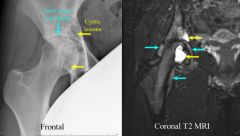
Findings and diagnosis
|
Findings
* The plain film demonstrates cystic areas in the femoral head and the acetabulum with joint space narrowing from secondary osteoarthritis. This is due to the diffuse type of PVNS. * There are T2 bright cystic areas in the femoral head and acetabulum with a joint effusion. Discussion Pigmented Villonodular Synovitis (PVNS) is a synovial proliferative process. Slow growing masses grow in the joint space and can cause erosion of the adjacent bone and pain. PVNS can be diffuse (as in this case) or focal and is most common in the knees, hips, and ankles, but can occur in any joint. |
|
|
Maisoneuvre Fracture
|
Slipping of distal tibio-talal joint; fracture of proximal fibula
|
|
|
Causes of chondrocalcinosis
|
Causes include DJD, CPPD (pseudo-gout), Gout, Acromegaly, Hemochromatosis, Hyperparathyroidism, Ochronosis, Wilson’s disease, Oxalosis
|
|
|
Hair-on-end periosteal reaction
|
Ewing's or osteomyelitis
|
|
|
Three causes of a solid periosteal reaction
|
osteoid osteoma, psoriasis arthritis, reactive arthritis
|
|
|
lytic epiphyseal tumor?
|
giant cell tumor
|
|
|
longitudinal ligament ossification in the spine is a sign of what
|
ankylosing spondylitis
|
|
|
What is DISH? What are the findings?
|
DISH occurs in the cervical and thoracic spine. Patients have back pain, back stiffness, and may have dysphagia. Cardinal features include ossification of the anterior longitudinal ligament bridging at least four vertebral bodies, lucency between anterior vertebral body and anterior longitudinal ligament, preservation of intervertebral joint space, and enthesophytes are frequently seen at tendonous insertions in the pelvis.
|
|
|
Gamekeeper's fracture
|
Gamekeeper's fracture is an avulsion of the ulnar collateral ligament (UCL) of the thumb. It is also called Skier’s Thumb and results from severe hyperabduction.
|